Features of the method
The Goldmann lens is a device consisting of a flat lens and three mirrors, each of which is rotated at a certain angle. These mirrors reflect the picture of the periphery of the fundus of the eyeball. The lens is named after its creator. Its invention occurred in the late 40s of the twentieth century.
Currently, this is the only device that allows us to note the slightest changes in the distant parts of the visual apparatus.
Mirror lenses are called gonioscopic. Each of them has its own name and functions:
| Name | Task |
| Small mirror | Study of the extreme periphery of the retina, as well as the angle of the anterior chamber of the eyes |
| Large mirror | Visualization of the bottom and mid-periphery |
| Middle mirror | Study of the areas of the periphery of the retina located in front of the equator |
The machine is used to examine the back of the eye, or fundus. The device allows you to cover all areas of the visual apparatus. It gives results even with constriction of the pupil.
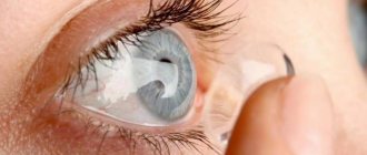
The lens is fixed on the eye, so this diagnostic method is classified as a contact one. Before the study begins, a drug with an anesthetic effect is dripped into the eyes.
The use of such a lens allows timely detection of retinal detachment and dystrophic changes in eye tissue.
Indications for fundus examination
The Goldmann lens is used to assess the condition of the fundus in the presence of the following indications:
- the need to assess the condition of the back of the eyes against the background of chronic ophthalmological diseases;
- diagnosis of common diseases that cause pathological changes (hypertension, diabetes mellitus, pathologies of the central nervous system);
- assessment of the results of surgical, conservative or laser eye treatment;
- a sharp deterioration in vision, pain in the eyes and the appearance of spots, flashing dots in front of them;
- myopia.
Ophthalmologists recommend diagnostic manipulation with a three-mirror lens for the following categories of people:
- elderly patients;
- athletes who engage in sports involving frequent impacts and injuries;
- women who are preparing for the birth of a child;
- patients who have recently injured eye tissue.
The individuals listed above are at risk of retinal detachment. Timely diagnosis of pathology helps preserve visual function.
Healthy people under the age of 40 are recommended to undergo an ophthalmological examination with a three-mirror lens every 2-4 years. At the age of 40 to 54 years - more often, once every 1-3 years. In the future, such manipulation is recommended once every 6–12 months.
The Goldmann lens is used during laser coagulation: it directs the laser beam to the affected area and prevents retinal detachment, which can lead to complete loss of vision.
There are no vital contraindications to manipulation with the Goldmann lens. An exception is patients with an unstable mental state, photophobia, or limited eye mobility.
Goldmann lens examination
Unlike traditional biomicroscopic examinations, examination with a Goldmann lens is aimed at examining the structure of the fundus.
Its advantage is that it allows you to examine it in all details (including peripheral areas) and obtain accurate data. This became possible thanks to the features of the device used - a three-mirror lens that reflects the image of the areas under study in such a way that the ophthalmologist can clearly see them. It is used to identify dystrophies and retinal detachment, as well as when examining patients suffering from refractive errors such as myopia.
Examination of the fundus with a Goldmann lens is carried out in conjunction with other diagnostic studies. An integrated approach allows you to get an accurate picture, make a correct diagnosis, and individually select treatment. Indications for the use of a three-mirror lens may be:
- pain symptoms of unknown etiology, localized in the eye;
- complaints of spots or fog before the eyes;
- traumatic damage to the eyeball.
It is carried out in the process of biomicroscopic studies, which make it possible to determine the condition of the retina, conjunctiva, lens, and anterior chamber. Laser coagulation of the retina is also prescribed. In the process of implementing the latter, the device allows the ophthalmologist to accurately direct the beam to the affected area and prevent detachment of the retina.
The groups of patients who are diagnosed using this ophthalmological device are as follows:
- pregnant women;
- patients whose professional activities involve a high risk of eye injury;
- elderly patients.
The technique has a number of contraindications, which are as follows:
- limited mobility of the eyeball;
- increased sensitivity to light.
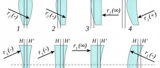
Examination of the eye with a Goldmann lens involves the use of an ophthalmological device, the design of which includes three mirrors. It has a flat shape, and its mirror edges are arranged in a special way: in a circle in increments of 120°, but with different tilt angles (59°, 66° and 73.5°).
It is this feature that allows you to get a high-quality overview of certain areas:
- a small mirror visualizes the edges of the retina and the angle of the anterior chamber;
- middle - all sections of the retina located in front of the equator;
- large - fundus and peripheral areas of the middle.
ProsCons
|
|
The procedure is contact because it requires contact between the lens and the ocular surface. Before performing it, the doctor instills drops into the patient’s eyes that reduce sensitivity. Depending on the purpose, pupil dilators may or may not be used.
Before the procedure, the ophthalmologist must sterilize the device. The process uses a special cleaning gel or hydrogen peroxide. Such measures eliminate the risk of infection of the eye structures. The patient is asked to assume a sitting position, a lens is applied to his eye, and the ocular structures are examined.
Thus, serious ophthalmological diseases are identified:
- Retinal detachment - if left untreated, will lead to loss of vision;
- Macular dystrophy - can provoke loss of central vision;
- Diabetic retinopathy - accompanied by retinal detachment and the development of glaucoma (increased intraocular pressure);
- Macular rupture.
Diagnostics will definitely be effective and highly informative if you undergo it at the Sfera ophthalmology clinic. We have a set of unique equipment and carry out diagnostics in accordance with international standards. Call: +7!
Source: https://www.sfe.ru/info/stati/osmotr-linzoy-goldmana/
Purpose of the procedure
The main goal of the manipulation is to obtain a general picture of the state of the visual apparatus.
An eye examination goes like this: a lens is placed on the cornea of the organ of vision. Thus, the specialist examines the peripheral parts of the eyes in detail. This diagnostic method allows you to detect dystrophy in high myopia, as well as retinal detachment.
Before the study begins, the patient is injected with a drug to dilate the pupil: this provides greater detail. Due to dilated pupils, after examining the fundus, you should not drive or engage in visual work.
Since the device is used for a contact diagnostic method, after each use the specialist must sterilize it to avoid the transfer of infections. To do this, use a mixture of alcohol and ether or hydrogen peroxide.
Advantages and disadvantages
The advantages of a diagnostic examination of the visual organs using a Goldmann lens include the following:
- the ability to identify pathologies of the visual apparatus at an early stage of development and begin timely treatment;
- ensuring a complete study of the organ of vision, including the far corners;
- obtaining a reliable diagnostic result;
- no need to prepare for the event.
Disadvantages of manipulation:
- contact of the lens with the membranes of the eye, which can cause infection if the device is not properly disinfected;
- inability to study the condition of the part of the fundus, which is located between the vascular arcades and the middle periphery.
The high performance of this device is confirmed by ophthalmologists.
Goldmann lens examination technique
To detect diseases at an early stage of development, various devices are used in ophthalmological practice. One of them is the three-mirror Goldmann apparatus.
This is an optical device equipped with three mirrors, which is used to examine the retina and fundus of the eye.
Thanks to this device, the ophthalmologist has the opportunity to thoroughly examine the condition of the visual organ, even if the pupil is constricted.
Three-mirror Goldmann apparatus
Details about the device
To understand how an examination with a Goldmann lens proceeds, it is recommended that you familiarize yourself with the design features of this device.
So, the Goldmann lens, named after the professor who invented this design, is used in ophthalmic practice for:
- Fundus examinations.
- Studying the condition of the internal tissues of the eye.
- To monitor structural changes in the structure of the ocular system.
Goldmann lens
The design of the device consists of three mirrors, each rotated at a certain angle, as shown in the photo. Thanks to this, the doctor has the opportunity to see the entire inner surface of the eye, studying in detail every millimeter of the visual organ. Let us note that in ophthalmological practice there are no other devices that can detect the slightest changes in the far corners of the eyes.
Research with a Goldmann lens is recommended for the following categories of patients:
- Athletes who engage in extreme sports.
- Pregnant women.
- For elderly people.
- Patients who have previously suffered eye injury.
Goldmann lens study
In what cases is it prescribed
Of course, not all patients are prescribed an examination of the visual organ using Goldmann lenses. There must be a basis for this examination.
The doctor may order an examination using a three-mirror lens if the patient complains of pain or significant deterioration in vision.
This examination is also prescribed when the patient experiences goosebumps, severe headaches after stress on the eye system, or after an injury.
Diagnostics
Advantages and disadvantages
The advantages of three-mirror examination of the eye organ include:
- Possibility of a complete study of the eye, including the farthest corners of the organ.
- There is no need for special preparation of the patient for the examination.
- The ability to get quick results and make an accurate diagnosis.
- Possibility of identifying pathologies at early stages of development.
The disadvantages of the technique include:
- Inability to study the retina, which is located between the vascular arcades and the middle periphery of the gas.
- Difficulties in making an accurate diagnosis in patients who have physiological limitations in eye mobility.
- High requirements for disinfection of the device, since this method is contact.
During a fundus examination using a Goldmann apparatus, the ophthalmologist drops pupil-dilating drops into the patient's eyes. After the examination, it is prohibited to drive a car or perform any work that puts stress on the visual organ.
About lens care
An ophthalmological examination with a Goldmann lens, shown in the photo, involves contact of the device with the patient’s visual organ.
Given the tenderness and fragility of this organ, it is important to properly sterilize all medical instruments that come into contact with the eyes. It’s no secret that various pathogenic bacteria can penetrate through the mucous membrane.
To protect patients from contracting any infectious diseases, after each procedure the doctor disinfects the three-mirror device.
A 6% peroxide composition is used for treatment. The outer surfaces are washed with a mixture of 3% peroxide and 0.5% washing gel.
The optical lenses themselves are cleaned using a mixture of 17% ether and 83% alcohol. First, clean the lenses from dust with a grease-free brush.
To avoid damaging the optics, it is important to ensure that they are not exposed to thermal and mechanical influences.
A doctor working with Goldmann lenses should know what is prohibited:
- Keep the device close to heat sources.
- Clean lenses with pure alcohol.
- Rinse off the detergent with cold or warm water.
In custody
Not all patients know what an ophthalmological examination using a Goldmann lens is.
Doctors claim that in order to identify a complete picture of the condition of the patient’s ocular system, an examination with a three-mirror apparatus is necessary.
Thanks to this device, the doctor is able to assess the condition of the eye, even in the farthest corners of the organ. Therefore, when prescribing this type of examination, you should not refuse.
Source: https://ya-viju.ru/metodika-goldmana