Every year in Russia and abroad the frequency of injuries to the maxillofacial region (MFA) increases; Moreover, these injuries are increasingly of a combined nature. The issues of diagnosis and treatment of bone fractures in the midface, as well as the prevention of complications, are still relevant today [1, 6]. Thus, victims with multiple and combined injuries of the facial skeleton make up from 10 to 28% of the total number of patients. In case of injuries to the middle zone of the facial skull, most often (in 20-25% of cases) we have to deal with fractures of the zygomatic bone and zygomatic arch [3, 8]. The largest group of patients are men of working age (from 18 to 40 years), so the problem of maxillofacial injuries is not only medical, but also socio-economic in nature [2, 4].
Surgical interventions on the middle zone of the facial skull involve solving specific problems related to recreating the architectonics of this area by comparing bone fragments, restoring lost functions, etc. During these manipulations, the surgeon, to one degree or another, uses technical techniques aimed at eliminating bone defects in the area of the medial and lower walls of the orbit, the walls of the maxillary sinus (MS), and the zygomaticalveolar ridge. For this, surgeons use auto- and allogeneic materials, synthetic implants (Teflon, tetrafluoroethylene). The use of implants made of titanium nickelide is promising [4, 5, 7, 9].
For the purpose of hemostasis and elimination of air clearance, the cavity of the upper jaw is traditionally filled with a gauze swab, which leads to a delay in the outflow of discharge and in 14.3% of cases - to the development of traumatic sinusitis [5, 9]. In addition, it is possible to develop neuralgia of the second branch of the trigeminal nerve, anesthesia of the mucous membrane (MS) of the mouth and teeth (usually disappears completely after 1-3 months); with extensive periosteal injuries and hematomas, there is a risk of developing abscesses and phlegmon in nearby cellular spaces. At the same time, the process of removing a gauze swab from the sinus is quite traumatic, and cases of repeated bleeding are not uncommon. Some authors prefer to install a silicone tubular drainage or sinus balloon into the formed anastomosis with the lower nasal meatus. In the specialized literature there are isolated reports that tamponade of the maxillary sinus is not at all advisable.
The purpose of the study was to increase the effectiveness of surgical treatment of patients with traumatic injuries to the mid-facial skull using a Foley catheter (Fig. 1, 2).
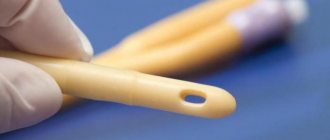
Rice. 1. Schematic illustration of a Foley catheter. 1 - main channel; 2 - small channel; 3 - balloon.
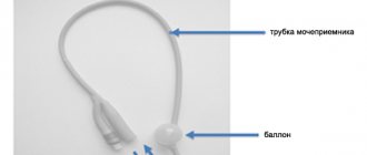
Rice. 2. General appearance and color marking of Foley catheters.
Material and methods
We have summarized the experience of using a Foley catheter in the treatment of patients with injuries and deformities of the middle zone of the facial skull. In 1990-1991 we used a Foley catheter in the maxillofacial surgery clinic of the Novokuznetsk Institute, and in 2007 we began using it in the maxillofacial surgery clinic of the First Moscow State Medical University named after. THEM. Sechenov. During this period, 352 victims with fractures of the middle zone of the facial skull were treated. The age of the patients ranged from 18 to 66 years, there were 314 men, 38 women (ratio 8.26:1). The distribution of patients by age is presented in Table. 1, depending on the diagnosis - in table. 2. Diagnosis of structural damage was carried out on the basis of a clinical examination, X-ray data in direct and semi-axial projections, computed tomography (CT) of the facial skeleton, consultations with doctors of related specialties (neurologist, ophthalmologist, otorhinolaryngologist). 184 (52.27%) patients were admitted to the clinic 1-3 days after the injury, 93 (26.42%) - after 4-7 days, 41 (11.64%) - after 8-14 days, 16 (4 .56%) - after 15-21 days, 18 (5.11%) - after 22 days and later.
Table 1. Distribution of patients by age
Table 2. Distribution of patients into groups depending on diagnosis
Upon admission, taking into account the timing from the moment of injury and the nature of the fracture, 36 (10.23%) patients underwent closed reduction of the zygomatic bone using a Limberg hook, the remaining 316 (89.77%) underwent open reduction and osteosynthesis with external titanium mini-plates , mini-braces made of shape memory alloys, or a combination of these techniques. Simultaneous reconstruction of the walls of the upper jaw, including the floor of the orbit, was performed in 107 victims. A Foley catheter was used in all 316 patients.
Operation technique
Under conditions of endotracheal anesthesia (orotracheal intubation), a skin incision of 10-15 mm in length was made in the projection of the zygomaticofrontal joint, 10-15 mm long, and skeletonized bone fragments. According to indications, the 2nd stage was a subciliary approach. The soft tissues were bluntly and sharply penetrated, the lower orbital margin and the lower wall of the orbit were exposed, and the latter was inspected under visual control. In cases of entrapment in the fracture line of the infraorbital nerve and motor muscles of the eyeball, decompression was performed. Next, from the side of the oral cavity, an incision was made in the mucous membrane along the transitional fold from the canine to the second molar. The anterolateral wall of the upper jaw and the zygomaticalveolar ridge were skeletonized, the fracture line was visualized, and the degree of displacement of the zygomatic bone was determined. Then the upper jaw was sanitized, abundantly irrigated with antiseptic solutions, small loose bone fragments, blood clots, foreign bodies, as well as pathologically altered SO were removed. From the side of the sinus, an inspection of the lower wall of the orbit was carried out. The next stage, under visual and manual control, was the reposition of the zygomatic bone, after which metal osteosynthesis was performed: in the area of the zygomaticofrontal suture and the lower edge of the orbit, we consider it possible to use both microplates and U-shaped mini-brackets made of titanium nickelide, in the area of the zygomaticalveolar ridge — titanium mini-plates or the above-mentioned brackets. Depending on the expected timing of installation of the Foley catheter into the cavity of the upper nasal cavity, its end was brought out through the vestibule of the oral cavity, or through the superimposed nasoantral anastomosis into the lower nasal meatus. In order to eliminate the bone tissue defect in the area of the anterolateral wall of the upper quadrant, replantation of bone fragments or endoprosthetics with mesh implants made of titanium nickelide was performed. If there were 1-3 large bone fragments, they were replanted and fixed with titanium microplates. If the thickness of the replanted fragments was small, they were fixed with non-resorbable suture material. Next, a Foley catheter was inserted into the sinus cavity; depending on the pneumatization of the sinus, the balloon was filled with physiological solution to the required volume (10-15 ml). Layer-by-layer suturing of surgical wounds was performed. It is better to close the wound along the transitional fold with interrupted sutures; in this case, a provisional U-shaped suture was placed at the site of the direct exit of the rubber tube of the catheter, which was tied after removing the catheter.
In cases where isolated fractures and the lower wall of the orbit were detected, surgical intervention was performed through the subciliary approach. An inspection of the orbital floor was performed, the upper jaw was sanitized according to indications, and endoprosthesis replacement was performed with porous or mesh implants made of titanium nickelide.
Clinical observation No. 1.
Patient R.
, 31 years old, was admitted to the clinic on December 11, 2012 with a diagnosis of a displaced fracture of the zygomatic-orbital complex on the left; fracture of the anterior and lateral walls of the left upper quadrant; fracture of the lower wall of the orbit; hemosinus; contusion of the left eyeball. Complaints: asymmetry of the left half of the face, numbness of the teeth on the left upper jaw. From the medical history: the injury was caused by a fall from the stairs 2 days before presentation. On 12/13/12 an operation was performed: reposition, metalosteosynthesis of the zygomaticoorbital complex on the left; rehabilitation of high-pressure joints; endoprosthesis replacement of the lower wall of the orbit with a mesh implant made of titanium nickelide. Fragments in the area of the zygomaticofrontal suture were fixed with mini-brackets made of titanium nickelide. Fixation in the area of the lower edge of the orbit is performed using a titanium micro-plate. The Foley catheter is removed from the oral cavity. The postoperative period was calm. The catheter was removed on the 2nd day. Sutures on the skin were removed on the 5th day, in the oral cavity - on the 10th day (Fig. 3-8).
Rice. 3. Patient R. Appearance upon admission.
Rice. 4. Custom mesh implant made of titanium nickelide mesh.
Rice. 5. Patient R. Computed tomography before surgery. a — three-dimensional reconstruction; b - axial section.
Rice. 6. Patient R. View of the postoperative wound in the area of the transitional fold.
Rice. 7. Patient R. Appearance at the time of discharge.
Rice.
8. Patient R. Control CT. a - three-dimensional reconstruction; b - axial section. Clinical observation No. 2.
Patient N.
, 27 years old, was admitted to the clinic on September 15, 2013 with a diagnosis of a displaced fracture of the zygomatic-orbital complex on the left; fracture of the lower wall of the orbit on the left; hemosinus on the left; paraorbital hematoma on the left; multiple abrasions on the left side of the face. Complaints: swelling and numbness of the skin in the left infraorbital region and upper lip, limited mouth opening. From the anamnesis: the injury was received as a result of an attack by unknown persons 2 days before hospitalization. On September 20, 2013, an operation was performed: reposition, metal osteosynthesis of the zygomaticoorbital complex on the left; sanitation of the left upper quadrant; endoprosthetics of the lower wall of the orbit on the left. Fragments in the area of the zygomaticofrontal suture and the lower edge of the orbit were fixed with mini-brackets made of titanium nickelide, in the area of the zygomaticalveolar ridge - with an L-shaped titanium mini-plate. The bone defect in the area of the lower wall of the orbit was corrected using a mesh implant made of titanium nickelide. The Foley catheter is withdrawn through the nasoantral anastomosis into the lower nasal meatus. The postoperative period was calm. The catheter was removed on the 4th day. Sutures on the skin were removed on the 5th day, in the oral cavity - on the 10th day (Fig. 9-12).
Rice. 9. Patient T. Appearance upon admission.
Rice. 10. Patient T. CT scan before surgery. a — three-dimensional reconstruction; b — frontal section.
Rice. 11. Patient T. Appearance 3 months after surgery.
Rice. 12. Patient T. Control CT. a - three-dimensional reconstruction; b — frontal section.
Rectal catheters
Rectal catheters, intended for gas removal, irrigation and lavage of the rectum with medications
Specifications
The catheter is an elastic transparent tube; the entry end is plugged, there are two oval holes on the side surface Catheter material – medical PVC plastic compound brand PM – 1/42 TU 6-05-1533-85 Catheter length – 500-1000 mm Catheters are produced in the following numbers (according to the Charrière scale): No. 18 ; 24; 27; thirty; 33; 36 with outer diameters respectively: 6.0; 8.0; 9.0; 10.0; 11.0; 12 mm
Results and discussion
The results of surgical treatment were assessed based on the patient’s complaints, external and intraoral examination, radiological diagnostic data (radiography in semi-axial and direct projections, CT), and repeated consultations with a neurologist and ophthalmologist were prescribed. In some cases, endoscopic examination of the paranasal sinuses was performed. Long-term follow-up periods ranged from 3 months to 6 years.
The decision on how to install a Foley catheter is often made intraoperatively, since it is not always possible to accurately determine the extent of the proposed reconstruction at the surgical planning stage. When the fracture is not accompanied by the formation of a large number of fragments (the formation of extensive bone defects cannot be expected in the future) to be replaced with implants, we consider it rational to install a Foley catheter into the cavity of the upper jaw for a short period of time (1-3 days) and bring its end into the oral cavity. Concomitant damage to the nasal bones, as well as severe deformation of the nasal septum, lead to certain difficulties when passing the Foley catheter through the nasoantral anastomosis, so its end should also be removed through the oral cavity. The withdrawn part of the Foley catheter was fixed with adhesive tape in the area of the lateral surface of the neck and the supraclavicular fossa. If a defect in the bone of the lower wall of the orbit or the walls of the upper jaw is detected, additional reconstructive techniques are used, for example, endoprosthetics using titanium nickelide implants, as well as replantation of bone fragments. Since the catheter must be held for a longer period (4-7 days), its end is removed through the nasoantral anastomosis into the lower nasal passage (Fig. 13). In addition, there are often cases of previously developed odontogenic or rhinogenic sinusitis, when it is necessary to perform a radical maxillary sinusotomy. We used two-way latex Foley catheters in sizes 12, 14, 16, 18 on the Charrière scale (Ch/Fr), since these catheters are optimal in their characteristics, easy to use, and easily and painlessly removed from the sinus.
Rice. 13. Methods of drainage of the upper jaw using a Foley catheter.
The Foley catheter installed in the cavity of the upper sinus, in addition to the drainage function, also performs a number of others, namely: it additionally supports the bone fragments of the sinus walls, held by the periosteum, preventing them from re-displacing into the sinus lumen. In addition, a Foley catheter was used during surgery in cases of extensive damage to the lateral wall of the upper quadrant in combination with prolapse of the Bichat fat pad into the lumen of the latter. During endoprosthetics, a filled balloon covers the inner surface of the implant; at the same time, it is isolated from the influence of environmental factors, which in turn reduces the risk of infection and the development of complications.
In most cases (258 observations), we obtained good and satisfactory results. Complications were recorded in 94 (26.9%) patients; most often they were diagnosed with neuralgia of the infraorbital nerve - in 46 (13.1%), contouring/cutting of the plate - in 23 (6.6%), dehiscence of sutures in the oral cavity - in 14 (4%), cicatricial deformation of the lower eyelid - in 7 (2%), persistent facial deformation in 4 (1.1%). There were no complications associated with the development of any inflammatory process.
Thus, the scope of the operation directly depends on the degree of displacement of bone fragments, the nature of the fracture, dysfunction of the eyeball, as well as the severity of pathological changes in the paranasal sinuses. A severe degree of damage to brain structures, severe impairment of the vital functions of organs and systems, as well as the presence of general somatic diseases affect the timing of surgical intervention. The use of fixing devices made from shape memory materials and their combination with titanium plates is effective when performing reconstructive operations on the middle zone of the facial skull. An important condition that increases the effectiveness of surgical treatment is the reconstruction of damaged anatomical structures to the fullest extent. Prevention of the development of traumatic sinusitis and restoration of nasal breathing function are mandatory. A multidisciplinary approach to diagnosing such patients and their treatment, in our opinion, is the key to the earliest and most complete rehabilitation, which is in line with modern trends.
Nasal catheters for secretory mucus suction
Nasal catheters for suction of mucus secretory are designed for suction of secretions from the nasopharynx
Specifications
The catheter is an elastic transparent tube, the entry part has one end and two through holes, the proximal end has a socket with a plug for connecting an adapter (t-shaped) Catheter material - medical PVC plastic compound grade PM - 1/42 TU 6-05-1533- 85 Adapter material medical grade polyethylene according to GOST 16338 - 85 Catheter length - 450 mm, adapter - 80 mm Catheters are produced in the following numbers (according to the Charrière scale): No. 6; 8;10; 12; 14; 15; 18 with outer diameters respectively: 2.00; 2.66; 3.33; 4.0; 4.67; 5.0; 6.0 mm
Urethral catheters for children
Urethral catheters for children are intended for emptying and washing the bladder in children.
Specifications
The catheter is an elastic transparent tube; the input end is plugged, there is one hole on the side surface; the proximal end has a socket (or expanded) for a syringe Material of the catheter – medical PVC plastic compound brand PM – 1/42 TU 6-05-1533-85 Catheter length – 360 mm Catheters are produced in the following numbers (according to the Charrière scale): No. 4.5 ,6,7,8,9,10,11 with outer diameters, respectively: 1.33; 1.66; 2.0; 2.33; 2.66; 3.0; 3.33; 3.66 mm
Nasopharyngeal secretory catheters
Nasopharyngeal secretory catheters are designed for suctioning mucus from the patient’s nasopharynx and bronchi through the nose.
Specifications
The catheter is an elastic transparent tube; the input end is plugged, there is one hole on the side surface; the proximal end has a socket for connecting an adapter (t-shaped) Catheter material - medical PVC plastic compound grade PM - 1/42 TU 6-05-1533-85 Adapter material medical grade polyethylene in accordance with GOST 16338-85 Catheter length - 460 mm Catheters are produced as follows numbers (according to the Charrière scale): No. 6, 8, 10, 11,12 with outer diameters, respectively: 2.0; 2.67; 3.33; 3.67; 4.00 mm
Urethral catheters for men
Urethral catheters for men are intended for emptying and washing the bladder
Specifications
The catheter is an elastic transparent tube; the input end is plugged, there is one hole on the side surface; the proximal end has a bell (or expanded) for a syringe Material of the catheter - medical PVC plastic compound brand PM - 1/42 TU 6-05-1533-85 Catheter length - 360 mm Catheters are produced in the following numbers (according to the Charrière scale): No. 12; 13 ; 14; 15; 16; 17; 18; 19; 20 with outer diameters respectively: 4.0; 4.33; 4.66; 5.00; 5.33; 5.66; 6.00;6.33; 6.66 mm
Gastric catheters for feeding children
Gastric catheters for children are intended for artificial feeding of premature, sick and postoperative children.
Specifications
The catheter is an elastic transparent tube; the input end is plugged, there is one hole on the side surface; at the proximal end there is a socket with a plug. Catheter material is medical PVC plastic compound brand PM - 1/42 TU 6-05-1533-85 Catheter length - 360-500 mm Catheters are produced in the following numbers (according to the Charrière scale): No. 4; 5; 6; 8;10;12 with outer diameters, respectively: 1.33; 1.67; 2.0; 2.67; 3.33; 4.0 mm Catheters are supplied complete and in separate numbers
Nasopharyngeal oxygen catheters
Nasopharyngeal oxygen catheters are designed for long-term and short-term oxygen supply
Specifications
The catheter is an elastic transparent tube; the input end is plugged, there is one hole on the side surface; the proximal end has a socket for connecting an adapter (straight) Catheter material - medical PVC plastic compound brand PM - 1/42 TU 6-05-1533-85 Adapter material medical grade polyethylene according to GOST 16338-85 Catheter length - 460 mm Catheters are produced in the following numbers ( on the Charrière scale): No. 6, 8, 10, 11,12 with outer diameters, respectively: 2.0; 2.67; 3.33; 3.67; 4.00 mm
Gastrointestinal catheters, 2-channel
Double-channel gastrointestinal catheters are designed for emptying and washing the digestive tract
Specifications
The catheter is a 2-channel (double-lumen) elastic and transparent tube; in the inlet end part there are holes and a number of holes in one of the channels (for irrigation of surrounding tissues), and at the proximal end of the same channel there is a cut, for the possibility of connecting another channel to the suction tip. Catheter material - medical PVC plastic compound, grade PM - 1/42 TU 6 -05-1533-85 Catheter length – 2000 mm Catheters are produced in the following numbers (according to the Charrière scale): No. 21; 24;27;30;33 with outer diameters respectively 7.0; 8.0; 9.0; 10.0; 11.0 mm