Meniere's disease is a disease associated with an increase in the amount of fluid (endolymph) in the inner ear.
Author:
- Filatova Evgenia Vladimirovna
otolaryngologist, rhinosurgeon
(Voted by: )
Meniere's disease is a disease associated with an increase in the amount of fluid (endolymph) in the inner ear. For the first time, a set of symptoms described in 1861 by the French physician Prosper Meniere, later received the name Meniere's disease.
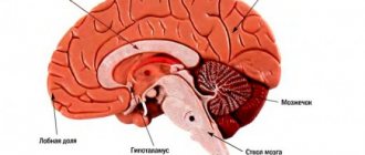
Anatomy of the inner ear
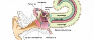
The inner ear is located inside the canals and cavities of the temporal bone, which are called the bony labyrinth. Inside the bone labyrinth there is a membranous labyrinth that follows its contours and dimensions.
The space between the membranous and bony labyrinth is filled with fluid - perilymph, which is similar in composition to cerebrospinal fluid. The space inside the membranous labyrinth is filled with endolymph, and it is completely isolated by its walls.
There are three parts of the inner ear that are different in structure and function - the vestibule, cochlea and semicircular canals. The cochlea is an organ of sound perception. It is here that the sensitive hair cells that give rise to the auditory nerve are located. The vestibule and semicircular canals are the organ of balance.
What causes the symptoms of Meniere's disease?
The appearance of symptoms is associated with an increase in the amount of endolymph. Endolymph, increasing in volume, stretches the walls of the membranous labyrinth. This condition is called endolymphatic hydrops or hydrops of the labyrinth. Since the membranous labyrinth has many sensitive receptor zones in all three sections, their irritation leads to the appearance of symptoms of the disease.
Meniere's disease is currently not fully understood, so there are several theories about where the excess amount of endolymph comes from:
- penetration of fluid from blood plasma through the vascular wall of capillaries;
- penetration of fluid from the perilymph through the wall of the membranous labyrinth;
- disruption of the mechanism of production and absorption (absorption) of endolymph;
- accumulation of ions and substances with high molecular weight in the endolymph, which leads to an increase in osmotic pressure;
- insufficient perilymph volume.
Symptoms
Meniere's disease is characterized by attacks with the following symptoms:
- dizziness;
- noise in ears;
- hearing loss.
This is a mandatory triad. In the absence of one of the symptoms, a careful differential diagnosis is needed, since dizziness, tinnitus, and hearing loss occur in many other diseases. In most cases, tinnitus and hearing loss are unilateral, that is, the pathological process occurs in only one ear.
Dizziness is quite intense, manifested in the form of rotation of the body or surrounding objects; this type of dizziness is called systemic. Often during an attack there is nausea and vomiting. Non-systemic dizziness (staggering when walking, darkening of the eyes, flashing “spots” before the eyes) is not typical for the typical picture of Meniere’s disease and can only occur in its final stage.
Also, at the time of an attack, there may be a feeling of fullness, bloating, or stuffiness in the affected ear.
There can be a different number of attacks - from once every few months to daily, which determines the severity of the disease.
Clinically, the stages of the disease are distinguished:
- Stage 1 (initial). Attacks of dizziness occur suddenly or after stress. Before an attack, there may be warning signs, for example, congestion in the affected ear. During an attack, noise in the ear appears, hearing decreases, after the end of the attack, hearing is restored to normal values within a few days. Outside of attacks, he feels good, there is no tinnitus.
- Stage 2 (pronounced clinical manifestations). Attacks of dizziness become more frequent and intensified, accompanied by nausea and vomiting. Tinnitus is also present outside of attacks. Hearing also steadily declines. There may be constant pressing headaches, a feeling of stuffiness, fullness in the sore ear.
- Stage 3 (final). At this stage, examination does not reveal typical signs of labyrinthine hydrocele. Irreversible death of receptor cells in both the auditory and vestibular parts of the inner ear occurs. Therefore, attacks of dizziness weaken - they become more rare and less pronounced, of a non-systemic nature - unsteadiness of gait, swaying when walking. Hearing in the affected ear is significantly and permanently reduced.
Diseases of the ENT organs
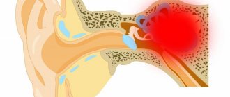
Pulsation in the ear can occur with diseases of the ENT organs:
- Any part of the ear can become inflamed, as well as the Eustachian tube connecting it to the nasopharynx. Pathological changes in the auditory analyzer distort sound waves and reduce their perception. With otitis, the outflow of serous-purulent exudate is disrupted, an “echo effect” is created, internal noise increases, and blood pulsates. The patient has throbbing in the left ear due to left-sided inflammation of the auditory analyzer. If otosclerosis develops, in which the mobility of the auditory ossicles in the middle ear is impaired, then patients experience decreased hearing, dizziness and tinnitus.
- Labyrinthitis is an inflammation of the inner ear caused by the penetration of infection from chronic foci in the body. The main manifestations of labyrinthitis are vestibular disorders. In patients, coordination of movements is impaired, everything floats before the eyes, ataxia, nystagmus, and autonomic disorders occur: pallor, hyperhidrosis and dizziness. Irritation and death of sound receptors leads to pulsation in the ears, reversible hearing loss and even deafness.
- Hypersecretion of earwax often results in the formation of cerumen plugs, which must be removed promptly. Earwax blocks the ear canals, which causes hearing loss, inflammation and the appearance of pathological pulsation.
Diagnostics
Routine examination of the outer ear and eardrum does not reveal any changes. To establish a diagnosis of Meniere's disease, a comprehensive examination of the inner ear is necessary. To do this, tests with tuning forks, a study of hearing thresholds using an audiogram, vestibular tests, X-ray examination of the temporal bones, CT and MRI of the head, and Doppler ultrasound of the great vessels of the head and neck are carried out. Consultations with a neurologist, otoneurologist, otosurgeon, ophthalmologist, therapist, and endocrinologist are necessary.
But, as already mentioned, at the initial stage of the disease, outside of an attack, all indicators may be within normal limits, and clinical symptoms resemble those of other diseases. To establish an accurate diagnosis, it is necessary to identify and confirm hydrocele of the labyrinth. Currently, there are two reliable ways to diagnose labyrinthine hydrops - electrocochleography and dehydration tests.
Method of dehydration tests. Initially, a hearing test is performed - an audiogram. Then the patient takes a solution of glycerol with lemon juice orally. The amount of glycerol is calculated based on the patient’s body weight. Then the audiogram is taken again 1, 2, 3, 24 and 48 hours after taking the solution. The test is considered positive if, after 2-3 hours, hearing improves by 5 dB over the entire frequency range or by 10 dB at three of the tested frequencies, and speech intelligibility improves by at least 12% of the original. The test is considered negative if after 2-3 hours the hearing decreases and speech intelligibility deteriorates. Other options are regarded as dubious.
Electrocochleography technique. Electrocochleography is the recording of electrical activity of the cochlea and auditory nerve after a sound stimulus. A sound such as clicks is sent to the ear being examined at a given interval, the device’s sensor amplifies the amplitude of the electrical signal arising in the sound-receiving hair cells of the cochlea, resulting in a graph resembling an ECG. Dropsy of the labyrinth has certain signs in this graph.
Overwork
In healthy people, pulsating tinnitus is a sign of fatigue, exhaustion of the nervous system and chronic stress. At the end of a busy day of work, spent in a noisy and uncomfortable environment, your ears begin to throb. Intrusive noise at night prevents a person from falling asleep, relaxing and resting. Even basic sounds: the ticking of a clock, raindrops, breathing begin to irritate and seem loud. In this state, the movement of blood through the vessels is perceived as a pulsating noise. Tired people listen to everything, become depressed and begin to invent non-existent illnesses. If drug therapy does not help to cope with the problem, the help of a psychotherapist is necessary.
What other diseases may resemble Meniere's disease?
Tinnitus, dizziness and hearing loss are not exclusive symptoms of Meniere's disease, therefore a comprehensive examination of the patient using the above methods is necessary to exclude other pathologies with similar manifestations: acute cerebrovascular accident, vertebrobasilar insufficiency, benign paroxysmal positional vertigo, brain tumors brain, skull trauma, labyrinthine fistula, inflammation of the vestibular nerve, multiple sclerosis, purulent complications of acute or chronic otitis media, psychogenic dizziness.
Acute otitis media
According to statistics, acute forms of otitis media account for 30% of the total number of ENT diseases. Most often it occurs in preschool children.
Symptoms of acute otitis media
The disease is characterized by an acute onset with the appearance of the following symptoms:
- earache;
- ear congestion or hearing loss;
- increased body temperature;
- anxiety;
- disturbance of appetite, sleep;
- headache and toothache.
Causes of development of acute otitis media
In most cases, the disease can be caused by various pathogenic microorganisms - viruses, microbes, fungi, etc. In exudate obtained from the middle ear, respiratory viruses are found in 30-50% of cases. The most common causes of otitis are parainfluenza viruses , influenza viruses, rhinoviruses, adenoviruses, enteroviruses, respiratory syncytial viruses, etc.
In 50-70% of patients with acute otitis media, bacteria are detected in the exudate from the middle ear (most often Streptococcus pneumoniae, Haemophilus influenzae, Moraxella catarrhalis).
Often the cause of otitis is a mixed (viral-bacterial) infection.
When making a diagnosis, a differential diagnosis is made with myringitis (inflammation of the eardrum) and exudative otitis media.
The occurrence of otitis media is directly related to the condition of the nose and nasopharynx: rhinitis and tonsillitis often provoke inflammation of the middle ear.
Otitis often occurs against the background of decreased immunity and immunodeficiency states.
1 Diagnosis of otitis in MedicCity
2 Diagnosis of otitis in MedicCity
3 Diagnosis of otitis in MedicCity
Routes of infection
The most common route of infection into the middle ear is through the auditory tube during rhinitis and sinusitis.
It is possible that infection can penetrate through the blood during influenza, scarlet fever and other infectious diseases.
In rare cases, the infection enters the middle ear through the ear canal due to injury (rupture) of the eardrum.
Stages of acute otitis
There are 5 stages of the disease:
- stage of acute eustachitis: feeling of congestion, noise in the ear, normal body temperature (if there is an infection, it may increase);
- stage of acute catarrhal inflammation in the middle ear: sharp pain in the ear, low-grade fever, inflammation of the mucous membrane of the middle ear, increasing noise and congestion in the ear;
- pre-perforative stage of acute purulent inflammation in the middle ear: sharp unbearable pain in the ear, which radiates to the eye, teeth, neck, pharynx, increased noise in the ear and decreased hearing, increased body temperature to 38-39 degrees, the blood picture becomes inflammatory in nature;
- post-perforation stage of acute purulent inflammation in the middle ear: pain in the ear becomes weaker, suppuration appears from the ear, noise in the ear and hearing loss do not go away, body temperature becomes normal;
- reparative stage : inflammation is stopped, perforation is closed with a scar.