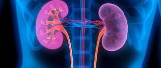
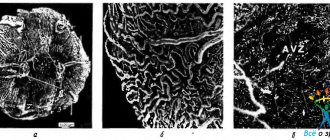
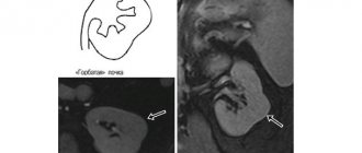
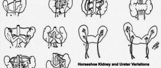
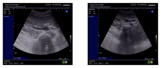
Duplex scanning of the renal arteries is prescribed for diagnosing the organs of the genitourinary system and the kidneys themselves. This study is non-invasive, gentle and painless, is highly effective and is classified as technically difficult. Based on the results of the study, the doctor will be able to assess the patient’s renal structure and choose the optimal treatment tactics.
What does it show
The scanning technique allows specialists to assess the specifics of the patient:
- patency of blood vessels;
- condition of the walls of blood vessels;
- blood movement inside the veins;
- functioning of the valve system.
In this way the following are diagnosed:
- anomalies of the renal veins and arteries;
- thrombosis and stenosis;
- renal failure;
- presence of neoplasms;
- almost any pathology related to the kidneys.
How the research is carried out
Doppler ultrasound in combination with conventional ultrasound of the kidneys makes it possible to conduct a detailed examination of this organ and explain the causes of disturbances in its activity.
To undergo ultrasound scanning, the patient is placed on a couch. The sensor is coated with gel and applied first to the front surface of the abdominal wall, then to one and the other side in the abdominal area, and finally, the kidneys are examined from the back.
In addition to the image received on the monitor, during the session the equipment produces various sounds: calm ones indicate that the blood flow is free, sharp ones indicate that it is impeded.
| No. | Service name | Price , rub. |
| 1 | Renal vessels and abdominal aorta (duplex scanning of veins and arteries). | 1 800 |
| 2 | Triplex scanning of the abdominal aorta and inferior vena cava | 1 500 |
Indications for duplex scanning of the renal arteries
This study is prescribed if the patient:
- suffers from kidney diseases (acute, chronic specific, nonspecific inflammatory);
- complains of pain in the lumbar region;
- often experiences increased blood pressure;
- found blood in the urine;
- received results of clinical urine tests that differ significantly from the results of the previous test;
- received an appointment for a clinical examination from your medical specialist;
- preparing for surgery;
- previously suffered from cancer.
Important! For pregnant women, this study is prescribed for late toxicosis.
Indications for Doppler ultrasound
Ultrasound ultrasound is required for:
- suspected renal failure;
- detection of tumors in the kidneys or adrenal glands on ultrasound;
- changes in urine tests (with an increase in the level of red blood cells and white blood cells);
- late toxicosis in pregnant women;
- diabetes mellitus;
- chronic kidney diseases (nephroptosis, pyelonephritis);
- before surgical interventions on the kidneys and adrenal glands.
The procedure may be necessary to study in more detail the cause of the following symptoms:
- persistent increase in blood pressure;
- urinary disorders;
- swelling;
- lower back pain;
- the presence of blood in the urine.
Triplex and duplex scanning are quite informative methods.
However, after studying the results obtained during them, to clarify the diagnosis, experienced medical doctors may decide on further examination, in particular, MRI or CT, which provide the opportunity to study the smallest vessels. Ultrasound scanning of the kidneys has no contraindications, but it should be postponed if there are injuries in the abdominal area (for example, after abdominal surgery) or rashes.
Preparing for Duplex Scanning
High-quality visualization of the renal arteries is only possible if the patient's intestines are not full of gases. In order to minimize flatulence, three days before the procedure, the patient should refuse:
- milk;
- juices;
- carbonated drinks;
- legumes;
- black bread;
- cakes;
- sauerkraut;
- raw fruits and vegetables.
If the patient is characterized by increased gas formation, it is recommended to supplement the diet with enterosorbents (2 tablets of espumizan or activated carbon three times a day).
Important! On the morning of the scan, the patient should refrain from taking any food or drinks, including plain water.
Observation after treatment
Call your doctor if you have:
- Fever,
- Breathing problems
- Rash,
- The area where the catheter was inserted becomes red, swollen and painful, or oozes blood.
- Pain in the abdomen or side on the side of the treated kidney.
- Very low pressure.
After renal artery stenting, you should avoid vigorous physical activity for at least 24 hours and drink plenty of fluids to wash out the contrast dye.
You can perform light activities the next day, but should avoid heavy physical labor or strenuous activities for five days. Stenting of the renal arteries is a modern high-tech method for the treatment of renovascular hypertension and is significantly superior in safety to open interventions on these vessels.
Where to do a duplex scan of the renal arteries
In our clinic, this study is carried out using the latest generation equipment: the center is equipped with three modern ultrasound machines, guaranteeing the most complete and detailed display of the condition of the arteries. Our specialists are highly qualified and have extensive experience, so their duplex scanning provides a comprehensive picture of the patient’s kidney condition. The presence of violations will become known at the earliest stages, and even the most inaccessible tissues will be examined with the utmost care.
Ultrasound of the kidneys with triplex scanning of the renal arteries
We invite you to sign up for a kidney ultrasound with triplex scanning of the renal arteries and veins
to our diagnostic center in Moscow.
The price for a kidney ultrasound procedure with triplex scanning of the renal arteries and veins is 2,600 rubles. The average price of a kidney ultrasound with triplex scanning of the renal arteries and veins in Moscow is 2,750 rubles. BC Clinic specialists have extensive experience and expert knowledge in the field of deciphering ultrasound readings. Ultrasound of the kidneys with triplex scanning of the renal arteries and veins
is a comprehensive examination that allows you to obtain accurate information about the condition and functioning of not only the kidneys, but also the lymphatic ducts and large vessels.
Many kidney diseases often lead to malfunction of the kidney vessels and disruption of good blood supply.
Diseases of these organs, as a rule, are accompanied by very diverse symptoms, so making an accurate diagnosis is possible only based on the results of hardware studies.
The value of the study lies in the ability to identify many dangerous changes at an early stage and prevent a severe course of the disease.
Ultrasound of the kidneys with triplex scanning of the renal arteries and veins reveals:
- pathologies of the structure and development of the kidneys, adrenal glands and blood vessels
- nephroptosis (prolapse of the kidneys)
- hydronephrosis (enlarged kidneys)
- stones in the kidneys, calyces, pelvis or ureters
- pyelonephritis (acute and chronic)
- benign and malignant neoplasms
- cysts, abscesses, fluid accumulations inside the kidney or in the perinephric tissue
- stenosis, torsion, kinks of vessels, accessory vessels
- occlusion (closing the lumen), identify blood clots in the renal veins and/or arteries.
Indications for an ultrasound appointment
An ultrasound of the kidneys with triplex scanning of the renal arteries and veins is necessary if the patient has the following symptoms:
- pain in the lumbar region, side, left side of the back
- recurrent renal colic
- sudden increase in body weight for no apparent reason
- high, “unbreakable” blood pressure
- swelling, puffiness
- urinary dysfunction (pain, incontinence)
- change in the volume and characteristics of urine excreted
- congenital diseases of the external genitalia
- infections and inflammatory processes
- toxicosis of pregnant women
- abdominal injury.
Also, ultrasound of the kidneys with triplex scanning of the renal arteries and veins is performed before and after surgery, if cancer is suspected, to monitor the patient’s condition during the treatment of diseases that cause vascular damage (diabetes mellitus, systemic connective tissue diseases), after radiation or surgery interventions.
Ultrasound diagnostic methods
When performing a kidney ultrasound with triplex scanning of the renal arteries and veins, the doctor examines all organs of the urinary system and looks at the following indicators:
- location relative to other organs
- anatomical structure, size, shape and contours of the organ
- uniformity
- echogenicity, wall density
- structure and condition of the parenchyma
- anatomical location and shape of the renal arteries and veins
- patency in the renal vessels and the state of blood flow.
Contraindications to the ultrasound procedure
This procedure has no contraindications, since the nature of the manipulation is safe, does not carry the risk of radiation exposure and does not involve surgical intervention. Even the presence of a pacemaker and insulin pump is not an obstacle to the procedure.
How is the ultrasound procedure performed?
The patient lies on his stomach, the doctor applies ultrasound gel to the area under study and moves the sensor over the surface, thus obtaining an image of the organ from different angles. During the examination, the doctor may ask you to turn on your right or left side. And also hold your breath, inflate your stomach, take a deep breath. The procedure lasts about 20-30 minutes. Upon completion, all data is entered into a study protocol, which the doctor, along with the images, hands over to the patient.
You can get a kidney ultrasound with triplex scanning of the renal arteries and veins in Moscow inexpensively and quickly by contacting the BS Clinic.
Advantages
The research procedure is absolutely painless and quite informative. It allows you to quickly obtain reliable data on the condition and functioning of the liver, thus identifying a dangerous disease at an early stage.
Preparation
The purpose of the preparation is to eliminate gas-forming processes in the intestines, since bloating makes it difficult to accurately identify the location of the organ. Before the procedure, it is recommended to exclude gas-forming foods: cabbage, legumes, confectionery, baked goods, milk, fresh vegetables. The procedure is carried out either on an empty stomach (in the morning), or after 5-10 hours of abstaining from food (in the middle of the day). The bladder must be emptied.
To sign up for a kidney ultrasound with triplex scanning of the renal arteries and veins in Moscow, call +7(499)389 44 55, or sign up online.
The price of a kidney ultrasound with triplex scanning of the renal arteries and veins in our center is 2,600 rubles.
Sign up
Duplex/Triplex scanning of blood vessels
Duplex/Triplex scanning of vessels (synonyms - duplex/triplex vessels, ultrasound duplex angioscanning (USAS)) is a non-invasive study of the arterial and venous systems, aimed at obtaining information about the condition of the vessel. During the examination, the following is assessed:
- condition of the vessel wall (its integrity, structure, thickness),
- patency of the vessel (presence, localization and extent of intraluminal formations - blood clots, atherosclerotic plaques) and the degree of patency impairment,
- its geometry, the anatomical course of the vessel (tortuosity, bending of vessels).
- measure the diameters of blood vessels,
- assess the speed and nature of venous and arterial blood flow.
- The method allows you to diagnose congenital and acquired changes in blood vessels (narrowing, local dilatation, abnormal development of the vessel).
Vascular examination using duplex (triplex) scanning is prescribed for a number of diseases:
- Atherosclerosis of peripheral arteries.
- Diabetic angiopathy of leg vessels.
- Aneurysm (local dilatation) of an artery.
- Identify and evaluate varicose veins.
- Vasculitis (inflammation of blood vessels)
- Vascular disease of the neck and brain
- When monitoring operations performed on various vessels.
- If vascular thrombosis is suspected.
- For postthrombophlebitic venous disease.
- With compression syndrome (external compression) of the vessel
preparation is required for the examination , except for examination of the vessels of the pelvis and abdominal cavity, where preparation similar to that for an ultrasound scan of the abdominal cavity is required.
The method is completely safe and has no contraindications.
The result is released immediately upon completion of the procedure
Advantages and disadvantages
The method has many advantages:
- non-invasiveness
- high information content and efficiency;
- painless, no anesthesia required;
- harmlessness due to the absence of radiation;
- there are practically no contraindications due to the fact that ultrasound does not have a negative effect on the body;
- carried out in real time.
There are some disadvantages:
- difficulties in examining small vessels;
- the result depends on the professionalism of the doctor;
- cholesterol plaques can make it difficult for ultrasound to pass through, which reduces the quality of the study.
- when studying cerebral vessels (transcranial duplex scanning - TCD), due to the impossibility of adequate visualization of the vascular wall and lumen of the vessel due to the technical features of modern ultrasound scanners, qualitative information about the state of the lumen of the vessel and its anatomical course is assessed by the nature of the change in blood flow (velocity indicators, nature of the spectrum and blood flow).
