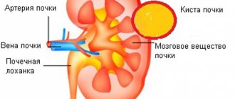
A kidney cyst is a benign tumor - a round fibrous capsule, in the cavity of which there is serous fluid. The renal sinus cyst is characterized by a typical location: it is localized in the sinus area near the renal pelvis or the vascular hilum of the kidney.
Most often, this disease is not accompanied by characteristic symptoms and is first discovered during a routine examination or examination for another disease.
The sinus cyst of the left kidney, like the sinus cyst of the right kidney, requires special treatment, which must be started immediately after the pathology is detected, which will prevent the development of serious consequences.
How does it arise?
Intrasinus renal cysts are formed due to compression of the tissues and vessels of the organ, resulting in atrophic changes, disruption of urine outflow and hemodynamic parameters. To treat the disease, the Yusupov Hospital uses predominantly minimally invasive surgical methods (treatment with needle aspiration, laparoscopic decortication). However, there are frequent cases when the sinus cyst of the right kidney, as well as the left one, recurs.
Congenital renal sinus cyst has a genetic origin. In addition, the development of this disease in children can be triggered by unfavorable environmental factors, intoxication, and infection in the prenatal period.
A kidney sinus cyst in adults can form as a result of certain diseases and conditions that are complicated by obstruction and blockage of the renal tubules:
- pyelonephritis, renal failure;
- infectious diseases;
- overweight;
- endocrine disorders;
- smoking, alcohol;
- hereditary predisposition;
- urolithiasis.
A kidney sinus cyst can be unilateral - then a sinus cyst of the left kidney or a sinus cyst of the right kidney is diagnosed. However, in some cases, sinus cysts of both kidneys are formed, which are characterized by symmetrical growth.
Intrasinus renal cysts: symptoms
Patients with large kidney sinus cysts complain of the following symptoms:
- pain syndrome;
- arterial hypertension;
- the presence of blood in the urine;
- renal failure, the main manifestations of which include weakness, swelling, thirst, daytime drowsiness and insomnia at night.
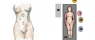
Pain sensations are localized in the hypochondrium, in the lower back and radiate to the groin.
Based on the location of the pain, doctors determine which of the two kidneys is affected by the cyst. A sinus cyst of the right kidney is manifested by aching or dull pain on the right side, and a sinus cyst of the left kidney is manifested by pain in the hypochondrium on the left.
The appearance of arterial hypertension is associated with excess production of renin, a hormone whose synthesis occurs in the kidneys. As a result of the interaction of this hormone with other hormones involved in the regulation of blood pressure, patients not only experience increased blood pressure, but also headache, weakness and nausea.
The presence of blood in the urine is an “alarm bell” that may indicate a serious impairment of kidney function. The development of hematuria is observed in various kidney diseases, incl. and sinus cysts. To detect blood in the urine, laboratory tests are carried out, although some patients can detect this symptom on their own - by a change in the color of the urine.
Stages of inflammation
Any inflammatory process has three stages. When a bacterium enters the mucous membrane of the urinary system, it begins to secrete toxins, as well as products of its vital activity. It happens that the local immunity of the ChLS itself solves the problem. The person does not feel anything, his condition does not change.
- If the immune response is weak, then the first stage of inflammation occurs. It is called alteration . As a result, the mucosal epithelium dies and defects appear.
- Then the second stage begins. It was given the name exudation . Leukocytes and other active cells “come” to the affected area to fight pathogenic microorganisms. And then the damaged area looks swollen because blood has rushed to it. And at such a moment, an ultrasound scan will show a thickening of the joints of both kidneys.
- The third stage is called proliferation . The tissue becomes even denser because the epithelium begins to grow to protect healthy areas from damaged ones.
Cells that have died are replaced by areas of connective tissue. They become sclerotic. For a more complete understanding, you need to imagine the scar left on the skin after wounds and scratches.
Diagnostic methods
Due to the anatomical features of the human body, the sinus cyst of the right kidney makes itself felt more often than the sinus cyst of the left kidney. However, the incidence of cysts in the left and right kidneys is the same. Due to the fact that the formation of a sinus cyst is often associated with intrauterine developmental anomalies, if a sinus cyst is detected in the left kidney, there is a high probability of finding a similar formation in the right kidney.
Diagnosis of sinus cysts in the kidneys at the Yusupov Hospital is carried out using modern informative research:
- ultrasound examination;
- X-ray examination (excretory urography of the kidneys);
- computed tomography;
- magnetic resonance imaging;
- laboratory tests of urine - allows you to determine the presence of protein and inflammatory elements in the urine;
- clinical blood test - to determine ESR;
- biochemical analysis - allows you to determine the level of creatinine, an increase in which may indicate renal failure.
A kidney sinus cyst must be differentiated from oncological processes, urolithiasis and other pathologies.
Prevention
Every person, especially if he is at risk of developing a sinus cyst on the kidneys, must follow simple rules:
- Reconsider your eating behavior. Eat only high-quality and healthy food.
- Drink plenty of fluids, preferably most of them being plain water.
- Try to avoid injuries to the lower back and pelvis.
- Control your weight. If you significantly exceed normal body weight, be sure to start losing those extra pounds.
- Quit bad habits (alcohol and tobacco).
- Dress according to the weather and do not overcool your body, especially the lumbar and back areas.
- Timely detect and treat diseases of the genitourinary system.
- Perform an ultrasound examination of the abdominal organs at least once a year.
- Take a course of treatment with diuretic and anti-inflammatory decoctions from plants.
You can also learn about kidney cysts from this video.
Treatment at the Yusupov Hospital
If patients with sinus cysts of the kidneys do not experience discomfort and bloody impurities in the urine, surgical intervention is not advisable. In such cases, it is sufficient to undergo a regular medical examination - at least once every six months.
Indications for surgical removal of a kidney sinus cyst are indicated in the presence of the following conditions:
- cyst rupture;
- cyst suppuration;
- suspicion of malignancy of the cystic formation;
- presence of blood in the urine;
- arterial hypertension provoked by an intrasinus renal cyst.
To remove a small sinus cyst of the left and right kidney, a percutaneous puncture can be performed, during which a sclerosing substance is injected into the cavity of the pathological formation.
The effective and most common method of surgical treatment of sinus cysts at the Yusupov Hospital is laparoscopic surgery.
Large cysts, suspicion of malignant degeneration of the formation, and cysts localized in close proximity to blood vessels require open surgery.
In addition, the open surgery method is used for patients suffering from concomitant diseases, such as urolithiasis.
Types and classification
Diffuse changes in the renal parenchyma, maxillary sinus or sinus with enlargement and thickening of the organ throughout its entire volume are often provoked by progressive urolithiasis, vascular destruction, and inflammation of the fat pad.
Diffuse changes in the kidneys are expressed by changes in the size and structure of the organ.
Renal diffuseness suggests the presence of fluid in the pelvis, early abscess, impaired metabolic processes and reabsorption involving sodium, structural changes in the renal vein, suspicion of thrombosis, varicose veins or stone formation. Based on the degree and nature of tissue damage, several types of renal diffuse changes are distinguished:
- an increase in organ size caused by inflammation;
- volume reduction caused by chronic pathology;
- thickening of the renal parenchyma;
- variability of value, deterioration of the sinus structure: uniform, focal.
Depending on the visualization on the monitor during ultrasound, diffuse changes may be:
- clear and unclear;
- weak or moderate;
- expressed.
Complications
The most dangerous complications of intrasinus renal cysts include renal failure and severe pyelonephritis.
With large cysts, there is a high risk of suppuration and rupture. As the kidney tissue grows, it is replaced by a cyst, as a result of which the patient may develop hydronephrosis of the kidney and impaired kidney function.
If a bacterial infection develops inside a kidney sinus cyst, pus accumulates in it. The absorption of toxins into the blood leads to the development of intoxication of the body.
Surgical intervention to remove a cyst can also be accompanied by complications:
- intraoperative – during the operation the kidneys themselves may be injured;
- postoperative - in some cases, cyst recurrences occur after surgery.
Diffuse changes in the kidney parenchyma
The kidney consists of parenchyma and a system for storing and excreting urine. The outer part of the parenchyma consists of glomeruli surrounded by a developed circulatory system, and the inner part is made up of renal tubules. The latter form so-called pyramids, through which the liquid enters the calyces and pelvis - components of the excretory system.
The thickness of the parenchyma changes with age - it becomes thinner. For young people, a thickness of 16–25 mm is considered the norm. In the older age group - more than 60 years, the thickness of the parenchyma rarely exceeds 1.1 cm.
Despite the fact that the kidney is protected by a fibrous capsule, the renal parenchyma is quite vulnerable. The blood entering it carries decay products, metabolic products, toxins, and so on, so the kidneys are often the first to react to changes in the body.
Diffuse change is not a specific disease or syndrome, but a condition of an organ in which physiological or physical changes are observed that affect the entire organ. Only after an examination can the cause of the changes be diagnosed and treatment begin.
Diffuse changes are usually accompanied by changes in the size of the organ itself, to the same extent in both children and adults. As a rule, in acute diseases the parenchyma thickens, and in chronic diseases it thins. Moreover, in old age, thinning is also observed due to purely age-related changes. Thinning of the parenchyma in childhood indicates the seriousness of the situation.
Diffuse changes in the kidneys of a newborn can be caused by a variety of reasons, both congenital defects - polycystic disease, congenital nephrotic syndrome, and acquired ones - pyelonephritis, secondary organ damage. Due to the characteristics of the newborn’s body, changes are rapid and especially dangerous.
On the other hand, up to 3 years of age, the child’s kidney has a lobular structure, which is visualized in a very specific way on ultrasound. If no signs of illness are observed, then the process is not pathological. If there are other signs of the disease, diagnosis is necessary.
Kidney parenchyma
Kidney examination
For a complete diagnosis, it is necessary to undergo a series of examinations.
Kidney ultrasound is one of the main examinations of renal pathology
Currently, ultrasound, computed tomography, and radioisotope studies are used for examination. But the most accessible and fastest is ultrasound - this method is always used at the initial stage of the examination.
Based on the results of the ultrasound examination and the doctor’s conclusion, it will be clear what pathology is present.
First of all, the screen will show the size and location of the kidneys, whether there are any anomalies (doubling of the organ), or whether there are deformities.
The main indicator of ultrasound is echogenicity - this is the ability of tissues to reflect a sound wave. The density of the tissue is determined on the monitor screen by the degree of lightening of the areas of the echostructures. The detection of stones is based on this. Those stones that can be detected using ultrasound will appear on the monitor as areas with increased echogenicity. The structure of such stones is dense, so they are easy to detect, calculate their size and location in the sinus of the kidney.