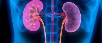
A kidney tumor is a dangerous condition in which kidney cells transform into pathological ones, which leads to a malfunction of the organ as a whole. A benign kidney tumor has a relatively positive prognosis - with its complete resection, it is possible to prolong the individual’s life for a long period. Cancer, on the other hand, has an unfavorable prognosis, and usually life can be prolonged only with a total removal of the kidney.
Benign and malignant kidney tumors are detected in individuals who have crossed the 60-year age limit (based on medical statistics). What is noteworthy is that representatives of the stronger sex suffer from the disease more often than women. Treatment of the tumor is invasive - excision of it together with the changed tissue is indicated.
Etiology
Scientists have not yet been able to say why exactly a tumor forms in the right or left kidney. But in the course of ongoing research, some predisposing factors have been identified that can give impetus to the development of oncology. These include:
- genetic factor;
- exposure to radiation or certain hazardous chemicals;
- addiction to drinking drinks containing alcohol, as well as smoking;
- a history of severe diseases with a hereditary transmission mechanism.
Diagnosis of kidney cancer
- Ultrasound research method.
More than half of kidney cancer cases are diagnosed with its use. In addition, ultrasound can detect metastases. A biopsy is also performed using ultrasound. - Liver biopsy. This procedure involves removing a small piece of the liver using a thin needle for later laboratory testing. This method is quite difficult. It has been used since the middle of the last century.
- CT method (computed tomography). Visualizes a large neoplasm in the kidney. Makes it possible to detect metastases and estimate their size. Has a high level of accuracy.
- MRI (magnetic resonance therapy). It is used as an additional study to CT. It also evaluates the extent of kidney tumor damage and the size of metastases.
- Urography method. Allows you to display changes in the outline of the kidney, its deformation and improper filling of the organ. This method allows you to assess the functionality of the kidney. But with its help it is impossible to identify the stage of the disease.
- Venography method. It is used before deciding on the advisability of surgical intervention.
- X-ray method. Allows you to assess the condition of the lungs in the presence of kidney cancer.
- There are additional examination methods that are used for varying severity of the disease.
Varieties
As already mentioned, both benign and malignant tumors can affect the kidneys.
Tumors from benign parenchyma:
- lipoma;
- fibroma;
- myxoma;
- myoma;
- oncocytoma;
- adenoma;
- dermoid
Malignant parenchymal tumors:
- lipoangiosarcoma;
- fibroangiosarcoma;
- myoangiosarcoma.
Kidney cancer or a benign tumor can also grow from the pelvis. In this case, a slightly different classification applies.
Benign types:
- leiomyoma;
- papilloma;
- angioma.
Malignant:
- sarcoma;
- transitional cell carcinoma;
- mucoglandular cancer;
- squamous cell carcinoma.
Kidney tumor
Kidney tumor. Kidney tumors are the growth of altered tissue in the kidney. All neoplasms are divided into benign and malignant. Benign neoplasms are not characterized by rapid growth, dissemination in the body (metastasis), or tumor disintegration.
Benign kidney tumors can be different in their tissue structure and among them are adenomas, angiomyolipomas, hemangiomas, leiomyomas, oncocytomas, etc. Before the use of ultrasound diagnostics and computed tomographs, benign kidney tumors were an accidental discovery when this organ was removed for another disease. Benign kidney tumors that differ in their tissue structure require the use of different methods for their detection and observation. Ultrasound, computed tomography, magnetic resonance imaging, vascular studies - this is an incomplete range of methods used by urologists. Initially, any identified kidney tumor is considered malignant and requires examination and observation accordingly. If it is impossible to establish a definitive diagnosis, a biopsy of the kidney tissue is performed, which makes it possible to clarify the nature of the tumor. Since the kidney is the most blood-supplied organ, blind biopsy and even ultrasound-guided biopsy are accompanied by moderate or heavy bleeding in more than 10% of cases. The optimal method of kidney biopsy is under visual control using endovideosurgical (laparoscopic) equipment. After establishing a diagnosis of a benign kidney tumor, observation is indicated at intervals specified by the urologist.
Malignant kidney tumors are detected more often than benign tumors and differ from tumors of other organs in their resistance to chemotherapy and radiation therapy, which brings to the fore surgical treatment - removal of the affected kidney or part of it along with the tumor. Immunotherapy for kidney cancer is currently available, but such treatment is only available in large international centers and is very expensive.
Surgical treatment involves removing the kidney. Access to the kidney can be open, with a skin incision, or laparoscopic (lumboscopic), which is less traumatic and does not require long-term rehabilitation. Sometimes, if technically possible, part of the kidney along with the tumor is removed. Such operations are used especially often in cases where the second kidney is compromised by some disease. In some types of malignant tumors, the ureter and even part of the bladder wall may be removed along with the kidney.
After removal of a malignant kidney tumor, the patient must be monitored by a urologist (urologist-oncologist) for life. I would like to note that such tumors are most often detected by ultrasound, which is now widely available, which should not be neglected and ultrasound should be performed once a year during clinical examination.
Features of manifestation
Symptoms of a kidney tumor, like any other type of oncology, do not appear for a long time, or their expression is so insignificant that the patient does not even take them into account. But at grades 2-3 the signs become quite pronounced. A person experiences:
- the appearance of blood impurities in the excreted urine;
- general condition suffers;
- body weight decreases;
- increase in blood pressure;
- temperature increase;
- renal colic;
- polycythemia;
- anemia;
- pain in the lower back;
- varicocele.
Why does kidney cancer occur?
It is difficult to say why kidney cancer occurs; it is impossible to say why kidney cancer occurs in a particular patient. There was evidence that kidney cancer was much more common among people employed in the production of aniline dyes. In this regard, some carcinogens produced in the production of aniline dyes have been accused of carcinogenic effects on the kidney. These same carcinogens have been implicated in bladder cancer. An increased risk of the disease is observed in patients with Hippel-Lindau disease, horseshoe kidneys, polycystic disease and acquired cysts, which are accompanied by increased levels of nitrogenous substances in the blood (uremia). The latter condition is the result of insufficient kidney function.
Treatment
The main treatment for kidney cancer is surgery. Surgical intervention in almost all cases where possible. During the operation, the kidney is removed, as well as the fatty tissue that surrounds it and the ureter (radical nephrectomy). Currently, organ-preserving operations for kidney cancer have also been developed. They are performed in the early stages of the tumor in cases where it is impossible for the patient to remove the kidney. However, we are not talking about the prevalence of the process. We are talking about those cases when the remaining kidney cannot take on all the functions of excreting metabolic products from the body. Such operations involve removing only part of the kidney. As scientific studies show, the long-term results of such operations differ little from operations to remove a kidney (nephrectomy). However, after breast-conserving surgery there is a higher risk of local recurrence.
The five-year survival rate for patients undergoing stage 1 radical nephrectomy is 70 to 80 percent. If the tumor affects the inferior vena cava, then after surgery 40–50 percent of patients survive for 5 or more years (stage 2). If the renal vein is involved (stage 2), the five-year survival rate is 50–60 percent. If the fatty tissue surrounding the kidney was involved (stage 3), the survival rate is 70–80 percent. With regional lymph node involvement (stage 3–4), the 5-year survival rate ranges from 5 to 20 percent. If the tumor has invaded neighboring organs or has distant metastases, the 5-year survival rate is no more than 5 percent.
Currently, most scientists recognize that it is advisable to perform surgery for single distant metastases of kidney cancer. Research results show that such operations improve the patient’s quality of life and prolong his life.
Diagnostics
Diagnosis is based, of course, on the patient’s complaints, as well as on examination data and general clinical research methods (clinical blood test, clinical urine test, etc.). Currently, the main methods of instrumental diagnosis of kidney cancer are ultrasound examination of the abdominal organs, urography (x-ray examination of the kidneys using contrast agents), radionuclide scanning, as well as computed tomography and magnetic resonance imaging. The last two methods make it possible to accurately determine the extent of the tumor. When examining a patient with suspected kidney cancer, it is mandatory to take an x-ray of the lungs, as well as x-rays of the pelvic bones and chest. If metastatic bone lesions are suspected, a radionuclide bone scan is necessary, which makes it possible to clarify the presence of metastases in the bones.
Treatment of malignant neoplasms
Treatment for kidney cancer depends on the age and general condition of the patient, as well as the stage of tumor development. The most commonly used methods are:
Surgery. Removing kidney tumors through surgery is the gold standard in medicine. Timely implementation of the procedure ensures a successful outcome in the vast majority of cases. The main condition is to remove the tumor at a time when it is in the initial stages. Such kidney operations are often organ-sparing in nature: only tumor resection is performed within healthy tissue. If the tumor is significant, the entire kidney is removed (nephrectomy). This operation can be performed either open or laparoscopic. Currently, kidney removal is often performed using an open technique. It is predicted that as endoscopic technologies develop, the proportion of laparoscopic operations will increase.
Conservative methods. Such methods can be both primary and auxiliary (as part of therapy after surgery). Kidney cancer, unlike most cancers, is almost resistant to radiation and chemotherapy. Therefore, if the patient has general contraindications to kidney surgery or surgical removal of a malignant neoplasm is impossible, other conservative methods are used. These include hormone therapy and immunotherapy. Arterial embolization is also used to reduce the size of the tumor in the kidney. The latter technique can also be used in preparation for surgical tumor removal.
Malignant neoplasms in the kidney are a serious diagnosis. But it is not a sentence at all. The most important thing in such cases is to start treatment as quickly as possible and choose truly qualified specialists for this. To get advice on removing a tumor in the kidney, you can call, request a call back, or leave a request through the website by filling out the form presented on the page.


