Pyelonephritis
Diabetes
Hepatitis
Rheumatism
42017 October 27
IMPORTANT!
The information in this section cannot be used for self-diagnosis and self-treatment.
In case of pain or other exacerbation of the disease, diagnostic tests should be prescribed only by the attending physician. To make a diagnosis and properly prescribe treatment, you should contact your doctor. We remind you that independent interpretation of the results is unacceptable; the information below is for reference only.
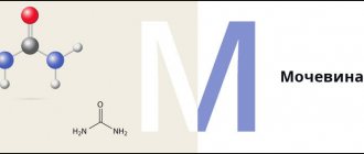
Urea in the blood: indications for prescription, rules for preparing for the test, interpretation of the results and normal indicators.
Indications for the purpose of the study
Urea in the blood is synthesized in the liver from ammonia and carbon dioxide, transported by the blood to the kidneys, there it is filtered through the glomerulus, and then excreted in the urine. Urea is an osmotically active substance, so its accumulation leads to swelling of the tissues of parenchymal organs (liver, kidneys, lungs, spleen, pancreas, thyroid gland), myocardium, central nervous system, subcutaneous tissue. An increase in urea concentration several times relative to the norm, accompanied, as a rule, by a pronounced clinical syndrome of intoxication, is called uremia.
Very often, against the background of kidney disease, simultaneously with an increase in the concentration of urea in the blood, its content in the urine decreases (a decrease in kidney function leads to an increase in urea in the blood).
The study is prescribed for coronary heart disease, systemic connective tissue diseases (rheumatoid arthritis, rheumatism, systemic scleroderma, etc.), arterial hypertension (regardless of the duration of its existence), when detecting abnormalities in a general urine test during a screening study, liver disease accompanied by dysfunction (hepatitis, cirrhosis), if inflammatory or infectious diseases of the kidneys are suspected, diseases of the gastrointestinal tract, as well as before and during drug therapy, before hospitalization of the patient due to an acute illness, after dialysis sessions for evaluation their effectiveness.
The concentration of urea in the blood characterizes:
- the state of the excretory function of the kidneys, that is, their ability to excrete substances unnecessary to the body with urine;
- the condition of muscle tissue (as a result of the breakdown of protein in the muscles, urea is formed);
- liver function, where ammonia is converted to urea.
Uric acid in daily urine
Uric acid in urine is uric acid from the blood filtered through the kidneys.
Synonyms Russian
Purine-2,6,8-trione, product of purine base metabolism, trihydroxypurine, 2,6,8-trioxypurine, heterocyclic urea ureide.
English synonyms
Urine Uric Acid, Urine Uric Acid Quantitative (24-Hour), Uric Acid in urine.
Research method
Enzymatic method (uricase).
Units
mmol/day (millimoles per day).
What biomaterial can be used for research?
Daily urine.
How to properly prepare for research?
- Eliminate alcohol from your diet the day before donating urine.
- Do not eat spicy, salty foods, or foods that change the color of urine (for example, beets, carrots) for 12 hours before the test.
- Do not take diuretics 2 days before the test (as agreed with your doctor).
- Avoid physical and emotional stress during the collection of daily urine (during the day).
General information about the study
Uric acid is formed as a result of cell renewal and also enters the body with food. Most of it leaves the body in urine, less in stool. With excessive formation of uric acid, its concentration in the urine can increase significantly, and if the kidneys are unable to filter blood in normal volumes, it can decrease.
Consistently high levels of uric acid cause the formation of uric acid crystals in the joint cavity. This painful pathological condition is called gout. If left untreated, uric acid crystals inside joints and adjacent tissues can form deposits that appear as hard bumps on the surface of the body.
Persistently high levels of uric acid in the urine can lead to stone formation.
Uric acid, which is dissolved in the blood, is delivered to the kidneys, where, after filtration, it is excreted in the urine. If the body produces too much uric acid over a long period of time or does not eliminate it well enough, a person may experience problems urinating, fever, chills, fatigue, and joint pain.
A condition in which the level of uric acid in the urine is elevated is called hyperuricosuria. This can cause kidney stones to form, blocking the normal flow of urine in the kidney tubules, ureter, and bladder.
What is the research used for?
- To assess uric acid metabolism.
- To identify disorders affecting uric acid production.
- To determine the severity of kidney damage.
When is the study scheduled?
- If necessary, find out the cause of the formation of kidney stones.
- When monitoring the condition of patients with gout.
What do the results mean?
Reference values: 1.48 - 4.43 mmol/day.
Causes of increased concentration of uric acid in urine:
- eating large amounts of food rich in purine bases (meat, especially offal),
- gout (increased production or insufficient excretion of uric acid),
- urolithiasis disease,
- polycythemia vera (excessive production of blood cells),
- Lösch–Nyhan syndrome (increased uric acid synthesis),
- Wilson–Konovalov disease,
- viral hepatitis,
- sickle cell anemia,
- malignant neoplasms with metastases, multiple myeloma, chronic myeloid leukemia (uncontrolled cell growth and division),
- Fanconi syndrome (decreased tubular reabsorption of uric acid due to a defect in tubular development).
Reasons for low concentration of uric acid in urine:
- chronic kidney diseases, such as chronic glomerulonephritis,
- xanthinuria (little uric acid is formed due to xanthine oxidase deficiency),
- lead intoxication (due to a pronounced decrease in kidney function),
- chronic alcoholism,
- folic acid deficiency.
What can influence the result?
Falsely inflated results are contributed to by:
- stress and intense physical activity,
- injuries,
- beta-blockers, caffeine, vitamin C, large doses of acetylsalicylic acid, calcitriol, asparaginase, diclofenac, isoniazid, ibuprofen, indomethacin, piroxicam, paracetamol, lithium salts, mannitol, mercaptopurine, methotrexate, nifedipine, prednisolone, verapamil.
A falsely low result can be caused by:
- allopurinol, glucocorticoids, imuran, contrast agents, vinblastine, azathioprine, methotrexate, spironolactone, insulin, small doses of acetylsalicylic acid, furosemide, ethambutol, pyrazinamide.
Important Notes
- 20-25% of people with nephrolithiasis develop hyperuricosuria.
Also recommended
- Serum uric acid
- Urea in serum
- Serum creatinine
- Urea in urine
- Creatinine in urine
Who orders the study?
Therapist, rheumatologist, gynecologist, hepatologist, oncologist, nephrologist.
What are the clinical manifestations of gout flares?
Persistent and elevated levels of uric acid in the body (hyperuricemia) is a common biochemical abnormality that results from excessive production of uric acid (urate) or when the kidneys do not eliminate uric acid .
Therefore, in all patients with gout during their illness, hyperuricemia (saturation of blood serum with urate) is always observed. However, most people with hyperuricemia never experience the clinical manifestations of gout associated with urate crystal deposition.
Clinical manifestations of gout include:
- Recurrent outbreaks of inflammatory arthritis (gout flare). Most often in autumn and spring.
- Do you have a sharply inflamed toe joint on your right foot? Read below what this is connected with.
- This is severe and acute pain in the joint, redness, the joint “burns,” swelling, and inability to walk. And usually the patient says that there was no injury! The picture itself usually appears within 12-24 hours, which is quite fast in itself and is typical for this attack. Complete resolution of the earliest outbreaks almost always occurs within a few days to a few weeks, even in untreated people.
- Gout flares are twice as likely to occur during the night and early morning than during the day. Patients often say that they cannot stand on their leg in the morning because, in their words, “the knee suddenly became inflamed overnight, but yesterday everything was fine, and I didn’t hit my leg anywhere.”
- Damage to the lower extremities. Most initial outbreaks affect at least one joint, most often at the base of the big toe (the first metatarsophalangeal joint, known as gout) or the knee.
- Recurrent joint problem, chronic arthropathy.
Can gout affect the joints of the fingers and elbows?
Yes maybe! Multi-joint gout flares – The multi-joint pattern is the initial presentation in a small number of patients with gout, but with increasing frequency in subsequent flares. Multi-joint symptoms are especially common in the later stages of untreated gout, when multiple relapses are common. Fingers, elbows, upper jaw.
Signs of inflammation that extend beyond the joint are caused by arthritis in several adjacent joints or tenosynovitis. Unusual involvement of the vertebral joints and sacroiliac joints, where gout may be much less common, which can cause diagnostic confusion. The area most often affected is the lumbar spine. However, most proven cases of gout, which manifests itself as acute or chronic back pain, have been associated with damage to the stomach. There may also be neurological signs and symptoms.
Accumulation of uric acid crystals in the form of external deposits.
Tophi are characteristic accumulations of solid urate, accompanied by chronic inflammatory and often destructive changes in the connective tissue. Tophi are often visible, can be touched and felt (palpated), and may be present on the ears or in soft tissues including joint structures, tendons, or periarticular bursae. Tophi (tophi) usually do not hurt and are hard in structure. They are visible on the skin and have a slightly yellow or white color. On the ear they appear in the form of balls and do not allow light to pass through.
Note for medical students and doctors
-Chronic inflammatory process extending beyond one joint due to uric acid deposition is reminiscent and similar to dactylitis seen in other diseases such as psoriatic arthritis, other spondyloarthritis and sarcoidosis. The expansive and destructive changes associated with gouty gout may be mistaken for osteomyelitis and sometimes lead to erroneous amputation of the involved digits.
Deposition of uric acid crystals in the kidneys. Nephrolithiasis.
Uric acid stones account for 5 to 10 percent of all kidney stones worldwide; however, they make up 40 percent or more of the stones in hot, dry climates. The most important biochemical risk factor for the development of urolithiasis is a persistently low urine pH. And low urine volume (with a high concentration of uric acid in the urine) and acidic urine pH promote the conversion of the relatively soluble urate salt to insoluble uric acid.
Patients usually have an attack of renal colic and complain of acute pain in the right or left side. Computed tomography (CT) without contrast/dye usually reveals the presence of a stone. For many patients, we recommend that the diagnosis be confirmed by chemical analysis of the stone. It is worth noting that on ordinary radio photographs, stones consisting of uric acid are transparent and almost impossible to notice.
Chronic nephropathy, which in gouty patients most often occurs due to concomitant conditions.
Chronic urate nephropathy is the last stage of chronic kidney disease in chronic gout, caused by the deposition of uric acid crystals in the kidneys (medullary interstitium). The crystals cause a chronic inflammatory response similar to that seen when tophi form in other parts of the body, ultimately leading to interstitial fibrosis and chronic kidney disease.
Clinical stages of the disease (“Gout flares,” “Recurrent gout flares,” “Chronic gout”) occur sequentially, with corresponding clinical severity that often coincides with the frequency of gout flares. Therefore, treatment with uric acid-lowering drugs (antihyperuricemics) is restraining and ensures effective recovery of the patient if the urate saturation level does not increase above <6 mg/dL or 357 micromol/L.
Go to top


