Author
Lukyanchikov Denis Vladimirovich
Leading doctor
Urologist
until July 31
We're giving away RUR 1,000 for all services per visit in July More details All promotions
Urinalysis according to Zimnitsky is a laboratory diagnostic method that allows you to study the functional state of the kidneys. Using the Zimnitsky urine test, the ability of the kidneys to concentrate and excrete urine is assessed.
Urine is produced in the kidneys by filtering blood. During the day, the kidneys pass 1800 liters of blood, while normally 1.5-2 liters of urine should be released. The body's waste products are eliminated through urine. Also, with the help of urine secretion, water balance is regulated. If the body receives little fluid, little urine is produced, but it becomes more concentrated. If there is a lot of fluid received (for example, when drinking a lot), the concentration of urine drops. When kidney function is impaired, this mechanism stops working, as a result, the water balance is disrupted, the composition of the blood changes, and this affects the general condition of the body.
Urinalysis according to Zimnitsky allows you to determine how much urine is excreted per day and what the concentration of urine is.
Interference:
- CREATININE - nephrotoxic drugs. UREA - corticosteroids, nephrotoxic drugs, tetracycline, excess thyroxine. URIC ACID - beta blockers, cisplatin, corticosteroids, cyclosporine, diazoxide, didanosine, diuretics, epinephrine, ethanol, ethambutol, filgrastim, fructose (iv), niacin (in large doses), norepinephrine, pyrazinamide, salicylates (in small doses) doses), fludarabine, hydroxyurea, idarubicin, mechlorethamine, theophylline (iv). ALBUMIN - progesterone. SODIUM - ACTH, anabolic steroids, androgens, carbenicillin, carbenoxalone, clonidine, corticosteroids, diazoxide, enoxolone, estrogens, guanethidine analogues, lactulose, mycorise, methoxyflurane, methyldopa, oral contraceptives, oxyphenbutazone, phenylbutazone, reserpine, sodium bicarbonate. POTASSIUM - beta blockers (rare), amiloride, aminocaproic acid, ACE inhibitors, cyclophosphamide, vincristine, arginine, cephaloridine, cyclosporine, digoxin, adrenaline (initial effect), foscarnet, heparin, histamine (iv), isoniazid, lithium, mannitol, methicillin, non-steroidal anti-inflammatory drugs, penicillin (potassium salt), phenformin (lactic acidosis), salt substitutes, spironolactone, succinylcholine, tetracycline, triamterene, tromethamine. TOTAL CALCIUM - alkaline antacids, androgens, calcium salts, calusterone, danazol, diethylstilbestrol (rapid increase within 24 hours in patients with breast cancer), dihydrotachysterol, chronic use of diuretics (including chlothalidone, ethacrynic acid, furosemide, thiazides), ergocalciferol, isotretinoin, lithium, progesterone, parathyroid hormone, testolactone
- CREATININE - Glucocorticoids URIC ACID - Acetogesamide, allopurinol, azathioprine, bishydroxycoumarin, chlorprothixene, clofibrate, contrast agents (diatrizoate, iodipamide, iopanoic acid, iopodate), ethacrynic acid (iv), fenofibrate, furosemide (iv), probenecid, salicylates (in large doses). ALBUMIN - Allopurinol, asparaginase, azathioprine, chlorpropamide, cisplatin, dapsone, dextran, estrogens, ibuprofen, isoniazid, nitrofurantoin, oral contraceptives, phenytoin, prednisolone (high doses), sargramostim, valproic acid. SODIUM - Aminoglutethimide, aminoglycosides, ammonium chloride, amphotericin B, ACE inhibitors, captopril, carbamazepine, carboplatin, chlorpropamide, cholestyramine, cisplatin, clofibrate, cyclophosphamide, desmopressin, diuretics, fluoxetine, hypertonic glucose solution, haloperidol, heparin, indomethacin , ketoconazole, lithium , lorcainide, miconazole, non-steroidal anti-inflammatory drugs, oxytocin, phenothiazines, thienylic acid, tolbutamide, tricyclic antidepressants, vasopressin, vinblastine, vincristine. POTASSIUM - beta-adrenergic agonists (salbutamol, terbutaline), albuterol, aminoglycosides, p-aminosalicylate, aminosalicylic acid (rare), amphotericin, azlocillin, bisacodyl, capreomycin, carbenicillin, carbenoxolone, cholestyramine, cisplatin, clopamide, corticosteroids, corticotropin, cyanocobalamin, anhydride dextrose, diclofenamide, diuretics, EDTA, enoxolone, fluconazole, glucagon, glucose, ifosfamide, insulin, levodopa, licorice, mezlocillin, nafcillin, penicillin G (sodium salt), phenolphthalein, piperacillin, polymyxin B, salicylates, sodium bicarbonate, sodium chloride , ticarcillin, theophylline. TOTAL CALCIUM - Albuterol, alprostadiol, aminoglycosides, asparaginase, barbiturates in the elderly, calcitonin, carbamazepine, carbenoloxone, carboplatin, corticosteroids, diuretics (triggering effect), ergocalciferol, estrogens (after menopause), fluorides, gastrin, glucagon, glucose, indapamide, insulin, isoniazid, laxatives (if used excessively), magnesium salts, methicillin, phenytoin, phosphates, plicamycin, isotonic sodium chloride solution (for hypercalcemia), tetracycline (in pregnant women).
Kidney function (screening)
A comprehensive laboratory test of blood and urine, which allows you to evaluate the excretory and filtration abilities of the kidneys and diagnose disorders of their function.
The research results are provided with a free doctor’s commentary.
English synonyms
Renal Function Tests.
Research method
Kinetic method (Jaffe method), UV kinetic test, dry chemistry method, microscopy.
Units
μmol/L (micromoles per liter), mmol/L (millimoles per liter), mg/dL (milligrams per deciliter), unit/dL (unit per deciliter).
What biomaterial can be used for research?
An average portion of morning urine, venous blood.
How to properly prepare for research?
- Do not eat for 8 hours before the test; you can drink clean still water.
- Avoid taking diuretics for 48 hours before urine collection (in consultation with your doctor).
- Women are advised to donate urine before menstruation or 2 days after it ends.
- Avoid physical and emotional stress 30 minutes before the test.
- Do not smoke for 30 minutes before the test.
General information about the study
The kidneys are paired organs located in the retroperitoneum on both sides of the lumbar spine. They ensure blood filtration from excess and toxic metabolic products, removing them in the urine, and, thanks to the processes of filtration, secretion and reabsorption, regulate the level of fluid, electrolytes and trace elements, acid-base balance in the body. They synthesize biologically active substances: renin, which plays an important role in the regulation of blood pressure, and erythropoietin, which stimulates the proliferation of red blood cells in the bone marrow.
Kidney functions are regulated by hormones of the hypothalamic-pituitary system and adrenal glands, so changes in the level of antidiuretic hormone, aldosterone, and cortisol greatly affect the content of electrolytes and fluid in the blood, their ability to reabsorb and filter. In addition, under the influence of parathyroid hormone and vitamin D, the kidneys participate in the regulation of phosphorus and calcium metabolism.
With kidney pathology, the release of toxins and fluids from the body is disrupted, and the balance of osmotically active substances in the blood changes. Severe kidney disease without adequate treatment, regular hemodialysis or, if necessary, donor organ transplantation leads to death.
There are more than 100 diseases and pathological conditions that can cause progressive impairment of kidney function. The most common are diabetes mellitus, hypertension, infections, autoimmune pathology (systemic lupus erythematosus, vasculitis), neoplasms, developmental abnormalities of the urinary tract, and pathologies of mineral metabolism.
Renal dysfunction usually occurs unnoticed. Kidney disease can be suspected when puffiness and swelling appear around the eyes, in the ankles and legs, chest and abdomen, when there are changes in the color, transparency and amount of urine, when urination increases, especially at night, when there is pain in the lower back, in the area where the kidneys are projected. The progression of kidney pathology is often accompanied by itching, loss of appetite, nausea, vomiting, swelling and numbness of the arms and legs, fatigue, decreased attention, darkening of the skin, and convulsions. However, changes in laboratory parameters appear much earlier than the first clinical symptoms.
Thus, an increased number of leukocytes in the urine indicates an inflammatory process and a possible urinary tract infection. Protein and red blood cells in the urine may indicate damage to the glomerulus, and increased concentrations of urea (azotemia) and creatinine in the blood are signs of kidney failure.
Detection of acute or chronic renal failure requires urgent treatment measures to avoid progression of the disease.
Due to the variety of causes of kidney disease, a comprehensive examination of the patient is necessary, taking into account clinical data, the results of laboratory and instrumental research methods, and in some cases, a kidney biopsy.
What is the research used for?
- To assess the functional state of the kidneys.
- To evaluate renal damage in certain systemic diseases (eg, diabetes mellitus, hypertension, systemic lupus erythematosus, amyloidosis, vasculitis, myeloma) and trauma.
- For differential diagnosis of urinary tract diseases.
- For monitoring patients with chronic kidney disease, renal failure.
- For the examination of patients on dialysis.
- For preventive examination before prescribing drugs with possible nephrotoxic effects.
- To monitor the treatment of kidney pathology.
When is the study scheduled?
- For symptoms of kidney pathology (changes in color, smell, clarity and amount of urine, frequency of urination, pain in the lumbar region, swelling in the face and other parts of the body, high blood pressure).
- If the results of a general urine test are questionable or there are deviations in individual biochemical parameters.
- For kidney pathology according to instrumental diagnostic methods (ultrasound, CT).
- For systemic diseases with a high risk of damage to kidney function.
- During preventive examinations.
- Before prescribing drugs that affect kidney function and while taking them.
- When monitoring the treatment of kidney disease.
What do the results mean?
Reference values
- BUN/creatinine ratio: 0 - 100.
- Serum creatinine
| Floor | Age | Reference values |
| Male | Less than 5 days | 27 - 88 µmol/l |
| 5 days – 1 month | 27 - 88 µmol/l | |
| 1 month – 1 year | 18 – 35 µmol/l | |
| 1-2 years | 27 - 62 µmol/l | |
| 2-12 years | 27 - 62 µmol/l | |
| 12-17 years old | 44 - 88 µmol/l | |
| 17-51 years | 74 - 110 µmol/l | |
| Over 51 years old | 72 - 127 µmol/l | |
| Female | Less than 5 days | 27 - 88 µmol/l |
| 5 days – 1 month | 27 - 88 µmol/l | |
| 1 month – 1 year | 18 – 35 µmol/l | |
| 1-2 years | 27 - 62 µmol/l | |
| 2-12 years | 27 - 62 µmol/l | |
| 12-17 years old | 44 - 88 µmol/l | |
| More than 17 years | 58 – 96 µmol/l |
- GFR (glomerular filtration rate): 60 and above.
- Urea in serum
| Age | Reference values |
| Less than 1 day | 1.4 - 4.3 mmol/l |
| 1 day – 14 years | 1.8 - 6.4 mmol/l |
| 14-16 years old | 2.8 - 7.2 mmol/l |
| More than 16 years | 2.8 - 7.2 mmol/l |
- Urine sediment examination:
| Index | Reference values |
| Reaction | 5.0 — 7.5 |
| Bilirubin | None or small amount |
| Specific gravity | 1.010 — 1.025 |
| Protein | Missing or traces |
| Glucose | Up to 30 mg/dl |
| Urobilinoids | 0.2 - 2.0 units/dl |
| Ketones | Up to 15 mg/dl |
| Nitrites | Not detected |
| Reaction to blood | Negative or traces |
| Leukocytes | Not found or traces |
| Color | From pale yellow to deep yellow |
| Smell | Unsharp specific |
| Transparency | Transparent |
| Epithelium: flat | For men: 0 - 3 in sight For women: 0 - 5 in the field of view |
| Epithelium: flat (in view) | For men: 0 - 3 in sight For women: 0 - 5 in the field of view |
| Leukocytes | For men: 0 - 2 in sight For women: 0 - 4 in the field of view |
| Red blood cells: unchanged | 0 - 3 in view |
| Red blood cells: altered | 0 - 3 in view |
| Cylinders: hyaline | 0 - 1 in view |
| Cylinders: others | 0 - 1 in view |
Serum creatinine
Causes of increased serum creatinine levels:
- renal failure;
- chronic kidney disease;
- chronic nephritis;
- disturbance of urine outflow;
- pathology of muscle tissue (gigantism, myasthenia gravis, muscular dystrophy, poliomyelitis);
- rhabdomyolysis;
- dehydration;
- congestive heart failure;
- shock.
Reasons for decreased serum creatinine levels:
- short stature, insufficient muscle mass;
- severe liver pathology;
- lack of protein in the diet;
- pregnancy.
1) Density (allows you to evaluate the ability of the kidneys to concentrate urine)
Causes of increased density (hypersthenuria):
- acute nephritis;
- diabetes;
- renal failure;
- excessive fluid loss (with vomiting, diarrhea, fever, dehydration);
- stress, surgery, excessive release of antidiuretic hormone;
- gestosis during pregnancy.
Causes of decreased density (hyposthenuria):
- acute glomerulonephritis;
- acute and chronic tubulointerstitial nephritis;
- diabetes insipidus (antidiuretic hormone deficiency);
- Conn's syndrome;
- hyperparathyroidism;
- drinking large amounts of liquid;
- variant of the norm in children of the first year of life.
2) pH (acidity)
Reasons for increased pH (alkalinization of urine):
- urinary tract infection caused by bacteria of the genera Proteus or Pseudomonas;
- chronic renal failure;
- metabolic acidosis (with vomiting);
- respiratory alkalosis (with hyperventilation);
- hypokalemia.
Reasons for decreased pH (urine oxidation):
- metabolic acidosis, diabetic ketoacidosis, diarrhea, fasting, uremia;
- urinary tract infection caused by the bacterium Echerichia coli;
- respiratory acidosis;
- kidney tuberculosis;
- fever.
3) Protein
Causes of protein excretion in urine (proteinuria):
- violation of the filtration barrier - loss of albumin (glomerulonephritis, nephrotic syndrome, amyloidosis, malignant hypertension, lupus nephritis, diabetes mellitus, polycystic kidney disease);
- decreased reabsorption - loss of globulins (acute interstitial nephritis, acute renal necrosis, Fanconi syndrome);
- increased production of proteins capable of filtering (multiple myeloma, myoglobinuria);
- isolated proteinuria without renal impairment (due to fever, exercise, prolonged standing, congestive heart failure, or idiopathic causes).
4) Nitrites
Reasons for increased nitrite levels:
- presence of bacteria in urine.
5) Glucose
Reasons for increased glucose levels:
- diabetes mellitus, gestational diabetes;
- other endocrine disorders (thyrotoxicosis, Cushing's syndrome, acromegaly);
- impairment of tubular reabsorption in the kidneys (Fanconi syndrome).
6) Epithelial cells of the renal tubules
Reasons for increased levels of renal tubular epithelial cells:
- kidney infarction;
- acute glomerulonephritis;
- pyelonephritis;
- viral infection (for example, cytomegalovirus);
- rejection of a transplanted kidney;
- poisoning with heavy metals, toxins;
- overdose of salicylates.
7) Red blood cells
Causes of hematuria (blood in urine):
- glomerulonephritis;
- systemic lupus erythematosus, lupus nephritis;
- urolithiasis or crystalluria;
- neoplasms (kidney cancer, prostate cancer, bladder cancer);
- infection (cystitis, urethritis, prostatitis);
- polycystic kidney disease;
- kidney infarction;
- vasculitis;
- renal vein thrombosis;
- trauma, damage to the urethra by a urinary catheter;
- kidney tuberculosis;
- benign familial hematuria, benign recurrent hematuria;
- congestive heart failure;
- subacute infective endocarditis.
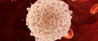
 Leukocytes
Causes of increased white blood cell levels (leukocyturia):
- urinary tract infection;
- interstitial nephritis, pyelonephritis;
- glomerulonephritis;
- kidney tuberculosis;
- fever.
9) Cylinders (indicate dysfunction of the glomerulus and tubules)
Reasons for the appearance of cylinders:
- kidney infarction;
- glomerulonephritis;
- nephrotic syndrome and proteinuria;
- tubulointerstitial nephritis, pyelonephritis;
- chronic renal failure;
- congestive heart failure;
- diabetic nephropathy;
- malignant hypertension;
- fever with dehydration, overheating;
- intense physical activity, emotional stress;
- heavy metal poisoning;
- kidney amyloidosis;
- kidney tuberculosis;
- kidney transplant rejection;
- lipoid nephrosis;
- paraproteinuria in myeloma.
10) Crystals appear in cases of mineral metabolism disorders and indicate the presence of stones or an increased risk of developing urolithiasis, nephrolithiasis.
11) Bacteria indicate a bacterial urinary tract infection.
Urea in serum
Causes of increased serum urea levels (azotemia):
- insufficient renal function in congestive heart failure, water and electrolyte deficiency, shock, stress, acute myocardial infarction;
- chronic kidney disease (chronic pyelonephritis, glomerulonephritis);
- disturbance of urine outflow;
- gastrointestinal bleeding;
- diabetes mellitus with ketoacidosis;
- excessive protein intake or increased protein catabolism (for example, with burns, neoplasms);
- taking anabolic steroids.
Reasons for low serum urea levels:
- liver failure;
- nephrotic syndrome;
- malabsorption, malnutrition, reduced protein intake;
- acromegaly;
- antidiuretic hormone deficiency syndrome.
BUN/creatinine ratio
Reasons for increasing the coefficient:
- renal failure;
- disturbance of urine outflow;
- dehydration, deficiency of electrolytes in the blood;
- heart failure;
- catabolic state with tissue damage;
- gastrointestinal bleeding;
- combination of azotemia and kidney pathology.
Reasons for lowering the coefficient:
- acute tubular necrosis;
- rhabdomyolysis;
- severe liver pathology, fasting;
- antidiuretic hormone deficiency;
- pregnancy;
- taking phenacemide.
What can influence the result?
- Long-term (several hours) storage of urine analysis until delivery to the laboratory.
- Excessive consumption of liquid, mineral waters, salt, alcohol, coffee, green tea before taking the test, violation of dietary recommendations.
- Parenteral administration of saline solutions, glucose solutions, contrast agents shortly before delivery of the material.
- Intense physical activity, emotional stress, pregnancy.
- Trauma to the urethra with a urinary catheter.
- Contamination of a urine sample with secretions from the genital tract, blood from hemorrhoids.
- Taking medications that affect certain study parameters (diuretics, insulin, hypoglycemic drugs, antibiotics, laxatives, anabolic steroids, glucocorticoids, antiepileptic drugs and others).
Important Notes
If changes in laboratory parameters are detected, it is necessary to conduct additional laboratory and instrumental (ultrasound of the kidneys, CT scan of the kidneys) studies to clarify the pathological process in the kidneys. A comprehensive assessment of the patient’s examination results, diagnosis and treatment should be carried out only by the attending physician.
Also recommended
- General urine analysis with sediment microscopy
- Leukocyte formula
- Erythrocyte sedimentation rate (ESR)
- Laboratory examination for pyelonephritis
- Diabetes monitoring
- Laboratory examination for arterial hypertension
- Urinalysis according to Nechiporenko
- Rehberg test (endogenous creatinine clearance)
- Potassium, sodium, chlorine in serum
- Total protein in whey
- Serum calcium
- Serum phosphorus
- Total protein in urine
- Albumin in urine (microalbuminuria)
- Urea in daily urine
- Creatinine in daily urine
- Erythropoietin
- Renin
- Bence Jones protein, quantitative (urine immunofixation)
- Diagnosis of autoimmune kidney damage
- Antibodies to glomerular basement membrane
- Circulating immune complexes (CIC)
- Culture of flora with determination of sensitivity to antibiotics
Who orders the study?
Nephrologist, urologist, therapist, general practitioner.
Literature
- Fischbach FT, Dunning MB A Manual of Laboratory and Diagnostic Tests, 8th Ed. Lippincott Williams & Wilkins, 2008: 1344 p.
- Wilson D. McGraw-Hill Manual of Laboratory and Diagnostic Tests 1st Ed. Normal, Illinois, 2007: 666 p.
- Sabatine. M. Pocket Medicine. 3d ed. USA, Philadelphia: LWW; 2008.
Renal blood parameters, interpretation
After the analysis, a medical report is issued on the presence of chemicals and BAs in the blood. Often, documentation is presented in the form of a table, where the name of the indicator is indicated in the first column, the individual value in the 2nd column, and the normal limits in the 3rd. Using this plate, the specialist assesses the deviation from the norm and, on the basis of this, can draw conclusions about disturbances in the functioning of internal organs.
The normal range for indicators may vary. Based on the medical history, the medical specialist makes the final decision on whether the indicators should be considered within the normal range.
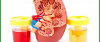
Collection of urine for analysis according to Zimnitsky
Urine collection for analysis according to Zimnitsky is carried out within 24 hours. To do this you will need 8 sterile containers (jars).
Urine collection begins in the morning. The first portion of urine after waking up is not collected, but goes down the toilet. Next, the urine is collected in jars, for which a separate jar is used every three hours:
- from 9-00 to 12-00 am;
- from 12-00 to 15-00;
- from 15-00 to 18-00;
- from 18-00 to 21-00;
- from 21-00 to 24-00;
- from 0-00 to 3-00;
- from 3-00 to 6-00 am;
- from 6-00 to 9-00 am.
Collected urine samples should be stored in the refrigerator. After collecting the last portion of urine, all material should be delivered to the laboratory.