Biliary
(or
bile
)
pigments
, also
bilins
- biological pigments, linear tetrapyrroles[1], formally derivatives of bilan (bilinogen) with oxidized terminal pyrrole cores, formed during heme catabolism.
They were first isolated from bile, which is given a characteristic color, which is where they got their name; The color of various bile pigments ranges from yellow-orange to blue-green. They are formed in many organisms as a product of the metabolism of certain porphyrins. Bilin (also called bilichrome
) has been named as the bile pigment of mammals, but it can also be found in lower vertebrates, invertebrates, as well as in red algae, green plants and cyanobacteria. The color of bilins can vary from red, orange, yellow and brown to blue and green.
Chemically speaking, bilins are a linear structure of four pyrrole rings (tetrapyrroles). In human metabolism they are represented by bilirubin, a product of heme destruction. Hydroxymethylbilane is one of the popular anabolic agents, obtained by the biosynthesis reaction of porphobilinogen (PBG) and uroporphobilinogen I (the reaction is widely known as porphobilinogen deaminase).
Bilins have been found in animals, and phycocinobilins in the chromophores of the photosynthetic pigment phycocyanin in algae and plants. In plants, bilins also serve as photopigments for the photoreceptor protein phytochrome. An example of an invertebrate bilin is micromatabilin, which gives the green color to the spider Micrommata virescens
[2].
Biosynthesis and biological role
Bile pigments are formed in vertebrates as a result of the metabolization of hemoglobin, myoglobin and heme-containing proteins, with the oxidative cleavage of the α-methine bond of heme (the prosthetic group of hemoglobin).
The process is catalyzed by heme oxygenase (1.14.99.3), with nicotinamide adenine dinucleotide phosphate (NADPH) acting as a hydrogen donor:
heme + 3 AH2 + 3 O2 \to biliverdin + Fe2+ + CO + 3 A + 3 H2O
The process also involves hemoprotein reductase (1.14.99.3), which reduces heme oxygenase.
In the first stage of the reaction, the α-methine group is hydroxylated to form 5-hydroxyheme, after which the hydroxylated bridge is oxidized to release carbon monoxide and form verdoheme. In turn, verdogem is oxidized to an unstable complex of biliverdin with ferrous iron, which decomposes to release Fe2+ and biliverdin [3].
Subsequently, biliverdin (blue-green crystals, yellow-green color in solutions) during reduction catalyzed by biliverdin reductase is converted into bilirubin (brown crystals, yellow-orange color in solutions):
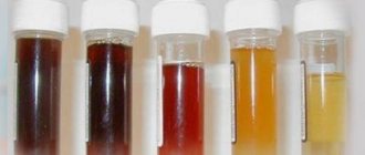
Bilirubin predominates in the bile of humans and carnivorous mammals; biliverdin predominates in the bile of herbivorous mammals, birds, reptiles and fish.
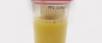
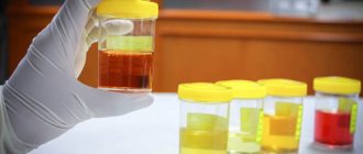
Metabolization of heme with the formation of bile pigments occurs in the cells of the reticuloendothelial system, which phagocytose old or damaged red blood cells - mainly in the spleen and Kupffer cells of the liver. In the intestine, bilirubin undergoes bacterial reduction with the formation of urobilins and urobilinogens, in particular, stercobilinogen (in humans - 40-280 mg per day). Stercobilinogen is oxidized under the influence of light to stercobilin.
Pathological conditions in which disturbances in the metabolism of hemoglobin and bile pigments lead to the accumulation of excess amounts of bilirubin in the blood (hyperbilirubinemia), leading to icteric discoloration of the skin, mucous membranes and sclera - jaundice.
Conversion of bile pigments in the intestine
Bilirubin entering the bile, reaching the distal small intestine and especially the large intestine, is exposed to normal intestinal microflora. Under the influence of its enzymes, it is deconjugated and then sequentially reduced with the addition of hydrogen atoms at the site of one or the other double bond, which is also accompanied by dehydrogenation processes. The initial stages of the recovery process may involve a certain amount of bilirubin, which still retains its conjugated form. The main role in these transformations is played by enzymes released from anaerobic microorganisms in the cecum. Experiments with fecal flora in vitro have established that it is capable of completely reducing bilirubin to the final product of its metabolism.
In the 60-70s. 20th century the study of this issue has become much more fruitful, which is connected, ch. arr., using the oxidation method with ferric chloride, followed by fractionation and study of the resulting products by mass spectrometry (see). A number of intermediate compounds have been identified. During recovery, bilirubin gradually loses its color, turning into colorless chromogens. However, at each stage of the process, dehydrogenation results in the formation of liquids colored in one color or another (see Scheme 2).
Scheme 2. Bilirubin gradually loses its color during recovery, turning into colorless chromogens. However, at each stage of the process, bile pigments colored in one color or another are formed by dehydrogenation
All these products of bilirubin conversion are called bilirubinoids (if the double bonds at positions 2′ and 7′ remain unsaturated) or urobilinoids (if these bonds are saturated). In addition, all of them can be divided into 4 types depending on the number of double bonds in the bridges connecting the pyrrole groups. These include: bilans that do not contain double bonds in bridges (mesobilirubinogen and further bilirubin reduction products); bilenes containing one double bond (eg, urobilins, stercobilin); bilidienes - with two double bonds (for example, mesobilirubin, bilirubin itself also belongs to bilidienes) and bilitrienes - with three double bonds (biliverdin, glaucobilin). The end product of bilirubin transformation is L-stercobilinogen, a colorless compound. Upon contact with air, it turns into L-stercobilin, an orange-yellow pigment, which, among other liquids, most determines the color of feces (see Stercobilin):
Apart from this main path, there is also another path which is considered as a bypass path. During the transformations of fatty acids along this path, dihydrobilirubinogen gives rise to D-urobilinogen, which in turn is converted into D-urobilin (see Urobilin):
Such a shunt path operates to some extent under normal conditions, but ch. arr. turns on when the enzymatic activity of the intestinal microflora changes, in particular when it is suppressed by broad-spectrum antibiotics (tetracycline, erythromycin). In people receiving these antibiotics, D-forms of stomach acid, predominantly D-urobilin, predominate in the intestinal contents and feces, whereas normally racemic i-urobilin and levorotatory urobilinoids predominate.
A distinctive feature of the shunt pathway for the transformation of fatty acids in the intestine is the preservation of the vinyl group (V) at the first pyrrole ring and, thus, the formation of monovinyl derivatives. It is assumed that the hydrogenation of the double bond at this position is catalyzed by a highly specific enzyme, which is lost or very weakly formed when the microflora is suppressed by antibiotics. Under these conditions, the hydrogenation of methine bridges occurs more easily than the vinyl group at the first pyrrole ring. At the same time, the ability of microorganisms to produce additional energy necessary for the conversion of D-urobilinogen into its L-derivatives decreases. Urobilinoid molecules do not have a linear structure, but are folded due to the formation of hydrogen bonds inside the molecule (see the structural formula of L-stercobilin). To change their configuration, it is necessary to break hydrogen bonds, unfold the molecule, and then fold it again in a different position. At the same time, further hydrogenation of the compound must occur. Naturally, under conditions of changes in the activity of microflora, such processes associated with the expenditure of additional energy are weakened. Therefore, the inclusion of a monovinyl shunt leads to a sharp predominance of D-urobilinoids in the total amount of liquid.
It is also noted that when the activity of intestinal microflora is suppressed by antibiotics, the conversion of bilirubin may be delayed or stopped at some intermediate stage. In this case, the unchanged bilirubin present in the feces can be oxidized in air into biliverdin, which gives them a green tint.
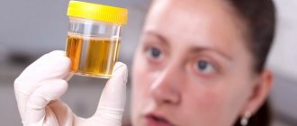
Various products of the transformation of bilirubin in very small quantities are absorbed into the blood and re-excreted by the liver as part of bile. Traces of them can appear in the general bloodstream and be excreted in the urine. All these processes are intensified in pathological conditions accompanied by an increase in the secretion of bilirubin with bile. Urobilinogen, even normally, passes into the blood of the portal vein in noticeable quantities and, oxidizing in the liver to bilirubin, is again excreted, and partly excreted with bile in an unoxidized form.
It has been established that a certain amount of bilirubin, after deconjugation in the intestine, passes into the blood of the portal vein, is conjugated in the liver and excreted with bile.
The fact of absorption of a significant amount of free bilirubin and, apparently, urobilinogen and their resecretion after appropriate transformations in the composition of bile indicates the participation of bile pigments in the hepatic-intestinal circulation. Although a relatively small part of all bilirubin entering the intestine is involved in this process, in contrast to the circulation of bile acids (see), nevertheless, the very existence of the bile acid circulation raises the question of whether they have any significance in the secretory liver activity. In hepatic bile, bilirubin is part of its lipid complex, which is endogenous lipid micelles. Such micelles can include various compounds having polar and nonpolar groups. It is possible that bilirubin, especially conjugated with acidic disaccharides, which give it pronounced polar properties, when included in this lipid complex, stabilizes its micellar structure and thereby promotes the transport of various substances in the composition of bile.
Nomenclature
| Parent structure | Number of methine groups | Color | Name | Compound |
| Bilan (5,10,15,21,23,24-Hexahydrobilin) | 0 | Colorless | Mesobilirubinogen (Urobilinogen I) | |
| Stercobilinogen | ||||
| Urobilinogen D (Mesobilirubinogen D) | ||||
| Bilene (5,15,21,24-Tetrahydrobilin) | 1 | from yellow to orange | Stercobilin | |
| Urobilin D (Mesobilin D) | ||||
| Urobilin I (Mesobilin I) | ||||
| Biladienes (10,23-Dihydrobilin, 5,21-Dihydrobilin) | 2 | orange-red | Bilirubin | |
| Mesobilirubin | ||||
| Phycoerythrobilin (see Phycobilins) | ||||
| Bilin (bilatriene) | 3 | from green to blue-green | Biliverdin | |
| Phycocyanobilin (see Phycobilins) |
Notes
- [goldbook.iupac.org/T06291.html tetrapyrroles // IUPAC Gold Book]
- Oxford, G. S. & Gillespie, R. G. (1998). Evolution and Ecology of Spider Coloration. Annual Review of Entomology 43:619-643.doi: [dx.doi.org/10.1146%2Fannurev.ento.43.1.619 10.1146/annurev.ento.43.1.619 ]
- [www.chem.qmul.ac.uk/iubmb/enzyme/EC1/14/99/3.html EC 1.14.99.3 // IUBMB Enzyme Nomenclature]
| This is a draft article on biochemistry. You can help the project by adding to it. |
| This is a preparation for an article on physiology. You can help the project by adding to it. |
An excerpt characterizing bile pigments
- Why not? - said the princess. No one answered, and Princess Marya, looking around the crowd, noticed that now all the eyes she met immediately dropped. - Why don’t you want to? – she asked again. Nobody answered. Princess Marya felt heavy from this silence; she tried to catch someone's gaze. - Why don’t you talk? - the princess turned to the old man, who, leaning on a stick, stood in front of her. - Tell me if you think anything else is needed. “I’ll do everything,” she said, catching his gaze. But he, as if angry about this, lowered his head completely and said: “Why agree, we don’t need bread.” - Well, should we give it all up? Do not agree. We don’t agree... We don’t agree. We feel sorry for you, but we do not agree. Go on your own, alone...” was heard in the crowd from different directions. And again the same expression appeared on all the faces of this crowd, and now it was probably no longer an expression of curiosity and gratitude, but an expression of embittered determination. “You didn’t understand, right,” said Princess Marya with a sad smile. - Why don’t you want to go? I promise to house you and feed you. And here the enemy will ruin you... But her voice was drowned out by the voices of the crowd. “We don’t have our consent, let him ruin it!” We don’t take your bread, we don’t have our consent! Princess Marya again tried to catch someone's gaze from the crowd, but not a single glance was directed at her; the eyes obviously avoided her. She felt strange and awkward. - See, she taught me cleverly, follow her to the fortress! Destroy your home and go into bondage and go. Why! I'll give you the bread, they say! – voices were heard in the crowd. Princess Marya, lowering her head, left the circle and went into the house. Having repeated the order to Drona that there should be horses for departure tomorrow, she went to her room and was left alone with her thoughts. For a long time that night, Princess Marya sat at the open window in her room, listening to the sounds of men talking coming from the village, but she did not think about them. She felt that no matter how much she thought about them, she could not understand them. She kept thinking about one thing - about her grief, which now, after the break caused by worries about the present, had already become past for her. She could now remember, she could cry and she could pray. As the sun set, the wind died down. The night was quiet and fresh. At twelve o'clock the voices began to fade, the rooster crowed, the full moon began to emerge from behind the linden trees, a fresh, white mist of dew rose, and silence reigned over the village and over the house. One after another, pictures of the close past appeared to her - illness and her father’s last minutes. And with sad joy she now dwelled on these images, driving away from herself with horror only one last image of his death, which - she felt - she was unable to contemplate even in her imagination at this quiet and mysterious hour of the night. And these pictures appeared to her with such clarity and with such detail that they seemed to her now like reality, now the past, now the future. Then she vividly imagined that moment when he had a stroke and was dragged out of the garden in the Bald Mountains by the arms and he muttered something with an impotent tongue, twitched his gray eyebrows and looked at her restlessly and timidly. “Even then he wanted to tell me what he told me on the day of his death,” she thought. “He always meant what he told me.” And so she remembered in all its details that night in Bald Mountains on the eve of the blow that happened to him, when Princess Marya, sensing trouble, remained with him against his will. She did not sleep and at night she tiptoed downstairs and, going up to the door to the flower shop where her father spent the night that night, listened to his voice. He said something to Tikhon in an exhausted, tired voice. He obviously wanted to talk. “And why didn’t he call me? Why didn’t he allow me to be here in Tikhon’s place? - Princess Marya thought then and now. “He will never tell anyone now everything that was in his soul.” This moment will never return for him and for me, when he would say everything he wanted to say, and I, and not Tikhon, would listen and understand him. Why didn’t I enter the room then? - she thought. “Maybe he would have told me then what he said on the day of his death.” Even then, in a conversation with Tikhon, he asked about me twice. He wanted to see me, but I stood here, outside the door. He was sad, it was hard to talk with Tikhon, who did not understand him. I remember how he spoke to him about Lisa, as if she were alive - he forgot that she died, and Tikhon reminded him that she was no longer there, and he shouted: “Fool.” It was hard for him. I heard from behind the door how he lay down on the bed, groaning, and shouted loudly: “My God! Why didn’t I get up then?” What would he do to me? What would I have to lose? And maybe then he would have been consoled, he would have said this word to me.” And Princess Marya said out loud the kind word that he said to her on the day of his death. “Darling! - Princess Marya repeated this word and began to sob with tears that relieved her soul. She now saw his face in front of her. And not the face that she had known since she could remember, and which she had always seen from afar; and that face is timid and weak, which on the last day, bending down to his mouth to hear what he said, she examined up close for the first time with all its wrinkles and details. “Darling,” she repeated. “What was he thinking when he said that word? What is he thinking now? - suddenly a question came to her, and in response to this she saw him in front of her with the same expression on his face that he had in the coffin, on his face tied with a white scarf. And the horror that gripped her when she touched him and became convinced that it was not only not him, but something mysterious and repulsive, gripped her now. She wanted to think about other things, wanted to pray, but could do nothing. She looked with large open eyes at the moonlight and shadows, every second she expected to see his dead face and felt that the silence that stood over the house and in the house shackled her.
Deposition of bile pigments in organs
Deposition of bile in organs, tissues and cells occurs with different types of jaundice (see), especially with obstructive jaundice, with hemolytic disease of newborns (see), bilirubin infarction (see), in the case of bile staining of necrotic tissue, and also after hemorrhages.
Fatty acids visible in cells and tissues under a light microscope and detected by histochemical methods include bilirubin, biliverdin, and hematoidin. The latter pigment is identical or similar in structure to bilirubin. Chem. and physical properties of Zh. items make it quite easy to distinguish them histochemically [AGE Pearse, 1962; Lillie (RD Lillie), 1969]. They do not exhibit primary luminescence, are insoluble in most fatty solvents and do not discolor under the influence of oxidizing agents. Bilirubin and hematoidin are characterized by a positive Gmelin reaction, intense reduction of ferricyanide and blackening after treatment with ammonia silver at 250° for 5-16 hours. The Gmelin histochemical reaction (see Gmelin test) is carried out by introducing conc. nitrogen, followed by a quick examination of the section under a microscope. Areas containing bilirubin and hematoidin crystals show a color transition from red to green, purple and blue. This is due to the oxidation of pigments into greenish bilirubin (bilathriene) and reddish-violet bilipurpurines. In the final stage of the reaction, the blue compounds are oxidized into yellow choletins. In a number of histochemical methods, oxidizing agents are used to convert yellowish-brown bilirubin into green biliverdin. These methods include the Stein method (J. Stein) using iodine (Lugol solution and tincture of iodine) as an oxidizing agent, the Glenner method (G. Glenner) with oxidation with potassium dichromate in an acidic environment (pH 2.2), oxidation by Kutlik (IE Kutlik, 1957) with ferric iron compounds (FeCl3, etc.). To check whether the pigment present in the tissue is indeed bilirubin, when performing these reactions, it is necessary to ensure that in control sections that were not exposed to the oxidizing agent, the pigment is brown and after oxidation this color turns to green. The use of ferrous potassium syneride makes it possible to bring the process of bilirubin oxidation even further than with the above reactions, namely to the stage of bilipurpurins.
Deposition of bile pigments (bilirubin and biliverdin) under pathological conditions can be observed in the tissues of the liver, kidneys, brain, intestines and other organs. Noticeable accumulation of fatty acids in the liver occurs with extrahepatic and intrahepatic cholestasis (see). Condensed masses of liquid matter in the form of the so-called. bile thrombi accumulate in the lumen of small bile ducts and dilated bile capillaries. The conversion of some bilirubin to biliverdin gives bile clots a greenish tint. In case of cholestasis, liver granules are also found in the cytoplasm of liver cells and stellate endothelial cells (Kupffer cells). Electron microscopy reveals liver cells in damaged liver cells in the form of large amorphous clusters of spherical shape, partially or completely surrounded by a single-layer membrane, or in the form of needle-shaped electron-dense bodies surrounded by a membrane of disintegrated mitochondria; in the intercellular spaces and bile capillaries, bile ducts have the appearance of electron-dense and needle-shaped formations, not surrounded by membranes. Increased secretion of fluids by the kidneys during obstructive jaundice is accompanied by their accumulation in hyaline and granular cylinders of the renal tubules (so-called icteric nephrosis). With hemolytic disease of newborns, kernicterus occurs with staining of the gastrointestinal tract of the nuclei of the cerebral hemispheres and the brain stem. This causes irreversible damage to the ganglion cells of the brain. Necrotic tissues easily adsorb bile along with the liver. This explains the greenish-yellow staining of Peyer's patches and solitary follicles in typhoid fever and the imbibition of the liver in foci of necrosis of the liver parenchyma in biliary cirrhosis. The yellow color of the liver in acute massive necrosis or acute toxic dystrophy, which occurs as a complication of Botkin’s disease, is also due to staining of necrotic tissue of the liver. According to I. V. Davydovsky, bile pigments, together with bile acids, penetrating into the tissues, can have an effect on them necrotizing effect.
Hematoidin is found in the area of extensive old hemorrhages and hemorrhagic infarctions. The pigment has the appearance of golden-yellow grains or crystals in the shape of stars, needles, and rhombic plates. It is formed intracellularly in macrophages that have absorbed hemoglobin breakdown products. After the destruction of macrophages, the pigment is located extracellularly in the center of the hematoma.
Bibliography:
Davydovsky I.V. General human pathology, M., 1969; Lilly R. Pathohistological techniques and practical histochemistry, trans. from English, M., 1969; Nesterin M. F. Narodetskaya R. V. and Shlygin G. K. Separation by the liver of a lipoprotein complex compound in the bile, Physiol, journal. USSR vol. 51, Ko 12, p. 1487, 1965; Pierce E. Histochemistry, trans. from English, Moscow 1962; Shlygin G.K. Modern ideas about the direct participation of the gastrointestinal tract in metabolism, Vestn. USSR Academy of Medical Sciences No. 12 p. 27, 1968; Klatskin G. Bile pigment metabolism, Ann. Rev. Med v. 12, p. 211, 1961, bibliogr.
G. K. Shlygin; V. B. Zolotarevsky (hist.).