Author of the article
Anatoly Shishigin
Reading time: 4 minutes
AA
Diseases of the genitourinary system often provoke the development of pathology, which disrupts the functioning of the urinary tract. Acquired or congenital defects form narrowings in the ureteral lumen, and in some cases completely block it.
Installing a stent in the ureter allows one to avoid complications with this problem, since all fragments are forced to expand with a narrowing in the urinary duct. Urine exits normally without stagnation.
Stenting in pregnant women
As you know, a pregnant woman is at risk of developing inflammatory processes in the urinary tract.
This is due to the pressure of the uterus on the bladder, the lack of hormonal balance and other aspects of bearing a child. When stagnation occurs, pathogenic microflora multiplies more actively, so gestational pyelonephritis in pregnant women is not uncommon. This pathology can be a threat to both the fetus and the mother. When a situation arises that threatens a woman's life, pregnancy, as a rule, does not continue, especially if there is a need to remove the kidney.
If a woman has the following pathologies, the risk of complications increases:
- urolithiasis disease;
- AIDS;
- diabetes;
- polycystic disease;
- pyelonephritis in chronic form;
- pathologies of urinary tract development.
Therefore, a stent in the kidneys during pregnancy has become extremely popular among pregnant women. It helps to avoid serious complications, successfully carry and give birth to a child. It is removed a few days after birth, and antibacterial therapy is required. Since there is no longer any reason to limit the use of antibiotics, the health of the mother is now in the foreground. Lactation during the treatment period is, of course, undesirable, but after the course of treatment you can begin breastfeeding.
The main advantage of the method is its low invasiveness. Installation of a stent does not require extensive surgery. The risk of complications after stenting is minimal, but it is absolutely necessary to monitor the patient’s health after installation of the endoprosthesis.
A stent in the kidney is necessary to restore the normal flow of urine from the kidneys to the bladder. This is a kind of assistant for the ureters. It is installed when the outflow of urine is impaired due to inflammatory diseases, or when obstructions form in the ureters.
What is a ureteral stent and why is it needed?
The stent looks like a narrow flexible tube (made of silicone or polyurethane) with spirally twisted edges. This structure allows the stent to be held in the desired position and prevents its displacement.
Stenting is performed if urine cannot be excreted from the body naturally, as well as before certain operations on the organs of the urinary system.
There are many reasons that can disrupt the natural flow of urine. Most often, the ureter is blocked by stones or blood clots.
The causes of urinary disorders can be:
- urolithiasis, if the stone penetrates the ureter and blocks it;
- benign and malignant tumors in the ureters or kidneys;
- prostate adenoma;
- complications of inflammatory diseases of the urinary system;
- swelling of the mucous membrane of the ureter;
- adhesions and strictures - narrowing of the lumen of the ureter.
Operation technology
Why and how is MRI examination of blood vessels performed?
As already mentioned, stenting is a minimally invasive procedure that is most often performed under local anesthesia. The procedure lasts from 15 minutes to half an hour.
The procedure is carried out as follows:
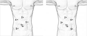
First, the doctor injects novocaine or lidocaine into the urethra to numb the pain. Then, using a special device, he examines the internal organs and finds the orifices of the ureters
If you imagine that they are located on a watch dial, then their location is at the numbers 5 and 7. Carefully installs the catheter. Using a stethoscope, the doctor makes sure that the stent is as close as possible to the opening of the ureter. Then the catheter is inserted 30 cm deep. The bladder is emptied, and the device with which all manipulations were performed is removed.
In some cases, radiography is used to ensure implantation is as accurate as possible. A day later, the patient is examined again by X-ray in order to detect stent displacement in time and correct it.
Removing a stent from the ureter
Vascular stenting
In the absence of undesirable reactions and inflammation, the drainage system is removed after two weeks, but no later than six months from the date of installation.
On average, the tube is replaced after two months.
If lifelong stenting is indicated, the device is changed every 120 days.
Frequent tube changes are necessary to avoid salt occlusions, organ infections, and damage to the ureteral mucosa.
The maximum duration of action of the stent is set by the manufacturer. The doctor takes into account the patient’s age and related factors.
The urinary structure is removed on an outpatient basis in 5 minutes under local anesthesia. This quick process is performed with a cystoscope.
A gel is placed into the urethra to facilitate passage of the device.
Under the control of X-ray equipment, the guide wire is inserted as deep as possible and the tube is straightened.
The outer end of the expander is grabbed and pulled out. The drainage system must be changed every 3–4 months. In people prone to stone formation, the tube is replaced after 3-4 weeks.
When the system is removed, the patient may experience a short-term burning sensation and tolerable pain. After the tube is removed, diagnostics are carried out for four days in order to select further treatment tactics. The patient feels discomfort when urinating for several days after removing the dilator.
Sometimes the stent needs to be removed and reinserted. But basically, while wearing the device, doctors remove the causes of blockage of the canal, and the patient can return to normal life.
Reviews about removing a stent from the ureter are as follows:
Svetlana. 55 years. Friends! I want to calm everyone down. I had a drainage structure removed from my ureter without any pain relief. You need to wait five minutes. It's unpleasant, but tolerable.
Irina is 59 years old. I was very afraid, but it turned out to be in vain. First, the nurse cleaned my genitals. I stocked up Katedzhel in advance. I recommend it to everyone before the procedure, it provided excellent pain relief. The doctor asked me to relax everything. In a second, he inserted the syringe and injected the gel. Unpleasant, but not painful. Then they stuck a cystoscope in, I even gasped. The doctor said that this is the most unpleasant thing. When the tube was pulled out, there was very slight pain for a few seconds. After the procedure, it burned a little, that’s all. The main thing is, go with the catedgel and don’t be afraid.
Sanatorium treatment
Physiology of the ureter
Sanatorium treatment of stones is prescribed to patients with urolithiasis at the initial stage or after removal of large stones. Therapy is aimed at preventing relapse and eliminating small salt deposits naturally. The main effect on the patient’s body is exerted by mineral waters, which have diuretic properties.
If the patient has uric acid or calcium oxalate deposits, it is recommended to visit resorts with slightly mineralized alkaline mineral waters. These could be Esentuki, Pyatigorsk, Zheleznovodsk. In Ukraine, the patient can visit the Truskavets resort.
You can relax and restore your health in the sanatorium at any time of the year. Similar bottled mineral waters, which can be bought in any store, will not replace a stay at the resort. Doctors recommend drinking no more than 500 ml. mineral water per day. At the same time, doctors strictly monitor the patient’s condition and conduct all relevant tests.
Urinary tract stenting
By analogy with the implantation of a stent in the arteries of the kidneys, stenting of the urinary tract - the ureteropelvic segment, the ureter - is also carried out. This is performed in cases of kidney stone removal to improve urine flow and prevent stagnation. The procedure is also indicated after ultrasonic crushing of a stone in the ureter, when remaining small stones and sand can cause spasm of the ureter and renal colic. This procedure often happens during pregnancy if a woman has urolithiasis.
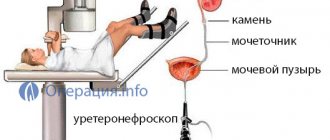
With a stent in the ureter, a woman can fully carry a child, give birth, and not be afraid of an attack of colic or the development of a kidney block, which is very dangerous during pregnancy. Subsequently, a few weeks after birth, the implant is removed from the ureter using a catheter-ureteroscope.
The procedure is carried out instrumentally and under the control of an X-ray scanner. A long flexible catheter with a rolled-up stent is inserted through the urethra and implanted into the lumen of the ureter.
Advice: if you become pregnant with urolithiasis, you need to be examined by a urologist and, if necessary, stenting. We must remember that an enlarged uterus aggravates problems with urinary tract pathology.
Modern endovasal and endoscopic technologies in many cases make it possible to avoid surgical intervention, restore the patency of blood vessels and other tubular hollow organs, and prevent the development of severe complications.
Kidney stenting is performed to normalize the functioning of the organ, which has been disrupted due to the effects of kidney diseases or injuries. A special stent is inserted into the kidney, which is a tube made of bendable plastic. Its length is about 30 centimeters. One end of the stent is placed in the pelvis and the other in the bladder.
Stenitation of the kidney is a method of restoring the functioning of the organ, characterized by minor surgical intervention.
What is a ureteral stent?
During normal functioning of the human body, the fluid secreted by the kidneys is discharged, as a rule, through ducts into the bladder. It is from this that it flows out when urinating. The ureters, those same ducts, are elastic and can expand in lumen diameter from 0.3 cm to 10 centimeters. Because of this, some pathologies narrow the entire lumen or only part of it, creating a barrier to urine filtered in the kidney.
Removing the stent from the ureter
The ureteral stent is a thin tubular-type structure, up to 0.6 centimeters wide and up to 60 centimeters long. The material used is usually polyurethane or silicone. The difference in length is due to the minimum value required to expand the fused fragment of the duct and the maximum distance from the renal pelvis to the bladder, at the apex of which the ureteral orifice is located.
Based on the length of the stent, its end (or both) is often curved in a spiral, which gives better fixation within the organ (bladder or kidney). This way the device will not move, or only by a small distance.
Postoperative period and possible complications
In the first few days after kidney stenting, the patient may feel some discomfort in the abdomen or lower back, experience pain and a frequent urge to urinate. To eliminate these symptoms, sufficient fluids and antispasmodics are prescribed.
Success in the treatment of urinary tract obstruction using stenting is determined by the frequency and type of complications of manipulation, which may be associated with the technique of implantation or removal of the stent or occur while wearing the stent in the ureter. Highlight:
- Perforation of the ureter - possible during the insertion of a stent due to inaccurate or violent movements of the surgeon’s hand, trying to overcome obstacles in the form of scars, stones, etc.;
- Injury to the renal pelvis - in the presence of stones and rough actions of the surgeon;
- Vesicoureteral reflux - develops in more than half of patients after stenting due to poorly selected drainage, disorder of urinary tract tone;
- Inflammatory processes - in the presence of bacteria in the urine, reflux, severe and prolonged stenosis of the urinary tract, concomitant chronic inflammation of the kidneys (antibiotic therapy after stenting and prophylactic administration of antibiotics before the procedure are indicated);
- Movement of the internal stent - if it is implanted incorrectly, or if smooth stents are used, as if they are sliding into the bladder;
- Urinary disorders - a feeling of discomfort in the early postoperative period, which may require changing the position of the stent;
- Deposition of salts and the appearance of stones on the stent with its closure, perforation or fragmentation inside the ureter (with prolonged stay in the ureter, the presence of crystals in the patient’s urine, infection).
Preventive measures during stent implantation include prescribing antibiotics before and after the procedure according to the flora present in the urine, stimulating diuresis, maintaining a drinking regime, careful endoscopic and radiological monitoring during surgery, regular catheter changes in case of salt deposition and stone formation.
The stent is removed quickly under local anesthesia, using a cystoscope and under X-ray control. Before removing the drain, the patient undergoes the same examination and preparation as before its implantation. Immediately after removing the catheter, pain and burning in the urethra and abdomen are possible. Stent removal using an endoscope is less painful and more comfortable for the patient.
After the stent is removed, the patient is under the supervision of a urologist for several days. He undergoes urine tests, is given antibiotics to prevent infectious complications, uroseptics, and his drinking regime is regulated. Discomfort when urinating goes away after 2-3 days.
Thus, internal ureteral stenting seems to be a fairly convenient and relatively safe way to restore the patency of the upper urinary tract, which ensures the preservation of the patient’s quality of life and the physiological path of urine outflow
For successful treatment of pathology by stenting, it is important to be careful during surgery, select the right stents, and evaluate the indications and contraindications for surgery.
What types of stents are there?
Devices in the ureter can have different designs, but they are all needed to eliminate the narrowing in the canal. Stents differ in type of design:
- of various diameters for ducts with any lumen;
- typical with a length of 3-32 centimeters, equipped with a pair of endings in the form of spirals;
- slight elongation, up to 61 cm, with a single ending in the form of a spiral;
- pyeloplastic, used in surgical interventions for plastic surgery in the urological field;
- transcutaneous, characterized by a special shape and changing length or appearance based on the installation requirements according to indications;
- stents with extensions along the entire length;
- with a special shape for comfortable and safe removal of stones outside after the crushing procedure.
More on the topic: How is a rapid urine test performed?
All elongated structures are placed most often during pregnancy, when the fetus puts pressure on the ureteral canal. Then the stent should be fixed at only one end, while the second should be installed with a margin of distance due to the displacement of organs during pregnancy.
According to the target orientation, stents are either uncoated or with a hydrophilic coating. The presence of a coating is required in cases where drainage must be installed in the duct for a long time, which is dangerous due to the occurrence of infection. The coating of the stent surfaces resists infectious agents, reducing adhesive processes on the canal walls. With this option, you can use the system for quite a long time. All devices are supplied in various configurations, but always include a pusher, the tent itself and a conductor with a fixed or movable core.
To determine whether the stent is installed correctly, it is made of polyurethane with radiopaque properties, which is clearly displayed on the pictures.
Stent in the kidney - what is stenting
Kidney stenting is a minimally invasive procedure (that is, carried out without extensive disruption of the integrity of the skin), which involves inserting a stent into the urethra. A stent is a tube about 30 cm long that is placed through the urethra into the patient's kidney to facilitate drainage of the urinary tract that is obstructed by a medical condition.
What is kidney stenting?
We recommend! For the treatment of PYELONEPHRITIS and other KIDNEY DISEASES, our readers successfully use Elena Malysheva’s method. After carefully studying this method, we decided to bring it to your attention.
- blockage of the urinary tract by blood clots or the formation of adhesions;
- consequences of infections and inflammatory processes;
- sand and kidney stones;
- lymphomas and tumors of various types;
- swelling of the mucous membrane of the internal walls.
The stent is installed for various purposes and for various periods. It can remain in the patient’s body for a period from a week to a year.
However, you should regularly monitor the condition of the stent using hardware diagnostic methods, and also, if necessary, get rid of salt deposits.
In addition, the patient needs to be attentive to his feelings in order to promptly inform the doctor about the discomfort he is experiencing. The following symptoms may be reasons to contact a specialist:
- frequent urge to urinate,
- change in urine color or blood in it,
- pain in the lower back or abdominal area.
To avoid accidental displacement of the stent, it is not advisable for the patient to engage in active sports throughout the entire period that the stent is in the body.
Renal artery stenting is performed to normalize blood pressure. It is known that patients with narrowing of the lumen of the renal artery develop stable hypertension, which provokes various pathological conditions of the body, including the development of myocardial infarction or hemorrhagic stroke.
With the development of such hypertension, drug methods of reducing pressure are ineffective, but stenting allows stabilizing the functioning of the kidneys and cardiovascular system of the body.
The main indications for stenting the renal artery are:
- atherosclerotic kidney damage;
- refractory form of arterial hypertension;
- atherosclerotic plaques in the renal artery.
There are two unknown words - stent, stenting, so a little theory in general. In medicine, a stent is a cylindrical frame structure.
Simply put, it is a special tube made of metal or plastic.
The purpose of the design is to expand the area that has narrowed due to pathological processes in the body, so stents are placed only in hollow organs, in their lumen.
What is stenting?
A stent in the ureter is a small tube, 8-60 cm in size and no more than 0.6 cm wide. It is made of special materials - silicone or polyurethane. It is used for various diseases of the kidneys and ureter. Due to the large length, the ends of this structure may not be the same. A coil stent is mainly used for better fixation in any organ.
There are also other types of it:
- stents of any diameter;
- standard designs with a length of 30-32 cm and spiral ends;
- long up to 60 cm with one spiral end;
- pyeloplastic – used in urological operations;
- transcutaneous – can change length and shape if necessary;
- having a certain shape for better passage of kidney stones.
If a person needs long-term use of this design, then special types of stents are installed - with and without hydrophilic coating. The first type is determined by particularly long-term use, as well as in the presence of infectious diseases. Thanks to the coating, the impact of microbes is weakened and pathology does not develop.
Removing the tube from the ureter
| Procedures and manipulations | price, rub. |
| Stent installation (without stent cost) cystoscopy | 8800 |
| Stent removal cystoscopy | 9900 |
Removal of a stent from the ureter in both men and adolescent boys occurs under local anesthesia. For this purpose, a special gel is used, which simultaneously improves the gliding of the system during extraction. Removal of the ureteral stent is performed after normalization of ureteral functions.
Removal occurs using a cystoscope, which the doctor inserts into the urethra. Next, the free end of the system is grabbed and pulled out. After removal of the stent, the patient feels discomfort, as well as during its installation. Usually after 2-3 days the discomfort goes away on its own.
The process of removing a stent from the ureter is simpler than installing it, but does not exclude the use of diagnostics, during which its position in the ureteral canal is assessed. During the process of removing the stent from the ureter and after the procedure, mandatory use of antibiotics is indicated to prevent the development of an infectious process.
You should not get carried away with the stenting procedure; you need to remember that this is a foreign body that should not be permanently present in the human body.
It is installed only to facilitate the process of urination. After the situation improves, mandatory removal is carried out, the duration of use is no more than six months.
Removal of the structure is carried out under local anesthesia. To do this, a cytoscope and gel are inserted into the urethral area; it allows the tube to come out more easily.
Grab the tip of the structure, which is located outward, and carefully pull out the entire stent. If the patient is indicated for a long-term use of a stent, then it must be remembered that it is replaced once every three months, otherwise unpleasant complications will develop.
After removal of the structure, the patient may feel slight discomfort for several days or an increase in body temperature.
Ureteral stents should be removed promptly, either as soon as they are no longer needed or every 9-12 weeks.
Otherwise, mucosal damage, tube occlusion, or urinary tract infection may occur. The procedure is performed under local anesthesia in an outpatient setting.
A cystoscope and a special gel are inserted into the urethra to facilitate the passage of the tube. The outer end of the stent is grabbed by the instrument and pulled out.
A ureteral stent is a device designed to widen a duct that allows urine to be drained from the kidneys to the bladder. It is installed for ureteral obstruction, as well as before lithotripsy of kidney stones. When the procedure is carried out correctly and high-quality material is used, the stent does not cause long-term discomfort
It is very important to change it every 3 months
For recipes for effective folk remedies for cleansing the kidneys, see the following material.
The stent should not be subject to the damaging effects of salts and at the same time be contrast. It is necessary to monitor its condition and location. If the tube is displaced or ruptured, immediate surgery is required.
Stents come in different lengths and are made of different materials; The implants also differ in the end of the tubes. Some have curved “tails” on both sides for better retention, while others have only one side of the implant.
Possible complications
Each patient has a different reaction to the invasion of a foreign body into the tissue. The following complications may occur after stent placement:
| № | Helpful information |
| 1 | bleeding in urine (hematuria) |
| 2 | burning and pain of varying intensity |
| 3 | dysuria |
| 4 | swelling of the ducts or bladder |
For lower back pain, relief occurs after a while, but the need to monitor the patient's condition remains quite acute during the use of the stent. If symptoms worsen, it may be necessary to remove the device, including:
- for infection and inflammation;
- when the drainage is displaced and incorrectly installed;
- with swelling and spasms of the lumen due to the stent;
- salt deposits on the walls of the device;
- damage to the walls of the ureter when installing a drainage system;
- with reflux from the ureter into the bladder.
The stent should be removed in case of gross hematuria, as well as when there is an allergy to the implant or when the patient has a prolonged increase in body temperature.
Temperature after removal of ureteral stent
If the temperature rises after removal of the stent in the ureter, the patient's temperature may rise and last for several days. In most cases, the device is removed or there is a need for repeated manipulation. An increase in temperature may occur due to infection of the organ or stagnation of urine due to reflux. The doctor prescribes a course of antibacterial drugs to avoid the development of complications.
In some cases, the temperature after removal of the stent from the ureter increases due to an infection that entered the body due to poor sterilization of instruments or the stent itself before immediate installation. The main symptom of the development of infection is pain during deurination.
More on the topic: How do specialists treat stress urinary incontinence?
Pain accompanied by distension of the lower back against a background of elevated temperature indicates vesicoureteral reflux, when during deurination the patient feels chills, intense pain in the lower back and distension in the abdomen. This condition is very dangerous for humans and requires emergency hospitalization.
Types of stenting
Kidney stenting is a surgical intervention that is performed in a minimally invasive manner using anesthesia, and a stand is placed in the kidney. The attending physician decides which type of anesthesia to use in a particular case. If the procedure is performed on a child, general anesthesia is indicated for safety reasons. This method of surgical intervention is performed when difficulties arise with the circulation of urine through the ureters (with neoplasms of various etiologies and stones). Kidney stenting can be of the following types:
- retrograde, when the tube is inserted through the bladder;
- anterograde, in which the doctor makes a small hole in the abdominal cavity, attaches a nephrostomy and inserts a catheter;
- stent in the renal arteries.
Renal artery stenting is performed when the artery of the organ is narrowed, which causes an increase in blood pressure. Stenting of the renal artery is performed by introducing a stent, which is initially in a compressed form. It is placed at the site of the stenosis, then an angiography is performed, which will show the correct placement of the tube. If everything is done without errors, the stent is deployed inside the artery using high pressure.
Indications
High blood pressure due to renal artery stenosis is an indication for stent placement.
The stand is installed in the following cases:
- high blood pressure, in which drug therapy does not produce results, and diagnosis indicates renal artery stenosis;
- high blood pressure in young people, in which pathological renal failure is observed.
Let's consider the diseases for which stenting is performed:
- adhesions and scars on the kidneys or in the ureter after inflammatory diseases or resulting from surgery;
- the presence of kidney stones;
- formation of a malignant or benign neoplasm on organs, when the organ is damaged by metastases;
- lymphomas;
- surgery to remove stones using the endoscopic method;
- abdominal surgery on the abdominal cavity;
- radiological therapy of the abdominal organs;
- infection.
Contraindications
A stent is not placed in the following cases:
- renal artery damage;
- respiratory problems;
- development of renal failure;
- problems with blood clotting;
- allergic reaction to medications used during surgery.
Stent during pregnancy
In the case when a woman develops a stone in the kidney and ureter during pregnancy, stenting is used. This allows you to avoid the development of undesirable consequences, as well as preserve the health of the mother and unborn child. Because during this period, taking antibiotics and other drugs is contraindicated for women, as they can harm the fetus.
After childbirth, the stent is removed and complex therapy is then carried out using medications. A stent in the kidney during pregnancy is prescribed only for certain pathologies and after a complete diagnosis.
Installation
A stent is a narrow metal, polymer or silicone tube that easily expands to fit the shape of the ureter. The length of the structure ranges from 10 cm to 60 cm.
Ureteral stent
A silicone dilator is considered optimal for short wearing periods, since such material is less affected by urine salts. The disadvantage of this type of stent is that it is difficult to fix.
If the therapy is planned to be used for a long time, then it is preferable to introduce a metal dilator, since rapid coverage of the material with epithelium prevents movement of the device.
The construct is inserted into the ureter in a sterile hospital environment in two ways:
- retrograde;
- antegrade.
Retrograde method
The method is used for compaction of the walls of the ureter, stones, tumors, and pathological pregnancy.
The stent barrel is inserted into the duct through the bladder.
For pregnant women, often in later stages, stenting is prescribed in case of poor urine drainage and the threat of nephrosis, paying attention to the hypoallergenic nature of the structure. The tube is monitored monthly with ultrasound
The stent is removed 30 days after delivery.
Installation of a stent in the ureter is accompanied by slight discomfort. The patient does not require general anesthesia or preoperative procedures, except for restricting fluid and food intake the day before.
Local anesthesia is assumed using dicaine, lidocaine or novocaine. It is enough to achieve relaxation of the sphincters of the urinary system. For children, stenting is performed under general anesthesia.
If blood or pus is released during the process, the procedure is stopped and the patient is further examined, since impurities in the urine make visualization of the ureters impossible.
To control the insertion of the stent into the lumen of the ureter and assess the blockage of the canal, the urologist uses a cystoscope device inserted through the urethra.
After the procedure, the cystoscope is removed and an x-ray of the ureter is taken to monitor the position of the dilator. You can leave the clinic on the same day.
Please note that after any anesthesia you should not drive a car. On the day of surgery, wear comfortable, loose clothing.
Antegrade method
If the urinary organs are injured, the urethra is not passable and insertion by the first method is impossible, the alternative stenting method is used.
The construct is inserted into the kidney through an incision with a catheter installed in the lumbar region.
For further outflow of urine, one end of the tube descends into the external reservoir. The installation is controlled by x-rays.
In case of adverse reactions or rejection, the catheter is left closed for three days after surgery. This method requires general anesthesia and a 2-day hospital stay.
The duration of installation of the expander is from 15 to 25 minutes. The timing of fixation of the urinary structure depends on the patient’s condition.
It must be emphasized that the operation to insert and secure the stent is usually simple and is generally completed successfully.
Features of the installation procedure
A stent is placed in the ureter after procedures for therapeutic and diagnostic purposes, which is extremely necessary to reduce the risks of complications. Diagnosis is performed using ultrasound equipment, x-rays (excretory type urography), cystoscopy and MRI. These examinations determine the parameters and dimensions of the ureter, its structural features, areas with narrowing and their location. In excretory urography, due to the contrast agent that the kidneys secrete when filtering blood, a picture of the urine excretion pathways is obtained.
More on the topic: How are strips designed to determine acetone in urine?
Drainage is placed under anesthesia, which is done through the mouths of the canals in a retrograde manner. If a stent is placed in children, general anesthesia is given. If there are pathologies that interfere with non-invasive methods, a stent is placed through an incision in the body called a nephrostomy. This method is called antegrade.
A cystoscope with fiber-optic equipment is inserted through the urethra into the cavity of the bladder, and an image of its internal walls and the place where the ureter enters is displayed on the screen. A stent is inserted into the lumen and fixed, after which the cystoscope is removed. After installation is completed, diagnostics are carried out to determine the final location of the drainage.
The entire operation lasts 25 minutes, but due to the anesthesia used, the patient needs to monitor the condition for several more days. All this time, it is important to take a large amount of fluid to avoid stagnation in the drainage and kidneys.
If inflammation is present, before installing a stent it is necessary to undergo a course of antibacterial treatment with medications as prescribed by a doctor.
Installation technique
The operation to install a stent belongs to the category of minimally invasive procedures, but its implementation involves the use of anesthesia.
During the day before stenting, it is forbidden to consume food and liquid. A prerequisite for the operation is a completely empty bladder. Cleansing the intestinal walls and eliminating excess gases is carried out using an enema.
The duration of the stent installation procedure is on average up to twenty minutes. Stages of stenting:
- preparatory measures (conversation with the patient, enemas, general recommendations);
- To anesthetize the procedure, the patient is given Lidocaine or Novocaine;
- before performing the operation, a thorough examination of the ureteral orifices is necessary;
- installation of a catheter (when a stent is inserted through an incision in the abdominal cavity);
- securing one end of the stent and bringing the second out;
- the procedure is controlled using a stethoscope;
- after fixing the stent, all additional devices are removed;
- The outcome of the operation is studied using an x-ray (after 24 hours, the x-ray is taken again).
Throughout the operation to install the stent, the doctor monitors the procedure through a computer monitor.
X-ray support of surgical intervention is a prerequisite for stenting.
This procedure is necessary to determine the exact location of the stent and check its condition. If the tube does not fit tightly to the orifices of the ureter, there is a risk of developing serious complications.