Until recently, the main method of treating kidney and ureteral stones was open surgery (at the moment their percentage is negligible, and in our clinic it does not exceed 1%). Constant attempts to create less traumatic operations through small accesses
or
natural openings (urethra)
led to the invention of special endoscopic instruments, which, having been significantly improved, are successfully used in urology.
How to prepare for surgery?
Before surgery, each patient undergoes an examination aimed at determining contraindications for surgery. Consultation with a therapist and anesthesiologist is mandatory. A list of medications that the patient uses is determined, since some medications can cause complications during surgery. The patient must report all existing diseases.
Before the operation, a thorough bowel preparation is carried out, and broad-spectrum antimicrobial drugs are prescribed to prevent infectious complications in the postoperative period.
Stages preceding surgery
Consultation with a specialist. The choice of surgical treatment is based not only on laboratory and instrumental examination data, but also on complaints and medical history. A physical examination is also performed and a number of diagnostic tests are ordered. Based on their results, the doctor determines the absence or presence of indications for removing kidney stones using percutaneous nephrolithotripsy and sets the exact date of the operation.
Diagnostic studies. Before removing kidney stones, the patient must undergo a series of tests. The standard list (which can be supplemented with other laboratory and instrumental diagnostic methods) includes:
- general urine analysis;
- blood tests: analysis for Rh factor and blood group, general analysis (with platelets and leukocyte formula), biochemical analysis (total bilirubin, protein, creatinine, AST, ALT, glucose), blood test for syphilis, hepatitis C and B;
- coagulogram (prothrombin time, fibrinogen, INR and APTT);
- ECG with a detailed description, completed no later than a month before the preoperative examination.
Preparing for surgery. At least five days before the procedure to remove kidney stones, the patient is advised to stop taking certain medications. These include medications that affect blood clotting: aspirin, Prodaxa, warfarin, etc. In addition, in the period preceding stone removal, the patient must provide the doctor with a complete list of the medications he is using. This is necessary in order to promptly stop taking medications that can cause complications or affect the effectiveness of removing kidney stones.
How is CULT performed?
Most often, the operation is performed under epidural anesthesia, which makes it completely painless. At the same time, at the request of the patient, he can remain conscious.
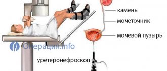
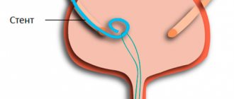
The patient is placed on his back with his legs apart. The operation begins with ureteroscopy and cystoscopy (examination of the urethra and bladder), followed by ureteroscopy (examination of the ureter). Once a ureteral stone is detected, its destruction begins using various types of energy (laser, pneumatic, ultrasonic and electrohydraulic lithotripters). To prevent stone fragments from migrating, special “baskets” are used to remove stones from the ureter. In some cases, special thin forceps are used to remove ureteral stones. At the end of the operation, if necessary, a stent is placed on the ureter on the side of the operation (the tube connecting the renal pelvis to the bladder), which is removed 1-2 weeks after the operation under local anesthesia.
Percutaneous nephrolithotripsy
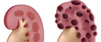
The essence of the method. Percutaneous nephrolithotripsy is one of the most modern types of operations to remove kidney stones. This type of X-ray endoscopic treatment is currently widely used in urology. If a patient is diagnosed with large coral-shaped kidney stones, surgery using this method will be much more effective in comparison with ESWL (external shock wave lithotripsy).
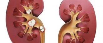
Indications. The main indication for stone removal using percutaneous nephrolithotripsy is the individual characteristics of the patient, excluding impact techniques, as well as the presence of large hard stones consisting of calcium oxolate and brushite. The localization zone of the formation is also important. The procedure is indicated in cases where the calculus is located in the lower poles of the organ.
Advantages. The main indication for performing percutaneous nephrolithotripsy is the size of the stone exceeding 2 cm, as well as the density of the stones (1500 HU), especially “hard” stones consisting of calcium oxalate. This operation eliminates the negative impact of external shock wave lithotripsy on the kidney parenchyma.
What complications can occur during and after contact ureterolithotripsy?
Complications during this manipulation can occur at any stage, but their number currently does not exceed 5%. Complications that may occur during surgery include:
- migration of stone fragments into the collecting system during surgery. To prevent migration, various types of devices (baskets) are used;
- Ureteral perforation is essentially a “perforation” of the ureter with a ureteroscope. (in order to avoid this complication, the doctor must insert a safety wire into the ureter through which he then inserts the ureteroscope);
- ureteral avulsion is an extremely rare complication that occurs when the ureteroscope is aggressively passed through the ureter, especially if there are narrowings (strictures) or “impacted” stones.
It is also possible to develop complications in the postoperative period:
- attack of acute pyelonephritis;
- stricture (narrowing) of the ureter.
Where to go
Urological Center of Branch No. 1 of the Federal State Budgetary Institution "GVKG named after. Academician N. N. Burdenko" provides a full range of services related to the diagnosis and treatment of pathologies of the genitourinary system. These also include the removal of kidney stones and operations related to the removal of stones from other parts of the excretory system. The advantages of our center include:
- modern technical equipment that allows stone removal using minimally invasive methods;
- many years of experience in the treatment and diagnosis of various urological diseases;
- staff of qualified specialists of the highest category.
What to expect after contact ureterolithotripsy?
On average, contact ureterolithotripsy lasts from 30 minutes to 2 hours. Its duration depends on the size, density, number of stones removed, and the condition of the ureter. After the operation, you will be transferred to the intensive care unit (ICU) or to a ward (depending on your condition).
On average, after surgery, hospitalization lasts about 2-3 days. A shorter hospital stay is also possible, it all depends on the patient’s condition and the effectiveness of the operation.
The urethral catheter is removed the day after surgery.
We have developed a system for proper stone formation prevention
Surgical treatment removes the product of urolithiasis, but cannot eliminate the cause. Therefore, we subject the removed stones to X-ray isotope analysis. Having determined its composition, we understand what metabolic disorder or disease leads to the deposition of salts in the form of stone. We prescribe the necessary diet to remove stone-forming substances from the body. If necessary, we order additional studies. The formation of stones can be affected by fractures of the pelvic and spinal bones, changes in the urinary tract, infectious and many other diseases. When the cause is established, we carry out complex personalized therapy aimed at combating the specific disease or condition leading to stone formation.
Instrumentation for removing ureteral stones.
Extractors.
Based on the low mortality rates, ureteroscopic removal of small ureteral stones is a relatively quick procedure compared with lithotripsy. This method should first be used to target small stones in the lower third of the ureter. New forms of baskets for endoscopic stone removal have already appeared. A tipless nitinol basket is much more effective than a similar wire basket because the wire core of the basket can be damaged during laser or electrohydraulic lithotripsy.
Ureteroscopes.
Semi-rigid and thin ureteroscopes are used. In more than 50% of cases, thanks to the small size of the instrument, it is possible to avoid expansion of the intramural ureter and associated complications.
The small diameter of the ureteroscope (6.0-8.5 F) facilitates access to the upper third of the ureter, which was appreciated in our clinic.
With the help of flexible ureteroscopes (7-7.5 B), it became possible to penetrate the upper third of the ureter and the urinary system of the kidney without resorting to dilation of the ureter in more than 75% of cases. But a flexible urethroscope is not suitable for penetrating the lower third of the ureter due to the risk of falling out into the bladder.
Irrigation fluid.
To ensure good visualization during ureteroscopy, the choice of fluid is very important. In most cases, it is necessary to use saline to avoid hypotonic absorption of fluid. The use of low-pressure systems (the height of the irrigation solution above the patient’s body is 60-70 cm reduces the risk of developing sepsis, while a careless increase in pressure in closed systems, especially during ureteroscopy, can lead to permanent damage to the fornix of the kidney.
Stone crushing devices.
Today, contact lithotripters with different principles of generating shock waves have appeared in the urologist's arsenal.
For mechanical destruction of stones, either air compressors (LithoClast) or miniature devices with electromagnetic shockwave emitters (EMSE) are used to create driving force (EKL, Olympus). The force of destruction is proportional to the duration of the energy pulse and the amplitude of movement.
The subsequent jackhammer effect may result, especially in the setting of continuous irrigation, in an undesirable driving force that may result in the stone moving up the ureter and into the kidney, making the stone unreachable by the ureteroscope, or the stone may not be sufficiently fragmented.
You can try to neutralize this effect by using suction devices or balloon catheters placed above the stone together with the probe.
Stone crushing using a ballistic lithotripter (pneumatic or electro-pneumatic) with a 2.4 F probe in a semi-rigid urethroscope is extremely effective (90%). The main advantages of this device are its low cost, simplicity and safety of handling. Its efficiency is three times higher than that of a laser lithotripter.
Current methods to stimulate the removal of kidney stones
Infusion therapy
Infusion therapy is an intravenous drip infusion of a large amount of liquid, which stimulates diuresis and dilation of the urinary tract with the help of drugs (baralgin, buscopan, spasmalgon). Unfortunately, such therapy in practice is often not as effective as we would like.
Taking diuretics without stimulating the dilation of the urinary tract in case of urolithiasis is more likely to be harmful than beneficial. Kidney stones often become an obstacle to the passage of urine. And by increasing urine formation, we can aggravate the process and cause renal colic.
Physiotherapy
Until recently, sound and ultrasound effects were used as physiotherapeutic methods. An intraphone is a device based on a sound wave generator, sometimes used in the treatment of urolithiasis. It has limited distribution and insufficient therapeutic effect.
Benefits of the procedure
The main advantage of this surgical treatment for the problem of insufficient urine outflow is its minimal invasiveness. It is possible to install a stent without surgery as such, despite the fact that the procedure is surgical.
After installing the wall in the ureter, the patient needs to be regularly examined by a urologist to monitor the condition of the system, since over time it can accumulate salts. Observation involves ultrasound and urography.
Why is it dangerous?
Renal colic
- Renal colic is unbearable, often lasting pain caused by spasm of the urinary tract due to blockage by a stone. The pain can radiate to the groin, lower abdomen, and lower back. Urination becomes painful and frequent, and blood may appear in the urine. The pain may be accompanied by nausea and vomiting. The patient rushes about in search of a position that would bring relief, but does not find such a position. People who have had renal colic will never confuse this pain with anything else.
Problems with urination
- Painful urination
- Difficulty urinating
- Cloudy urine, sometimes mixed with blood
Development of chronic pyelonephritis
- There is a law in urology: an established diagnosis of urolithiasis is necessarily accompanied by a diagnosis of chronic pyelonephritis. And both diagnoses are for life. In any case, there is a periodic exacerbation of chronic pyelonephritis in a person with kidney stones. Aching nagging pain in the kidney area. There may be a feeling of cold, unexplained chills in the evenings, sweating, weakness, that is, signs of general chronic inflammation.
Impaired kidney function - poisoning of the body with toxins
- The kidneys are the most important organ, a filter that removes nitrogenous waste from the body. If there are factors that disrupt this function, then it is imperative to cleanse the body. And these small stones should not be allowed to turn into large stones that threaten renal colic and require more serious interventions.
For the time being, urolithiasis and small kidney stones may not manifest themselves in any way.
Whatever the real cause of urolithiasis, the reality is that if there are stones, they must be removed in one way or another
There is no other way