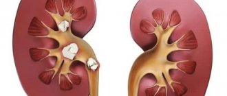
Symptoms of a ureteral stone
Getting into the ureter, the stone causes a disruption in the outflow of urine from the kidney and provokes painful sensations of varying intensity.
Most often, renal colic is severe pain in the lumbar region. It does not go away with changes in body position, but decreases somewhat after taking painkillers and antispasmodics.
A stone in the lower third of the ureter can irritate the bladder, causing you to urinate more frequently.
Stones up to 5 mm in size can pass on their own; stones larger than 5 mm usually require surgery.
The most dangerous condition – acute (obstructive) pyelonephritis – is manifested by severe fever and chills. The condition requires urgent medical intervention.
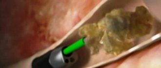
Ureteroscopic contact lithotripsy (contact ureterolithotripsy)
Contact lithotripsy is performed when the stone size is more than 5-6 mm or when stones have been standing in the ureter for a long time.
This method is most widely used for stones in the lower third of the ureter.
ureterolithotripsy
The method is based on the fact that an energy generator is brought directly to the stone by ureteroscopy through the bladder, the stone is destroyed and its fragments are removed using a special loop or basket.
The method of crushing stones using a holmium laser shows the best results compared to others, but it is also the most expensive.
Contraindications to contact ureterolithotripsy:
- Inflammatory processes in the urinary tract (pyelonephritis, urethritis, cystitis, prostatitis).
- Cicatricial deformities of the ureter.
- Large prostate adenoma.
Contact transurethral lithotripsy is completed with the installation of a ureteral stent, which is left in place for several days or, if indicated, up to 3-4 weeks.
The cost of contact transurethral lithotripsy ranges from 35 to 65 thousand rubles.
Video: ureteroscopic removal of stones from the lower third of the ureter
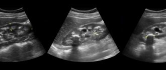
Diagnosis of ureteral stone
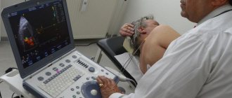
The primary screening is ultrasound. Going down the ureter, the stone interferes with the normal outflow of urine and is manifested by the expansion of the pelvis and calyces - the collecting system of the kidney that produces urine.
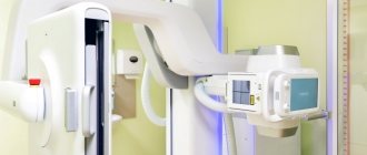
Then a plain X-ray of the urinary system is taken. If there is a shadow in the projection of the urinary tract, excretory urography is performed. The patient is injected intravenously with an X-ray contrast agent and after 7 minutes a series of new X-rays is taken. A stone in the ureter is considered proven if the movement of the dye is hampered at the site of the darkening detected on the first x-ray.
An alternative to ultrasound and urography is computed tomography; it allows you to accurately determine the size, location and density of the stone. It can be performed with or without contrast.
Symptoms of urolithiasis
In men, this pathology is detected three times more often than in women. The clinical manifestations of urolithiasis are the same in men and women.
The severity of symptoms depends on the size of the stones and where they are located.
In the presence of small stones, the disease is asymptomatic, or after heavy physical activity discomfort may appear in the lumbar region. At this stage, stones are most often diagnosed accidentally during examinations.
Localization of pain in urolithiasis
The most integral symptom is pain.
The pain can be constant or paroxysmal; aching or acute in nature; The severity of pain depends on the size of the stone and its location.
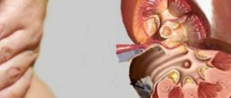
Pain from kidney stones
When stones are found in the kidneys or upper parts of the ureter, pain occurs in the lumbar region and is aching in nature.
However, if a stone causes obstruction (blockage) of the ureter, the outflow of urine is disrupted and the pain increases significantly. The patient develops renal colic. It is characterized by severe pain that does not go away when changing body position. The pain can last from a few minutes to several days. Patients rush about, and there is a frequent urge to urinate.
The pain is most often unilateral, but can rarely be bilateral.
As the stone moves through the urinary tract, the pain decreases.
Pain in the lower abdomen in men can spread to the external genitalia and scrotum. The pain resembles prostatitis, testicular torsion.
In women, pain in the lower abdomen radiates to the labia and vulva.
Urination becomes difficult, it becomes frequent and painful.
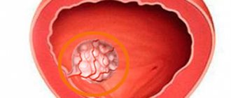
Pain from bladder stones
When there are stones in the bladder, the pain is localized in the suprapubic region; with small stones, the pain is aching. Irradiation of pain also into the area of the external genitalia.
Blood in urine and sand
The second most common symptom is hematuria (blood in the urine).
Hematuria occurs when stones move through the urinary tract due to damage to the mucous membranes. When passing small stones, blood can only be detected by examining urine. And when large stones pass, the patient himself may notice a pink coloration of the urine.
The patient may also see small stones (sand) in the urine sediment.
Treatment of ureteral stone
The Urology Clinic of the First Moscow State Medical University is the leading medical center in our country for the treatment of urolithiasis. There is a specialized department for nephrolithiasis, a department for lithotripsy and endoscopic treatment of urolithiasis. A special Siemens endo-X-ray operating room is equipped; all modern techniques are available for removing stones of any size.
Remote nephrolithotripsy is the crushing of stones using focused intense shock waves without introducing any instruments into the body under X-ray or ultrasound control. The stones break down into grains of sand, which come away when you urinate.
Contact laser lithotripsy is the destruction of stone with laser energy, which is carried out using the thinnest endoscopic instrument (3-5 mm in diameter) inserted through the urethra. If necessary, stone fragments are removed using special extractor baskets or micro forceps.
Laparoscopic stone removal (ureterolithotomy) - the surgeon removes the stones entirely through several punctures in the anterior or lateral abdominal wall. Performed for large stones in the upper and middle third of the ureter.
Ureteral stone Pyelonephritis Frequent urination Nephrolithotripsy Lithotripsy Laparoscopic removal
Diagnostics
If signs of urolithiasis are detected, you must consult a urologist or nephrologist and undergo the necessary examination.
General urine analysis:
- Allows you to detect hematuria - the appearance of red blood cells in the urine. In the presence of inflammation in the urinary system, an increased number of leukocytes and an increase in urine density are detected. Salts (oxalates, phosphates, urates) are found in urine sediment.
- If there are stones in the urine sediment, they are examined. The character of the stone is established.
Biochemical blood test:
- Aimed at identifying metabolic disorders. The level of uric acid, phosphates, oxalates, kidney function (creatinine, urea, glomerular filtration rate) are assessed.
General blood analysis.
- Anemia (decreased hemoglobin) can be detected with prolonged blood loss; an increase in the number of leukocytes and the erythrocyte sedimentation rate of ESR during the inflammatory process.
Ultrasound examination of the kidneys and bladder.
- Allows you to identify the presence of stones and signs of inflammation.
To detect stones in the ureter, clarify their location and the degree of obstruction of the urinary tract, excretory urography is performed. The study is carried out by introducing a radiopaque substance and then assessing its removal rate.
If there is a blockage in the lower urinary tract, retrograde ureteropyelography is performed. The contrast is injected not into the kidneys, but from bottom to top - along the ureters.
A computed tomography scan may also be prescribed to clarify the diagnosis. It allows you to clarify the size of the stone and its position.
“Stone path” as a complication of extracorporeal lithotripsy
G.A. Volkova, T.A. Bolshakova, F.P. Kapsargin, A.S. Repin, E.V. Repina, E.V. Gromov, V.F. Zhuravlev Krasnoyarsk
The importance of the problem of urolithiasis is great, since it is one of the most common urological diseases and is often prone to recurrence. This metabolic disease, caused by various endogenous and (or) exogenous causes, is often hereditary in nature and is manifested by the presence of a stone in the patient’s urinary system.
Among the numerous methods for removing stones from the urinary tract, extracorporeal lithotripsy (ESLT) has firmly taken its place in the treatment of urolithiasis. In our conditions, DLT was carried out using a domestic device "Urat P" in the MER mode of 0.7-0.9 mm, 1200 pulses on four capacitors, 1800 on six, with x-ray control every 150 pulses.
The purpose of DLT was to destroy stones in the kidney and urinary tract into small fragments and their spontaneous passage. However, in 12.1% of cases, according to our observations, in 649 people who received remote crushing of stones, a “stone path” of different localization, length, and different clinical picture of severity appeared. In 87.9% of patients in this group, stone fragments passed on their own without any significant symptoms within a few days.
The reasons for the emergence of “stone paths” turned out to be different.
According to our observations, 25 people had a history of surgical treatment on the corresponding side of the urinary tract, 22 patients had already undergone DLT in the anamnesis and 29 suffered from large (up to 2 cm or more) kidney stones, while catheterization of the ureter was not performed before DLT, three had a history of serious kidney injury In addition, the presence of a stone in the ureter for more than 4-6 weeks was not favorable for remote lithotripsy, due to granulations that occurred at the site of a long-term location of the stone or the occurrence of pyelouretereitis, pyelonephritis, and attention should also be paid to the crushing mode.
External lithotripsy is the method of choice in the treatment of urolithiasis. DLT was performed depending on the clinical manifestation, from the localization of the “stone path”, its length, period of standing, from inpatient observation and conservative stone-expelling therapy, catheterization or stenting of the ureter, percutaneous nephrostomy, ureteroscopy with lithoextraction or contact lithotripsy to open lumbotomy with sanitation of the kidney and drainage of its cavity and retroperitoneal space.
Thus, DLT expands the possibility of therapeutic care for patients suffering from urolithiasis, but requires an individual approach and a correct assessment of the medical history and condition of the urinary tract in order to prevent the occurrence of such a complication as a “stone path” with possible more severe consequences.
Topics and tags
Urolithiasis
External lithotripsy
Collection of scientific papers of the X regional scientific and practical conference of urologists of Western Siberia
Comments
To post comments you must log in or register
First aid
If the pain is very intense, and it is difficult to wait for an ambulance or a visit to the doctor, you can take emergency measures at home:
- Drink an antispasmodic. This will help relieve spasm and relax the muscles of the ureter, which facilitates the passage of urolith out.
- To reduce pain, take an effective pain reliever.
- Fill the bathtub with water at a temperature of 37–38°C and sit in it for half an hour. You need to position yourself so that your lower back is completely under water. This will not only help relieve pain, but will also relax the internal smooth muscles. While in the bathroom, you can drink plain water or herbal decoctions with a diuretic effect - dill, fennel or horsetail.
- Immediately after the bath you should move actively. If your condition allows, you can even jump to move the stone from its place.
If quick relief occurs, it means that the problem has been resolved, but otherwise, you just have to wait for medical help.
At high temperatures and the presence of blood impurities in the urine, warm baths are strictly prohibited!