Types of kidney stones
There are four main types of kidney stones.
Calcium stones
Calcium stones are the most common type of kidney stones and contain calcium oxalate, calcium phosphate, or a combination of both.
Calcium oxalate stones consist of calcium oxalate dihydrate and calcium oxalate monohydrate.
A calcium oxalate dihydrate stone will have jagged edges. However, a calcium oxalate monohydrate kidney stone, which is more common, will have a smooth surface.
Types of kidney stones, the main criteria for distinguishing them
The stone itself is a mixture of certain mineral and organic substances. Mostly stones are found that are homogeneous in composition. Much less often, they can contain several heterogeneous components at once. Thus, the first and main criterion by which stones should be distinguished is their chemical composition.
In order to ensure the effectiveness of the prescribed therapy, it is important to assess the morphological structure of the stone, as well as its size and density.
Video: what stones form in the kidneys
Uric acid stones
Uric acid stones account for 3-10% of kidney stones. Uric acid stones usually look like pebbles. Some of the stones may be hard on the outside but soft on the inside because they are composed of different types of uric acid and calcium oxalate monohydrate.
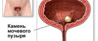
Struvite stones
Struvite stones are the next most common type of kidney stones (7-8% of cases) and are larger than the others.
Cystine stones
Cystine stones are the result of cystinuria
, which can be inherited. The kidneys produce cystine, a type of amino acid.
Cystine stones are compact, partially opaque, and amber in color.
Methods for researching kidney stones
To diagnose kidney stones, various research methods are used. First of all, these should include laboratory tests:
- General urine analysis. Using this method, the presence of suspensions and foreign inclusions in the urine is determined, which is an indirect sign of urolithiasis. The degree of acidity of urine, also detected through analysis, allows us to evaluate the composition of the stone.
- Biochemical blood test. Determines the content of phosphorus, calcium, uric acid in the blood, that is, those elements that take part in the formation of stones.
Laboratory research methods help determine the chemical composition of stones, but their morphological structure can be assessed after instrumental studies. These include:
- Radiography. This method is widely used in cases of suspected urolithiasis, but its level of information content remains low, since it can only visualize large stones.
- Excretory urography. At its core, this is a more advanced x-ray method. Before the image, a special contrast agent is injected into the urinary system, due to which a more detailed visualization of the renal structures and foreign inclusions, which are stones, occurs.
- Ultrasound. Examination of the kidneys using ultrasound is the most informative method for identifying stones, allowing the morphological structure to be assessed most accurately. This method is gradually replacing traditional radiography due to its effectiveness and complete safety.
Ultrasound can detect kidney stones and is a safe method of examination
- Spiral computed tomography (SCT). It is carried out in cases where it is impossible to determine the location and structure of stones using ultrasound. The method is considered very effective, but expensive. In essence, it is similar to computed tomography, but is performed by rotating the X-ray tube and the tomograph itself in a spiral, which allows you to obtain multidimensional X-ray images and detect stones up to 1 mm in size.
Computed tomography is the most accurate but expensive method for detecting kidney stones
How do kidney stones pass?
Kidney stones can pass without medical intervention. Smaller stones are more likely to pass, but larger ones may block the urinary tract. The stones can take up to 6 weeks to pass; the patient should drink plenty of water during this time.
People often experience pain when a kidney stone passes through the ureter. If the pain is unbearable, you should consult a doctor. The doctor may prescribe strong pain medications, anti-nausea medications, and a drug called tamsulosin, which relaxes the ureter, making it easier for the kidney stone to pass.
Passage of stone depending on size
The average size of the ureter is 3-4 millimeters wide. The chances of passing stones depend on their size:
| Stone size(mm) | Probable percentage of passage through the ureter (%) |
| Less than 4 | 80 |
| 4–6 | 60 |
| More than 6 | 20 |
Types of kidney stones by color
The color of a stone directly depends on its chemical composition. Its spectrum is quite wide: stones can be white, black, red, greenish. This is explained by the wide variety of stones that occur in the kidneys. However, the most common colors are white, yellowish, black, as well as their shades and combinations:
- White. This color is characteristic of carbonate and phosphate stones.
- Yellow. Characteristic of urates, struvites, cystine stones.
- Black. Oxalates are black or dark brown in color, as are cholesterol stones, which are quite rare.
Oxalates are crystals consisting of calcium oxalate and ammonium oxalate salts
Treatment of urolithiasis
If a patient has large kidney stones or stones that are blocking the urinary tract, the doctor may recommend surgical removal.
There are three types of surgical interventions:
- Shock wave lithotripsy
: Kidney stones are broken into smaller pieces, allowing them to pass through the urinary tract. - Cystoscopy and urethroscopy
: these procedures use a camera to detect stones in the urethra or bladder. Once the urologist finds kidney stones, he can remove them completely or break them into small pieces, which will then pass on their own. - Percutaneous nephrolithotomy
: The urologist inserts a camera directly into the kidney through the back wall to remove stones. If the stones are very large, a laser is used to break them into pieces.
Risk factor
Some factors that may increase your chance of developing kidney stones include:
- the person does not drink enough fluids
- doing too little or too much exercise
- is obese
- eats foods containing too much salt or sugar
- consumes foods containing fructose
People are more likely to develop kidney stones if they have:
- urinary tract abnormality
- obesity
- cystinuria
- gout
- recurrent urinary tract infections
- cystic kidney disease
- digestive problems
- hypercalciuria - an inherited condition in which the urine contains too much calcium
- hyperoxaluria - a condition in which the urine contains too much oxalate
- hyperparathyroidism, a condition in which the body produces too much parathyroid hormone, with extra calcium in the blood
- hyperuricosuria - a condition in which the urine contains too much uric acid
- renal tubular acidosis, a condition in which the kidneys do not excrete acid into the urine, leaving acid in the blood
Use of drugs
People taking the following medications are more likely to develop kidney stones:
- diuretics, which help the body get rid of excess water
- calcium-based antacids
- indinavir, a drug that treats HIV
- topiramate, antiepileptic drug
Stones in the kidneys
Gastritis
Ulcer
Gout
Pyelonephritis
Cystitis
15482 01 February
IMPORTANT!
The information in this section cannot be used for self-diagnosis and self-treatment.
In case of pain or other exacerbation of the disease, diagnostic tests should be prescribed only by the attending physician. To make a diagnosis and properly prescribe treatment, you should contact your doctor. Kidney stones: causes of occurrence, what diseases cause them, diagnosis and treatment methods.
Definition
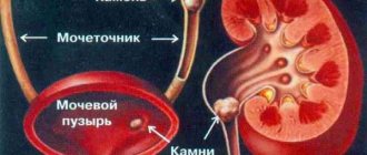
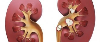
Disorders of mineral metabolism lead to the formation of kidney stones. Salts of various minerals accumulate in the renal pelvis, the inner cavity of the kidneys, gradually turning into dense stony agglomerates (dense accumulations of particles). The process most often affects only one kidney.
The sizes of the stones vary from a few millimeters to tens of centimeters.
The most serious complication that occurs as a result of the formation of stones is a violation of the outflow of urine. It is characterized by sudden, acute pain in the lumbar region, groin, and perineum. In addition to the pain syndrome, there is severe weakness, headache, nausea, bloating, painful urination or its absence in the presence of urge, and blood often appears in the urine.
Sometimes the pain reaches such intensity that the person loses consciousness.
In this case, you should immediately seek medical help. Types of kidney stones Kidney stones are classified according to their composition:
- Urate stones consist of uric acid, potassium and sodium salts. They are dense, yellow in color and have a smooth surface. Due to the lack of calcium, they are not visible during radiographic examination. The diagnosis is made based on ultrasound and urine analysis.
- Oxalate stones are the most common. They contain a lot of calcium, they have a high density, so they are easy to diagnose during instrumental examination. The stones are dark gray, uneven. Such stones severely injure the ureteral mucosa, which is accompanied by intense pain and the appearance of blood in the urine.
- Phosphate stones contain calcium salts of phosphate acid. They are smooth, crumble easily, and grayish-white in color. Most often occur against the background of chronic kidney infection.
- Struvite stones increase in size very quickly, have a soft consistency and a grayish color. Mostly diagnosed in women. The main danger of these stones is that in just a few months they grow to large sizes and can completely fill the renal pelvis.
- Cystine stones are rare and consist of the sulfur compound of the amino acid cystine. They are yellowish-white in color, round in shape, soft in consistency, with a smooth surface. The peculiarity of these stones is that they are found in children and young adults. The only cure for this pathology is a kidney transplant.
- Xanthine stones are more likely to form in patients with a hereditary predisposition. They are almost always X-ray negative (that is, they are not visualized on photographs), so ultrasound is used to diagnose them. Xanthine stones cannot be removed on their own; the only way to remove them is surgery.
To size:
- small (up to 3 mm);
- medium (3–10 mm);
- large (more than 10 mm).
Possible causes of kidney stones
Urate stones are most often found in people who eat a lot of meat, cheese and wine.
The appearance of oxalate stones is associated with excessive consumption of various citrus fruits, sweets, especially chocolate, as well as coffee, tea, beets and black currants. The formation of such stones can also be caused by a disturbance in calcium metabolism in the body.
Calcium metabolism, in turn, is closely related to vitamin D.
The risk group includes older people who, due to natural reasons, develop osteoporosis, that is, bones become fragile due to the fact that calcium ceases to settle in bone tissue and most of it circulates in the blood. Blood enriched with calcium is filtered by the kidneys, as a result of which excess calcium accumulates in the kidney cavity, leading to the formation of stones.
A similar process occurs in diseases of the thyroid and parathyroid glands. With an excess of hormones, calcium metabolism is disrupted; it begins to be actively washed out of bone tissue into the blood and then filtered by the kidneys, where it is gradually deposited.
Violation of normal absorption of nutrients is also observed in diseases of the gastrointestinal tract. This causes metabolic imbalance and provokes the occurrence of urolithiasis.
Infectious and inflammatory kidney diseases often lead to the formation of stones. Inflammation disrupts the processes of filtration and excretion of urine, some of the salts remain inside the renal pelvis, and a stone forms.
Urolithiasis can be caused by taking certain medications (especially uncontrolled use of ascorbic acid).
You should be careful about the composition of the drinking water you consume. Hard water with excess calcium salts increases the risk of kidney stones.
Urolithiasis is a common occurrence in bedridden patients. Lack of movement promotes the proliferation of microbes in the kidneys, which leads to impaired kidney function and the formation of stones.
Diseases leading to the formation of kidney stones
- Diseases of the thyroid gland and parathyroid glands.
- Stomach diseases (gastritis, peptic ulcer).
- Intestinal diseases (Crohn's disease, a condition after removal of part of the intestine).
- Osteoporosis.
- Gout.
- Diseases of the genitourinary system (pyelonephritis, prostatitis, cystitis, prostate adenoma).
- Deficiency of vitamins D and A.
- Dehydration.
Which doctors should you contact if you suspect kidney stones
? An attack of renal colic is a serious condition that requires immediate emergency medical attention.
A nephrologist treats kidney diseases. He will be able to prescribe the necessary tests and instrumental examination methods. To make a diagnosis and prescribe treatment, consultations with a gastroenterologist, endocrinologist, gynecologist, or urologist may be required.
Diagnosis and examinations for suspected kidney stones
A thorough history taking into account all the patient’s complaints, examination and additional diagnostic methods will help establish the correct diagnosis.
- A clinical blood test with a detailed leukocyte formula allows us to identify inflammatory changes in various infectious diseases.