Live broadcasts on Instagram every Sunday at 12:00
Subscribe so you don't miss out! Subscribe
- home
- Technologies
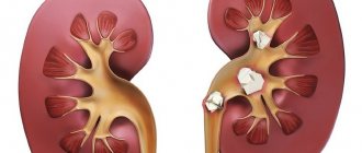
- Technique for laparoscopic kidney resection
Video of the operation using this technique by Prof. Puchkova you can see here
Regardless of the approach, the basic surgical principles when performing partial nephrectomy for a tumor remain the same:
- early control of renal vessels;
- protection of the function of the operated kidney from ischemic damage;
- removal of the tumor within healthy tissue;
- hermetic closure of the urinary tract;
- good hemostasis;
- closure of the kidney defect.
The key to successful kidney resection for cancer is good hemostasis, which can be temporary (at the time of organ resection) and final (suturing the kidney wound). For temporary hemostasis during open resection of the kidney, clamping of the vessels of the renal pedicle is used, as well as digital compression of the resection area. When a clamp is applied to the renal pedicle, thermal ischemia of the operated organ occurs, which limits the time of blocking the renal blood flow.
Thus, early control of the renal vessels is necessary for their subsequent clamping so that the operation takes place on a dry surgical field. Healing of surgical defects during operations on the urinary tract occurs under conditions of constant function and in contact with urine. Insufficient sealing of defects leads to a number of complications in the form of prolonged leakage of urine into the wound and the formation of urinary fistulas, urinary leaks that can develop into urinary phlegmon, therefore the issue of using additional techniques for sealing a renal wound is relevant (the adhesive compositions used are Floseal, Tissucol (fibrin gel) , BioGlue and Glubran (cyanoacrylate)).
Overlapping the renal vessels makes it possible to limit the time of warm ischemia to 15-30 minutes, which does not subsequently affect renal function.
We emphasize the need to protect kidney tissue from ischemic damage in case of blockage of renal blood flow, for which we use anti-ischemic drug protection of the kidney with intravenous infusion of Lasix and angioprotectors. Local hypothermia is necessary if the renal ischemia time exceeds 30 minutes.
Kidney resection refers to the removal of a tumor with an adjacent area of normal renal parenchyma, up to 1 cm wide. During enucleation, the tumor is enucleated within the true or pseudocapsule. A transitional option is ennucleoresection, when the tumor is removed along with the adjacent renal parenchyma, less than 1 cm wide (usually 5-7 mm).
It is believed that the greatest radicality of organ-preserving surgery is achieved by removing at least 1 cm of normal renal parenchyma, since this eliminates microscopic satellite tumors adjacent to the main node, and also eliminates the possibility of leaving extratumoral tumor invasion outside the pseudocapsule. At the same time, a number of authors show good long-term results when removing 5-7 mm of normal renal parenchyma (Graham and Glenn, 1979; Topley et al., 1984; Bazeed et al., 1986; Licht and Novick, 1993).
The following characteristics of the tumor process are of great importance in determining the possibility of technical implementation and type of kidney resection:
- location of the tumor (tumors located on the posterior surface of the organ near the renal hilum are more difficult to remove),
- direction of tumor growth (if the tumor grows predominantly extrarenally, resection is technically more easily feasible),
- the depth of invasion of the neoplasm into the renal parenchyma (with deep invasion, the pyelocaliceal system is always opened, which requires its careful suturing)
- tumor size (is not the main parameter for the possibility of kidney resection.
We share the opinion of a number of authors who perform a biopsy from the resected surface of the kidney to prove complete excision of the tumor (Giuliani L., 1989; Thrasher JB et al., 1994; Novick AC, 1997). A rapid morphological study of fresh frozen sections was not performed due to the high frequency of false-positive responses.
If the urinary tract was opened, then when using laparoscopic access, the defect, due to optical enhancement of the kidney wound, can be detected and sutured, and there was no need for drainage of the upper urinary tract. In case of opening of the urinary tract, the calyces were sutured with Polysorb 3/0 thread on an atraumatic round needle with separate interrupted sutures. Closure of the kidney defect was carried out as follows. The kidney wound was treated with the BioGlue adhesive composition, after which 3-4 closing sutures were applied with a thread made of synthetic absorbable material on an atraumatic needle. The suture line was then re-treated with BioGlue. After complete polymerization of the adhesive composition, the clamp from the main renal vessels was removed and renal blood flow was restored.
The wound of the renal parenchyma was closed using interrupted Z-sutures with Polysorb or Vicryl “0” thread on a round atraumatic needle in one row (as a rule, with a tumor size of up to 4 cm, 3-4 sutures are required). In our observations, when using the laparoscopic approach, the most effective was a separate interrupted suture with placing fragments of a hemostatic sponge under the loops and nodes to prevent their cutting through the renal parenchyma.
When a technical possibility presents itself, we carry out paranephric plasty, suturing its fragments above the resection area. Thus, there was no need for additional fixation of the kidney in our observations.
The following set of instruments is necessary and optimal for performing laparoscopic partial nephrectomy and lymphadenectomy:
- laparoscopic complex (video camera, illuminator, insufflator, coagulator, video monitor);
- laparoscope with a diameter of 10 mm - all with an end cut of 300;
- fiber optic cable;
- trocars with a diameter of 5.5 mm, 11 mm, 12 mm with stylets - blunt-ended and conical, and a set of “adapters” from one size to another - steel or plastic;
- atraumatic clamps - straight and curved - 5 mm in diameter, with the possibility of monopolar coagulation, with a set of handles - without ratchet and with ratchet;
- ultrasonic scissors;
- bipolar coagulation device “LigaSure” with instruments with a diameter of 5 and 10 mm;
- scissors – curved, with two moving jaws, with the possibility of monopolar coagulation, with a diameter of 5 mm;
- suction/irrigation cannula with a diameter of 5 mm;
- endoscopic needle holder with a diameter of 5 mm;
- endoclipper with a diameter of 10 mm (with large clips);
- endoscopic stapler 12 mm (with used cassette);
- endopush (to form an extracorporeal suture);
- suture material (Polysorb, Vicryl 3/0 and 0 on an atraumatic round needle;
- plastic container - Endocatch II;
- 10 mm endobebkok instrument;
- 10 mm Endorettractor instrument.
To perform laparoscopic surgical interventions on the kidneys, we use 5 approaches (Fig. 1): 4 of 10 mm and 1 of 5 mm.
Fig.1. The location of the ports during laparoscopic interventions for kidney cancer: a – tumor on the left, b – on the right.
The patient's position on the operating table is on the back with legs apart, with the head slightly elevated and the table rotated 15-20° to the side opposite the affected kidney. The surgeon is positioned on the opposite side, the monitor is located near the patient’s arm on the side of the tumor.
The main stages of organ-saving surgery using laparoscopic access include:
1. Laparoscopic access creates conditions for good visualization of the inferior vena cava and aorta (we perform paracaval and para-aortic lymphadenectomy from the crura of the diaphragm to the aortic bifurcation), mobilization of the renal arteries and vein, the upper third of the ureter. The steps of creating a laparoscopic approach and lymphadenectomy are common to nephrectomy and partial nephrectomy (Fig. 2).
Rice. 2. Lymphadenectomy during resection of the right kidney (stages a and b - the inferior vena cava, aorta, aortocaval space, right renal artery are visible).
Rice. 3. Preparation of ligatures (stitching fragments of the Surgisel hemostatic sponge with an atraumatic needle).
2. The main renal vessels can be mobilized with a fragment of paravasal tissue, which will serve as a shock absorber when applying a clamp to the renal pedicle. We also highlight the upper third of the ureter.
3. We mobilize the kidney as a single block with perinephric tissue and fascia. We open the paranephrium in a tumor-free area and isolate the remaining part of the kidney. We expose the fibrous capsule of the kidney up to 3-4 cm from the edge of the tumor. In this case, it is important, if possible, not to disrupt the integrity of the fibrous capsule of the kidney, which may subsequently complicate suturing the resection area. Isolation of a kidney from paranephrium can be carried out using either the LigaSure device (Covidien) or an ultrasonic scalpel. Perinephric tissue from the tumor is sent for morphological examination in a separate container.
4. Next to the resection area, we place pre-prepared ligatures (Polysorb or Vicryl 0) on an atraumatic needle with fragments of a hemostatic sponge strung on them (Fig. 3). Angioprotectors are administered intravenously.
1. We clamp the renal pedicle with an endoscopic stapler with a used cassette (without staples) (Fig. 4). We pass the instrument through an additional 12 mm port. It is desirable that the warm ischemia time does not exceed 15-30 minutes.
Rice. 4. Application of a stapler to the vascular pedicle (a – resection of the right kidney, b – resection of the left kidney)
with a fragment of fiber and blocking the renal blood flow.
2. Kidney resection is carried out within healthy tissue, 1 cm away from the edge of the tumor. For better visualization of the tissue in the resection area and more correct results of morphological examination, we use curved scissors without coagulation (Fig. 5). An ultrasonic scalpel may be used at this stage, although we did not see any benefit in using it. A biopsy from the resection area allows you to confirm the radicality of the operation.
Rice. 5. Wedge resection of the left kidney using endoscopic scissors.
3. In order to identify exposed elements of the pyelocaliceal system, we must conduct an inspection of the resection zone. If there are defects, they are carefully sutured with Polysorb or Vicryl 3/0 thread on an atraumatic round needle.
4. Suturing of the renal parenchyma is carried out using interrupted Z-sutures using Polysorb or Vicryl “0” thread on a round atraumatic needle, placing fragments of the hemostatic sponge Surgisel under the loops and nodes to prevent their eruption (Fig. 6, 7).
Rice. 6. Suturing the resection area (a – b – stages of suturing)
Rice. 7. View of the surgical field after suturing the kidney.
If the volume and mobility of the paranephrium is sufficient, we stitch its fragments over the operated organ, covering it with a fat capsule (Fig. 
Rice. 8. Plastic surgery of paranephrium of the left kidney after wedge resection.
1. We remove the lymph nodes of the renal pedicle if the fiber was left. We remove the tumor, as well as the lymph nodes in the container (Fig. 9). The abdominal cavity is drained by one safety drainage.
Rice. 9. Removal of the removed tumor and lymph nodes in a plastic container.
results
The duration of organ-saving surgery using laparoscopic access was 80+/-20 minutes. Warm ischemia time during partial nephrectomy was 14+/-3 minutes.
We believe that the operation of choice for localized stage T1aN0M0 and T3aN0M0 kidney cancer (tumor size up to 4 cm) is laparoscopic resection of the affected organ, if it can be performed within healthy tissue.
K. V. Puchkov, A. A. Krapivin, V. B. Filimonov. Laparoscopic surgery of kidney cancer K. V. Puchkov, D. S. Rodichenko. Hand suture in endoscopic surgery
LIST OF PUBLISHED WORKS ON THE TOPIC LAPAROSCOPIC NEPHRECTOMY AND KIDNEY RESECTION
- Puchkov K.V., Filimonov V.B. Laparoscopic radical nephrectomy for kidney cancer // Prospective directions in the diagnosis and treatment of kidney cancer: abstract. report – M., 2003. – P.116-117.
- Puchkov K.V., Filimonov V.B., Vasin I.V., Strelkov A.N., Katrovsky A.V. Laparoscopic radical nephrectomy for kidney cancer // Endoscopic surgery. - 2004. - T.7, No. 2. - P.25-30.
- K.V. Puchkov, V.B. Filimonov. Surgical treatment of kidney cancer: laparoscopic radical nephrectomy // 8th Moscow. international congr. in endoscopic surgery. RSCH RAMS, M., 2004 - pp. 287-288.
- Puchkov K.V., Filimonov V.B., Strelkov A.N. Laparoscopic radical nephrectomy for hypernephroid kidney cancer // Endoscopic surgery. -2004.- T.10, No. 1 (add.). – P. 122.
- Puchkov K.V., Filimonov V.B., Vasin I.V., Strelkov A.N., Katrovsky A.V. Kidney cancer: laparoscopic radical nephrectomy // Ryazan. honey. Vestn. - 2004. - No. 13 (153). – P.14-15.
- Puchkov K.V., Filimonov V.B. Laparoscopic access in the surgical treatment of kidney cancer // Materials of V Ros. scientific forum "Surgery-2004", Moscow, November 1-4, 2004 - M., 2004. - P. 157.
- Puchkov K.V., Kolesov V.Yu., Kolesnikova N.O. Radiation diagnostics today - search, solution, prospect // Magnetic resonance imaging in a multidisciplinary clinical hospital. - Ryazan: GUZ ROKB, 2004.- P.3-7.
- Puchkov K.V., Filimonov V.B. Laparoscopic regional lymphadenectomy for kidney cancer // Endoscopic surgery. – 2005.-T.11, No. 1. – P.108.
- Puchkov K.V., Filimonov V.B., Vasin I.V. Laparoscopic nephrectomy and regional lymphadenectomy // 9th Moscow. international congr. in endoscopic surgery, Moscow, April 6-8. 2005: abstract. report / ed. Yu.I. Galingera. – M., 2005.- P.301-302.
- Puchkov K.V., Ivanov V.V.. Technology of dosed ligating electrothermal effects at the stages of laparoscopic operations: monograph. - M.: ID MEDPRACTIKA, 2005. - 176 p.
- Puchkov K.V., Filimonov V.B., Vasin I.V. Laparoscopic radical nephrectomy // Issue. oncology.-2005.-T. 51, No. 4. - P.476-479.
- Puchkov K.V., Filimonov V.B., Vasin I.V., Vasin R.V., Dmitrienko S.V. Laparoscopic access in the surgical treatment of kidney cancer // Endoscopic surgery. – 2006.-T.12, No. 2. – P.111-112.
- Puchkov K.V., Filimonov V.B., Vasin R.V., Dmitrienko S.V. Laparoscopic surgery for kidney cancer // Minimally invasive methods of diagnosis and treatment in modern urology: abstract. report 3rd international Conf. - St. Petersburg, March 2-3. 2006 – St. Petersburg, 2006. -P.57-59.
- Puchkov K.V., Filimonov V.B., Vasin R.V. Method of preoperative calculation of optimal installation sites for ports during laparoscopic operations on retroperitoneal organs // Current issues of modern surgery. Regional (Southern Federal District) scientific-practical. conf. surgical doctors, Nalchik, May 26-27, 2006 - Nalchik, 2006.- pp. 276-278.
- Puchkov K.V., Filimonov V.B., Vasin I.V., Vasin R.V. Laparoscopic nephrectomy and regional lymphadenectomy in the surgical treatment of kidney tumors // Nizhny Novgorod Med. journal – 2006.-(Appendix Transplantology). – P.103-104.
- Puchkov KV, Filimonov VB, Vasin RV Laparoscopic access in surgical treatment of benign tumors of the retroperitoneal organs // Abstracts of The 10th World Congress of Endoscopic Surgery, 13-16 September, 2006, Berlin. – P.298.
- Puchkov KV, Ivanov VV, Barsukov VA, Filimonov VB Determining of safety of soldering of vessels in tissue mass of various types while using the technique of dosed ligating electrothermal influence of ligasure // Abstracts of The 10th World Congress of Endoscopic Surgery, 13 -16 September, 2006, Berlin. – P.288.
- Puchkov K.V., Filimonov V.B., Vasin I.V., Vasin R.V. Morphological structure of tumors and the frequency of detection of metastases in regional lymph nodes after radical laparoscopic nephrectomy with regional lymphadenectomy // Oncourology. - 2006. - (Materials of the 1st Congress of the Russian Society of Urological Oncology, October 4-5, 2006. - Moscow). – P.154-155.
- Puchkov K.V., Filimonov V.B., Vasin R.V., Vasin I.V. Surgical treatment of malignant diseases of the genitourinary organs using laparoscopic access // Trends, strategies and development of medical care in a multidisciplinary hospital: collection. scientific tr. – Ryazan, 2007. – pp. 244-247.
- Puchkov K.V., Filimonov V.B., Vasin R.V., Vasin I.V. Laparoscopic access in oncourology // Endoscopic surgery. – 2007.-T.13, No. 1. – P.67-68.
- Puchkov K.V., Krapivin A.A., Filimonov V.B. Organ-removing surgery of kidney cancer from laparoscopic access // Minimally invasive methods of diagnosis and treatment in modern urology. Materials of the IV International Scientific and Practical. conf. – St. Petersburg, 2007.- pp. 106-107.
- Puchkov K.V., Krapivin A.A. Laparoscopic kidney resection for cancer // Minimally invasive methods of diagnosis and treatment in modern urology. Materials of the IV International Scientific and Practical. conf. – St. Petersburg, 2007.- pp. 107-108.
- Puchkov K.V., Filimonov V.B., Krapivin A.A., Vasin I.V., Vasin R.V. Radical nephrectomy using laparoscopic access // Vestn. Transplantology and artificial organs. – 2007.- T. 33, No. 1. – P.63-71.
- Puchkov K.V., Filimonov V.B., Krapivin A.A., Vasin R.V., Vasin I.V. Minimally invasive technologies in surgery for renal cell cancer // Oncourology. – 2007.-(Materials of the II Congress of the Russian Society of Oncourologists, October 4-5, 2007.- Moscow). – P.137-138.
- Puchkov K.V., Filimonov V.B., Krapivin A.A., Vasin R.V., Vasin I.V. Surgical treatment of kidney cancer at the present stage // Current issues in specialized surgery: scientific and practical materials. conf. – Tashkent, 2007. – P.207-208.
- Puchkov K.V., Filimonov V.B., Krapivin A.A., Vasin R.V. Kidney resection for cancer using laparoscopic access // Materials of the Congress. XI Russian Oncology Congress, November 20-22. 2007 - Moscow). – P.230.
- Puchkov K.V., Filimonov V.B., Vasin R.V., Rodichenko D.S. Method for preoperative calculation of optimal installation sites for manipulation trocars during laparoscopic operations on retroperitoneal organs // Endoscopic surgery. – 2007.-T.13, No. 5. – P.17-24.
- Puchkov K.V., Krapivin A.A., Filimonov V.B.. Laparoscopic surgery for kidney cancer: Monograph. - M.: ID MEPRACTIKA - M. - 2008. - 164 p.
- Puchkov K.V., Filimonov V.B., Krapivin A.A., Vasin R.V., Vasin I.V. Surgical treatment of kidney cancer today: laparoscopic radical nephrectomy and kidney resection // Urology . – 2008. – No. 1. – P.52-58.
- Puchkov K.V., Ivanov V.V., Filimonov V.B., Khubezov D.A. Clinical aspects of the use of the “LigaSure” device during laparoscopic interventions // Bulletin of emergency medicine. – 2008.- No. 2. – P.74-80.
- Puchkov KV, Filimonov VB, Vasin RV Laparoscopic access in urooncology // Abstracts book of the 16th EAES Congress 2008, 11-14 July, 2008, Stockholm, Sweden. – P.208.
- Puchkov KV, Filimonov VB, Vasin RV Laparoscopic access in urooncology // Abstracts of The 11th World Congress of Endoscopic Surgery, September 2-5, 2008, Yokohama, Japan. – P.115.
- Puchkov KV, Filimonov VB, Vasin RV Lymphadenectomy as a necessary component of either nephrectomy or kidney resection // Abstracts book of the 17th EAES Congress 2009, 17-20 June, 2009, Prague, Czech Republic. – P.179.
- Puchkov K.V., Vinarov A.Z., Savelyev S.N., Balakleytsev I.I., Kurchatov O.P. The first experience of using BioGlue glue (Cryolife) during laparoscopic kidney resection // Moscow Surgical Journal. – 2009.- No. 4 (8). – P.16-23.
- Puchkov K.V., Filimonov V.B., Vasin R.V. Surgical treatment of kidney cancer at the present stage // Oncourology. – 2009.- (Materials of the 4th Congress of the Russian Society of Urological Oncology, October 1-2, 2009.- Moscow). – P.145-146.
- Filimonov V.B., Puchkov K.V., Vasin R.V. Comparative assessment of the quality of life after open and laparoscopic nephrectomy for kidney cancer // Oncourology. – 2009.-(Materials of the 4th Congress of the Russian Society of Urological Oncology, October 1-2, 2009.- Moscow). – P.148-149.
- Filimonov V.B., Puchkov K.V., Vasin R.V. Comparative assessment of long-term results of open and laparoscopic nephrectomy for renal cell cancer // Oncourology. – 2009.-(Materials of the 4th Congress of the Russian Society of Urological Oncology, October 1-2, 2009.- Moscow). – P.149-150.
- Puchkov K.V., Vinarov A.Z., Savelyev S.N., Balakleytsev I.I., Kurchatov O.P. The first experience of using BioGlue glue (Cryolife) during laparoscopic kidney resection for space-occupying lesions // Oncourology. – 2009.-(Materials of the 4th Congress of the Russian Society of Urological Oncology, October 1-2, 2009.- Moscow). – P.144-145.
- Puchkov K.V., Vinarov A.Z., Savelyev S.N., Balakleytsev I.I., Kurchatov O.P. The first experience of using laparoscopic ultrasound in the surgical treatment of a kidney tumor // Oncourology. – 2009.- (Materials of the 4th Congress of the Russian Society of Urological Oncology, October 1-2, 2009.- Moscow). – P.143-144.
- Puchkov K.V., Filimonov V.B., Vasin R.V. Laparoscopic access during operations on the organs of the retroperitoneal space: a method of preoperative calculation of the optimal installation sites for ports // Endoscopic surgery. – 2009.-T.15, No. 1. – P.220-221.
- Puchkov K.V., Ivanov V.V., Osipov A.V., Biryukov A.S. On the question of the methodology for using modern electrothermal systems // Endoscopic surgery. – 2009.-T.15, No. 1. – P.213-214.
- Ivanov V.V., Puchkov K.V., Osipov A.V. Tissue dissection in endosurgery for obesity: experimental analysis of ligating systems // Russian Medical and Biological Bulletin named after. Academician I.P. Pavlova. 2010. No. 4. P. 128-135.
- Puchkov K.V., Savelyev S.N., Balakleytsev I.I., Kurchatov O.P. Intraoperative ultrasound in the treatment of patients with kidney tumors using laparoscopic access // Almanac of the Institute of Surgery named after. A.V. Vishnevsky. T.7, No. 1 - 2012. “Materials of the XV Congress of the Society of Endoscopic Surgeons of Russia.” – Moscow, 2012. – P. 427-428.
- Puchkov K.V., Andreeva Yu.E., Dobychina A.V. Experience in performing simultaneous operations in surgery, urology and gynecology. Minimally invasive simultaneous operations // Almanac of the Institute of Surgery named after. A.V. Vishnevsky. T.7, No. 1 - 2012. “Materials of the XV Congress of the Society of Endoscopic Surgeons of Russia.” – Moscow, 2012. – pp. 16-17.
Laparoscopic nephrectomy, operated by Professor K.V. Puchkov (Dnepropetrovsk, Ukraine April 2010)
2. Reasons
It should be immediately stipulated that calculi (stones), as well as tumors, are not included in the concept of “foreign body”, although it is advisable not to introduce either one or the other into the genitourinary system, and if they appear (from the inside, we note) - then not launch. A foreign body means an object or substance that has somehow entered the body from the outside. However, the kidneys and ureters, unlike the urethra and even the bladder, are hidden very deeply in the visceral, internal space; they are protected by several layers of dense (though not hard) fabrics.
Accordingly, something very serious must happen for any foreign object to end up in the kidney or ureter.
Indeed, the main “supplier” of such a diagnosis is penetrating bullet, shrapnel, shot wounds (in a combat zone, hunting, in criminal situations), as well as severe industrial and transport accidents. However, with the development of minimally invasive, gentle medical technologies, primarily endoscopic and laparoscopic, it is necessary to mention sporadic cases of iatrogenic, i.e. damage caused by the medical intervention itself, including the introduction of foreign bodies into the upper urinary tract. This probability, we repeat, is extremely small, but still not zero: no one is insured, for example, from a technical defect in the form of an internal crack or cavity in the endoscope material, in the fastening of a catheter-stent (artificial duct), etc. It is ruptures or “slipping” of catheters into the ureter that are sometimes mentioned today as a reason (not the only one, however) for interventions to remove iatrogenic foreign bodies. Some authors also draw attention to the fact that such a stent, designed to ensure unimpeded passage of urine, if left in the ureter for too long, can become clogged with salt deposits, losing conductivity, becoming overgrown with stones and, thus, acquiring the character of an obliterating foreign body - but not immediately, but gradually.
Visit our Urology page