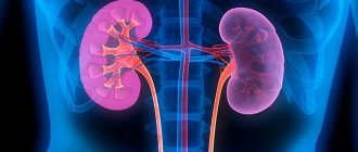
Kidney oncocytoma is a nodular-shaped benign tumor of renal tissue limited by a capsule, which has clear contours and consists of light epithelial cells (oncocytes) that have undergone dystrophic changes and have lost their functionality. These formations are the second most common among all benign kidney tumors. They can reach large sizes (more than 15 cm in diameter) and can lead to disruption of the functions of these paired organs. According to the observations of specialists, oncocytomas are not prone to malignant degeneration and metastasis.
Kidney oncocytomas can occur in one or two organs at once. The bilateral location of these neoplasms is especially dangerous, since due to damage to both kidneys, the excretory function is disrupted to an even greater extent.
According to the observations of specialists, kidney onocytomas are more often detected in men. In addition, oncourologists note that often (in about 10% of cases) oncocytomas occur in parallel with renal cell carcinomas.
Why do oncocytomas occur? What are the symptoms of these kidney tumors? How are they identified and treated?
Causes
Heredity is one of the factors that increases the risk of developing oncocytoma.
So far, scientists have not been able to establish the exact causes of the development of kidney oncocytomas. It is assumed that the following predisposing factors can provoke the growth of this benign neoplasm:
- heredity;
- chronic kidney disease;
- influence of toxic substances (taking certain medications, smoking, working in a hazardous enterprise);
- kidney injuries.
Renal angiomyolipoma and pregnancy
During pregnancy, a woman's body undergoes many changes. In particular, at this time the production of female sex hormones is activated.
It is believed that changes in hormonal levels contribute to the development of angiomyolipoma. In this case, the tumor can be detected during a routine ultrasound.
An existing angiomyolipoma develops more intensively during pregnancy. This tumor does not pose a threat to pregnancy failure and does not harm the baby.
Symptoms
Oncocytoma of the kidney can be completely asymptomatic for a long time, since the formation is small in size and does not have a negative effect on the tissues of the organ and its surrounding structures. At this stage of development, such tumors may be incidentally detected during ultrasound, MRI, or other imaging tests for other abdominal pathologies.
With large sizes of kidney oncocytoma, the patient develops the following complaints and symptoms:
- aching or dull pain in the lumbar region;
- general deterioration in health (pallor, lethargy, apathy, etc.);
- blood in the urine (change in its color or detection of red blood cells in a general analysis);
- blood pressure surges;
- swelling of the arms and legs;
- varicocele on the affected side (in men).
When palpating the kidneys, a seal of various sizes may be detected on the organ.
The appearance of the symptoms described above indicates the presence of a large oncocytoma, which puts pressure on the kidney parenchyma, ureters, blood vessels and nerve trunks. All emerging signs should be a reason for mandatory consultation with a doctor and a comprehensive examination.
Classification of tumors
Benign kidney tumors come in several types. Each of them has its own characteristics and symptoms.
Adenoma
Benign
Hypernephroma is formed in the fibrous capsule separating the low-quality tumor from healthy tissue. Hypernephroid kidney cancer has the most favorable prognosis for treatment among all types of organ cancer.
Mixed tumors are very aggressive, with a poor prognosis for survival during the first five years after treatment.
When diagnosing carcinomas, the international classification under the abbreviation TNM is used. It helps to clarify the location of the tumor and predict the likely outcome of therapy.
Diagnostics
All manifestations of kidney oncocytoma are nonspecific and can be observed in other diseases of the urinary system. That is why, for an accurate diagnosis, the following laboratory and instrumental studies must be performed:
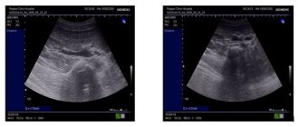
- Ultrasound of the kidneys - this simple and safe method allows you to detect the presence of a tumor in the parenchyma, determine its size, the degree of influence on surrounding tissues and location;
- and/or MRI using contrast - layer-by-layer images of an organ allow you to obtain even more accurate data on the parameters of the tumor and the degree of involvement of surrounding tissues and structures in the pathological process;
- targeted puncture biopsy and histological examination of the biopsy - this method makes it possible to accurately determine the type of tumor and distinguish it from other types of tumors (including malignant);
- urine tests - a test is carried out to assess the functioning of the kidneys;
- Blood tests (including adrenal hormone tests) – performed to assess general health, kidney function, and detect abnormalities in adrenal hormone levels.
After receiving all the examination data, a differential diagnosis of kidney oncocytoma with the following pathologies is performed:
- kidney cancer;
- other benign kidney tumors;
- urolithiasis disease;
- polycystic kidney disease.
Kidney tumors. Kidney cancer. Renal cell carcinoma (RCC)
Kidney tumors are represented by both benign (angiomyolipoma, oncocytoma, etc.) and malignant (renal cell carcinoma) neoplasms. More than 90% of all kidney tumors are malignant and are represented in most cases by a subtype of renal cell carcinoma (RCC) - clear cell renal cell carcinoma.
In the last decade, the incidence of kidney cancer in Russia has increased by 40%. Thanks to the widespread use of ultrasound and computed tomography in the diagnosis of various diseases, currently up to 75% of kidney tumors are detected by chance at an early stage, when surgical treatment provides excellent results.
Treatment of kidney cancer in the early stages can only be surgical. In the Russian Federation, the most common treatment method is nephrectomy, i.e. removal of the entire kidney affected by the tumor. Despite the high results of cancer treatment, this method has serious drawbacks: several years after surgery, patients have a significantly increased risk of developing chronic renal failure, myocardial infarction and stroke.
To avoid these complications, for tumors less than 4 cm, partial nephrectomy should be performed, i.e. remove only part of the organ along with the tumor. In expert-class clinics, kidney resection can also be performed for tumors up to 7 cm in size. Kidney resection is a much more complex procedure than nephrectomy and requires the surgeon to have extensive experience and impeccable surgical technique. Our surgical team has the most experience performing partial nephrectomy using the da Vinci robot in the Russian Federation.
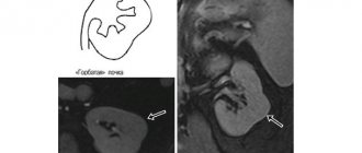
The main methods for diagnosing kidney cancer are computed tomography (CT) and magnetic resonance imaging (MRI), performed with intravenous contrast to accurately visualize the structure and size of the tumor, as well as the anatomical features of the vascular structure of the kidney. Any kidney tumor detected by ultrasound and/or confirmed by CT or MRI is an indication for surgery. The nature of the tumor (malignant or benign) is established after the intervention through morphological examination in the laboratory.
Robot-assisted partial nephrectomy is the procedure of choice for localized kidney cancer (ie, the tumor is limited to the kidney). The da Vinci robot provides unparalleled freedom of movement of instruments, surpassing the capabilities of the human hand, and also allows you to work at up to 10x magnification in 3D mode. These features help to perform the operation with minimal blood loss and minimal time of clamping of the renal vessels, which has a beneficial effect on the functioning of the kidneys in the postoperative period. The da Vinci robot also allows you to perform kidney resection in complex tumor localizations, for example in the upper segment or on the posterior surface of the kidney.
Nephrectomy remains the procedure of choice for large tumors and in cases where partial nephrectomy is not technically feasible. Nephrectomy is preferably performed using laparoscopic or robot-assisted methods, which provide comparable results. But they, in turn, surpass the results of open nephrectomy in such parameters as the average volume of blood loss, postoperative pain syndrome, and cosmetic effect.
Treatment
The only effective treatment for oncocytoma is surgery.
The main treatment for kidney oncocytoma is surgical removal of the tumor or the entire organ affected by it. When choosing intervention tactics, preference is given to organ-preserving and minimally invasive techniques. The operations are performed under general anesthesia. They can be performed laparotomically (that is, after making classical incisions) or using laparoscopic techniques.
The following types of surgical operations are performed to remove oncocytomas:
- nephrotomy - removal of the tumor and part of the organ tissue;
- nephrectomy – the affected kidney is completely removed.
After the operation, the tactics of which are completely determined by the clinical case, the patient is prescribed symptomatic and supportive therapy.
What symptoms can you use to recognize the disease?
At the initial stage of the disease, the tumor is small and often develops in one of the kidneys.
Symptoms do not appear at the initial stage of the disease.
The primary disorder of both organs appears quite rarely, only with a hereditary predisposition.
The tumor grows quickly and the blood vessels, having a strong muscle wall, but weak elastic plates, do not keep up with the increase in muscle fibers.
The manifestation of these symptoms is a reason to urgently consult a doctor and make an accurate diagnosis. Because the larger the kidney formation, the more serious its consequences will be.
A tumor of large dimensions can cause unexpected organ rupture and large-scale intra-abdominal bleeding.
The spread of the tumor to nearby lymph nodes or to a vein of an organ threatens the appearance of a large number of metastases.
Important!
To treat kidney diseases, our readers successfully use Galina Savina’s method.
Which doctor should I contact?
If you experience pain in the lumbar region, problems with urination, or blood in the urine, you should immediately contact a nephrologist or urologist. After an ultrasound, CT, MRI, blood and urine tests, and a biopsy of the identified tumor, the doctor will draw up a plan for further treatment.
Kidney oncocytoma is a benign tumor that consists of non-functioning cells of the renal parenchyma, is surrounded by a capsule and, when it reaches a large size, puts pressure on the tissues of the organ and its surrounding structures. Small neoplasms are asymptomatic and make themselves felt only after increasing in size. Treatment of these formations is carried out surgically. The surgical technique is determined for each patient individually and may involve partial or complete removal of the affected kidney.
Rating: (votes - 1 , average: 5.00 out of 5)
ONCOCYTOMA
ONCOCYTOMA
(
oncocytoma
; Greek onkos mass, tumor + kytos receptacle, here - cell + -oma) - a tumor formed from large light cells of the oncocyte type with eosinophilic granularity in the cytoplasm. The term reflects histogenetic and morphol, characteristics of the tumor, independent nozol, does not matter. Wedge, the picture depends on the location of the tumor.
Oncocytes (B cells, Hürthle cells) were first described in 1931.
Gamperlem (H. Natrerl) in the epithelium of the glands of the esophagus, and later found in many other organs and tissues: in the pituitary gland, thyroid, parathyroid and salivary glands, trachea, larynx, lungs, stomach, intestines, adrenal glands, kidneys. Oncocytes form small groups, or follicles, and can line the excretory ducts of the glands, cysts and papillary processes. For a long time it was believed that oncocytes are cells that have lost their function and activity. Subsequently, it was found that high activity of the enzyme succinate dehydrogenase is constantly determined in oncocytes; they produce and contain biogenic monoamines, in particular serotonin, and the totality of these cells forms the so-called. neuroendocrine system, or APUD system. Oncocytes can become a source of O.'s development in various organs. The O. of the salivary glands and the O. of the thyroid gland, which develops from the B-cells, are described in most detail.
Microscopically, the O. of the salivary glands usually has a solid structure and consists of groups and strands of oncocytes, divided into irregular lobules by delicate connective tissue septa. The tumor does not have a capsule and spreads diffusely in the surrounding gland tissue.
Microscopic specimen of oncocytoma (trabecular adenoma) of the thyroid gland: numerous oncocytes (indicated by arrows) with abundant granular cytoplasm are visible; hematoxylin-eosin staining; X 240.
In the thyroid gland, O. may have the character of a follicular, papillary or trabecular adenoma (Fig.), follicular and papillary adenocarcinoma or undifferentiated cancer (see). These varieties of O. have different structures, and their cellular elements are more or less similar to normal oncocytes. The unity of the histogenetic source of these tumors, i.e. the connection, for example, with B-cells of the thyroid gland, can be established by histochemical and electron microscopic studies.
Treatment is often surgical; for malignant O. sometimes combined treatment is used. For more details on treatment, see articles on tumors of individual organs, for example. Stomach, tumors; Lungs, tumors; Parotid gland, tumors, etc.
Bibliography:
Raikhlin H. T. and Smirnova E. A. Oncocytes - the serotoninocyte system, Arkh. pathol., t. 37, v. 2, p. 12, 1975, bibliogr.; Guide to pathological diagnosis of human tumors, ed. N. A. Kraevsky and A. V. Smolyannikov, p. 315, M., 1976; Ashley DJ V. Evans'histolo-gical appearances of tumors, p. 232 ao, Edinburgh ao, 1978.
Yu. N. Soloviev.
Diagnostics and therapy
At the 7th Congress of ROOU, the famous professor V.B. Matveeva presented a report that became a real breakthrough in the drug treatment of renal AML. Scientists have proven the fact that one of the targeted drugs, belonging to the group of mTOR inhibitors, reduces the volume of renal angiomyolipoma by 45-50% over a 12-month course.
Although there is currently no reliable data on the tolerability of this drug, it can be firmly stated that this is the greatest breakthrough in this field. After all, everyone knows that drug treatment is less traumatic for the human body.
It is important to know. If you have been diagnosed with kidney angiomyolipoma, it is strictly forbidden to resort to treatment with folk remedies.
Self-medication in such a case cannot be effective; moreover, it can lead to dire consequences, including
and to death. If you value your health, then first of all consult a doctor.
Only an experienced doctor will give you an accurate diagnosis and prescribe the correct treatment.