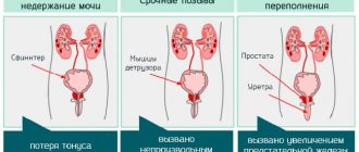
Hypothyroidism
A condition of the thyroid gland in which there is a decrease in hormone production.
The causes of this disease may be:
- • Removal of part of an organ;
- • Autoimmune damage;
- • Inability to absorb iodine;
- • Genetic disorder.
When the thyroid gland is healthy, it provides the body with a certain amount of secretion. If part of the gland does not participate in synthesis due to illness, then the hormones produced become less.
In the case of an autoimmune lesion, immune cells attack the organ, thereby destroying thyrocytes. This negatively affects the functioning of the organ, which leads to thyroid disorders.
In congenital disorders, the enzymes that are involved in protein synthesis have a defect due to which iodine is not fully absorbed. This causes the hormones to be defective (not functioning properly).
There are three forms of violation. The first is caused by a disease of the organ. The second is caused by disruption of the pituitary gland. The third form is associated with the activity of the hypothalamus.
Symptoms
The primary stage of hypothyroidism occurs in almost every person, however, the person is usually unaware of the pathology (due to the absence of symptoms). At the first stage of the process, a decrease in mood, apathy, and decreased performance may be observed. Against the background of stress in which a modern person usually finds himself, such symptoms are not alarming. at the end of the first stage, swelling appears on the face, dryness and flaking of the skin, changes in speech (several tones lower), hearing deteriorates, pain in the joints, discomfort in the chest, weakness in the muscles. The list of symptoms is very wide; if they appear, you should consult a doctor for diagnosis.
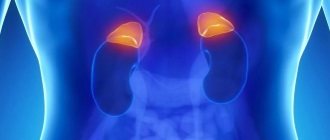
Parathyroid diseases and osteoporosis
The parathyroid glands are small glands (usually 4) located behind the thyroid gland.
The main function of the parathyroid glands is to maintain normal serum calcium levels with the help of parathyroid hormone (parathyroid hormone).
This hormone stimulates the release of calcium from bone tissue and reduces calcium loss in the urine. Diseases of the parathyroid glands can manifest as either increased (hyperparathyroidism) or decreased (hypoparathyroidism) production of parathyroid hormone.
Manifestations of hyperparathyroidism may include
- osteoporosis and fibrocystic bone formations
- fractures due to minor injuries
- urolithiasis, chronic renal failure
- drowsiness, fatigue, memory loss, psychosis and depression
- muscle weakness
- decreased appetite, nausea, vomiting, abdominal pain and constipation
Symptoms of hypoparathyroidism may include
- numbness of limbs, spasms, convulsions
- rapid heartbeat, heart enlargement
- dry skin, brittle nails
- mood swings, memory impairment
Since calcium is a vital element, a sharp decrease or increase in its level in the blood can lead to acute conditions - hypo- and hypercalcemic crises, which pose a danger to the patient’s life.
Therefore, timely diagnosis and adequate treatment measures for disorders of the parathyroid glands are one of the priorities of specialists at the Rassvet clinic.
Special attention should be paid to vitamin D deficiency. Currently, insufficiency, and to a greater extent, vitamin D deficiency affects the majority of the general population, including children and adolescents, adults, pregnant and lactating women, menopausal women, and the elderly.
Vitamin D deficiency primarily affects calcium-phosphorus and bone metabolism. Since vitamin D normally increases the absorption of calcium in the intestine, its deficiency leads to a decrease in its level in the blood and a compensatory increase in the secretion of parathyroid hormone, the so-called secondary hyperparathyroidism. An increased level of parathyroid hormone leads to the “washing out” of calcium from bone tissue and the development of osteoporosis. Vitamin D deficiency is also manifested by muscle weakness, difficulty walking and maintaining balance, and a tendency to fall, which naturally increases the risk of fractures. Data have been obtained on the relationship between vitamin D deficiency, autoimmune and cancer morbidity, and overall mortality.
The knowledge of Rassvet clinic specialists on this problem allows us to effectively identify vitamin D deficiency in patients and promptly recommend measures to restore and maintain its normal level.
Osteoporosis is a skeletal disease characterized by loss of bone mineral density. 50% of women and 20% of men over 50 already have signs of low bone density. The main danger of osteoporosis is bone fractures (femoral neck and vertebrae), which lead to high disability and mortality. In addition to fractures, in severe osteoporosis, microdamage to the bone tissue of the vertebrae can cause persistent back pain.
Risk factors for osteoporosis include
:
- menopause
- elderly age
- already suffered fractures
- fractures in relatives
- underweight
- hypodynamic lifestyle
- smoking and alcohol abuse
- unbalanced diet, insufficient consumption of dairy products
- use of a number of medications
Fractures can be prevented with prompt medical attention. At the Rassvet clinic, early diagnosis of osteoporosis is carried out and the optimal therapy for the prevention of fractures is individually selected.
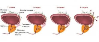
Hyperthyroidism
Hyperthyroidism is the opposite of hypothyroidism. The symptoms of the disease vary greatly, as does the mechanism of disease development. The basis is hyperfunction of the thyroid gland, in simple terms - it produces too many hormones. When they enter the blood in excess, they cause symptoms of poisoning, and metabolism accelerates several times.
Thyrotoxicosis is a complex of symptoms that is associated with serious health problems.
The causes of this disease may be:
- • Long-term use of hormonal drugs;
- • Various abnormalities of the thyroid gland;
- • Excess iodine;
- • Other reasons.
When you are prescribed hormonal drugs, you must strictly follow all recommendations for use and dosage, otherwise you can put your body into a state of artificial hyperthyroidism. Oncological damage to the gland, diffuse toxic goiter and Plummer's disorder also provoke excessive synthesis of hormones.
Other reasons include: cancer of the ovaries, pituitary gland, side effects of medications. Depression, stress, and pregnancy contribute to the deviation.
Symptoms
Unlike hypothyroidism, symptoms of hyperthyroidism are noticeable from the very beginning. These are excitability, aggressiveness, mood swings. There are other signs: absent-minded attention, sleep disturbances, and susceptibility to panic. When palpated, an enlarged gland is observed, which often causes difficulty swallowing.
Differences hypo and hyperthyroidism:
- • Increased heart rate, increased heart rate;
- • Visual impairment;
- • Weight loss with good appetite;
- • Interruptions in the menstrual cycle;
- • Sweating.
Against the background of intoxication, the body tries to get rid of excess dangerous substances. The heartbeat increases, the eyeball increases in volume (its rotation becomes difficult, and tearing also appears).
Due to the acceleration of metabolism, the absorption of nutrients occurs faster, so a person loses weight even when eating as usual. For women, this often results in difficulty conceiving or termination of pregnancy.
Hyperthyroidism and hypothyroidism are interrelated - they can replace each other as different stages of the autoimmune process.
PsyAndNeuro.ru
A positive dexamethasone test in patients with melancholic depression, indicating hyperactivation of the hypothalamic-pituitary-adrenal axis (HPA axis), has been known since my student days. Moreover, hyperactivation of the HPA axis is a powerful biological correlate of depression along with dysfunction of monoamine neurotransmitter systems and is considered one of the main links in its pathogenesis. However, over the past few decades, scientists' knowledge of how the HPA axis functions has expanded significantly. It has now become clear that in addition to the functioning of the hypothalamus, pituitary gland and adrenal glands, which occupy one of the central places in the pathogenesis of depression, a much larger number of areas of the brain and its neurotransmitter and neuropeptide systems are involved in the pathological process. This “expansion” of the theory of HPA axis hyperactivation in depression allows us to take a fresh look at the mechanisms of development of this disorder, as well as find new specific targets for drug therapy. It is also worth noting that hyperactivation of the HPA axis occurs only in 70% of patients with this disorder, and is not typical for such types of depression as menopausal, atypical or seasonal, which, on the contrary, are characterized by a decrease in the activity of the HPA axis.
Rice. 1 Functioning of the HPA axis is normal
The stimulus that caused the stressor reaches the HPA axis from the amygdala. In response to this, vasopressin and corticotropin-releasing hormone (CRH), the main regulator of the HPA axis, are released in the paraventricular nucleus of the hypothalamus. It consists of 41 amino acid residues, and its synthesis is regulated by the CRF gene. Together with vasopressin, CRH travels through the portal vein system to the anterior pituitary gland and stimulates the production of the prohormone proopiomelanocortin (POMC), which is then processed into adenocorticotropic hormone (ACTH), opioids and melanocortin. ACTH, in turn, stimulates the release of glucocorticoids (cortisol in humans and corticosterone in mice) in the zona fasciculata of the adrenal cortex, which subsequently exert negative feedback on the pituitary gland and hypothalamus through effects on mineralcorticoid (MR) and glucocorticoid receptors (GR), due to which the degree of activation of the HPA axis decreases (Marni N. Silverman, 2012).
Despite the widespread belief that the main manifestations of hyperactivation of the HPA axis are associated only with hypercortisolemia, there is increasing evidence that increased levels of corticotropin-releasing hormone (CRH), which is the main regulator of the HPA axis, have a much greater negative effect on functional systems brain.
The effects exhibited by CRH depend on the zones of its synthesis and the places of greatest concentration of its receptors. So, in addition to the hypothalamus, it is also secreted in the neurons of the amygdala, hippocampus and locus coeruleus. This neuropeptide has a local neuromodulatory effect on neurons within a few seconds after release, acting through two specific CRH receptors types 1 and 2 (CRHR1, CRHR2), which are widely distributed in various parts of the brain. It is also worth noting that the family of CRH neuropeptides, in addition to CRH itself, also includes urocortins (UCN 1, 2 3), which, like wozopressin, orexin and dynorphin, affect brain homeostasis.
Depending on the dose and time of exposure of CRH to targets, its effects also differ. For example, the release of CRH in optimal doses during acute stress in the central nucleus of the amygdala promotes memory consolidation, and in the hippocampus it increases synaptic plasticity. However, when exposed to high doses of CRH over a long period of time, hippocampal function deteriorates, manifested by neuronal damage and a decrease in the number of synapses.
Thus, during the entire episode of melancholic depression, patients exhibit elevated levels of CRH in plasma and cerebrospinal fluid. Postmortem studies of deceased people with depression have found evidence of CRH hyperactivity in the paraventricular nuclei of the hypothalamus, cortical areas, pontine nuclei, and locus coeruleus. At the same time, elevated CRH levels in depressed individuals decreased after a course of electroconvulsive or antidepressant therapy.
Moreover, in healthy individuals with a high genetic risk of developing depression in their families, the results of the combined DEX/CRH test (DEX/CRH: dexamethasone-suppression/corticotropin-releasinghormone-stimulationtest), which combines dexamethasone suppression with CRH stimulation, are between results of patients with depression and a control group of healthy volunteers. These results indicate that even small changes in HPA axis functioning have a genetic background that increases the risk of developing depression or other stress-related illnesses with age. Another study showed that individuals with depression had a significantly increased frequency of the G allele polymorphism (rs242939) of the CRHR1 gene compared to the control group. Taken together, these observations support the concept that HPA axis dysregulation, including elevated CRH levels, may be associated with genetic predisposition and represent a risk factor for the development of depression.
In depression, a decrease in the activity of neurotrophic factors is also observed. This leads to decreased synaptic connections in the hippocampus and prefrontal cortex, which correlates with depressive symptoms. A close relationship has been found between neurotrophic factors, in particular brain-derived neurotrophic factor (BDNF), and CRH, which modulates their production, production and activity.
It is also interesting that changes in the CRH receptor apparatus also affect the formation of certain symptom complexes. Thus, repeated exposure to stress leads to a change in the ratio of CRHR1 and CRHR2 towards an increase in CRHR1 in areas associated with depression. CRHR1 is able to modulate anxious behavior independently of the HPA axis, and its deficiency protects a person from the formation of negative consequences of stress, regardless of age. This is due to the fact that CRHR1 controls glutamatergic, noradrenergic and dopaminergic neural circuits, making a significant contribution to the manifestations of stress-related disorders.
This is supported by extensive evidence showing that the three major neurotransmitter systems (serotonin, norepinephrine and dopamine) closely interact and influence CRH levels. Thus, in the locus coeruleus (the most important nucleus of the noradrenalinergic system in the brain), the dorsal raphe nuclei (the most important nucleus of the serotonergic system) and in the ventral tegmental area (the most important nucleus of the mesocorticolimbic dopaminergic system) a high level of expression of CRHR1 and CRHR2 is found, and the overall level of expression of these receptors and their ratio is an important indicator of individual stress tolerance and the risk of developing depression.
For example, in the dorsal raphe nucleus, CRH has opposing effects on the serotonergic system depending on which receptor (CRHR1 or CRHR2) it acts on. Thus, activation of CRHR1 in this area leads to increased GABAergic inhibitory effects on the serotonin system, and activation of CRHR2, on the contrary, has a potentiating effect.
The influence of CRH on the formation of anhedonia, which is characterized by a decrease in activity in mesolimbic dopamine projections from the ventral tegmental area to the nucleus accumbens (ventral striatum) and further to the cerebral cortex, has also been proven. During periods of acute stress, CRH stimulates the release of dopamine in the nucleus accumbens, which is an important part of the “reward” system. However, after severe acute stress or with prolonged exposure, CRH does not stimulate the release of dopamine, which can be considered as one of the mechanisms for the formation of anhedonia.
An imbalance in the neurotransmitter systems also affects the HPA axis itself, since acetylcholine, dopamine and norepinephrine promote the secretion of CRH in the hypothalamus, and serotonin, in turn, inhibits the secretion of CRH in the hypothalamus and ACTH in the pituitary gland, which also contributes to HPA axis dysfunction. axes.
Another important feature of the physiology of depression is the disinhibition of the REM sleep phase (REM, Rapid Eye Movement), which is specifically associated with the central activity of CRH and a decrease in the level of somatotropin-releasing hormone (SRH), which regulates slow-wave sleep. dream. Thus, in one clinical study assessing the safety and tolerability of CRHR1 antagonists (R121919), EEG recordings were performed during sleep before and after 28 days of treatment. The results showed that the majority of patients with significant REM sleep stage disturbances showed improvement in depressive symptoms between 50 and 90% on the Hamilton Depression Rating Scale, while improvement in patients with normal sleep stages was consistently below 50%.
Rice. 2 Sleep disturbances in depression
Sleep disturbance in depression is characterized by disruption of slow wave sleep and increased REM sleep as a result of an imbalance between CRH and SRH (Steiger, 2003).
The generalized results suggest that disinhibition of the REM sleep phase can probably be considered as a specific indicator reflecting the central activity of CRH, with the help of which it is possible to identify patients with depression in whom it is advisable to undergo therapy with CRHR1 antagonists, which are widely used for the treatment of endocrinological diseases.
Source: Kasyanov E.D., Mazo G.E. Functioning of the hypothalamic-pituitary-adrenal axis in depression: current state of the problem. // Journal of Mental Health. – 2017. – No. 8. – P. 27 – 34.
Transition between states
As we have already mentioned, a transition from one state to another is possible. A situation is often observed when hyperthyroidism turns into hypofunction (after increased production of hormones, their production slows down). Over the course of a person's life, they may experience several different thyroid diseases.
In the case when hypothyroidism goes into a reverse process, this means that the body actively began to produce antibodies, and a switch to another autoimmune process occurred.
At the first signs of disruption in the functioning of your body, you should consult an endocrinologist. Our multidisciplinary clinic in Moscow “Sante Clinic” employs experienced endocrinologists. By contacting us you will save yourself from queues, tedious waiting and inattentive attitude. We are located next to the Polezhaevskaya, Khoroshevo and Oktyabrskoye Pole metro stations. We are waiting for you at Sante Clinic.
Endocrinology
The pituitary gland and its dysfunction
The endocrine system of the body has a complex hierarchical system, which, when functioning correctly, affects the metabolism of all metabolic substances.
It includes the hypothalamic-pituitary system, adrenal glands, ovaries in women and testes and testes in men, thyroid and pancreas. The most important gland is the pituitary gland. It is a small gland that is the size of a baby's fingernail, but at the same time it regulates all the processes of the endocrine glands of the body. Depending on the amount of hormones produced by the pituitary gland, hypofunction and hyperfunction of the pituitary gland are distinguished, which leads to various complications.
Pituitary gland dysfunction
With a lack of pituitary hormones, the following occurs:
- Hypothyroidism, which appears as a result of a lack of iodine and related hormones in the body;
- Lack of antidiuretic hormone, which leads to metabolic disorders or diabetes insipidus;
- Hypopituitarism. This is a complex disease that is associated with underdevelopment of the pituitary gland. As a result, this gland does not produce almost all hormones, which leads to delayed puberty in children, and in adults, decreased sexual desire, impaired reproductive function, and so on.
When there is an excess of pituitary hormones, the following disorders are observed:
- High levels of prolactin, which affects the menstrual cycle, infertility, and premature milk production. In men, prolactin suppresses sexual desire, and in large doses causes erectile dysfunction;
- Increased levels of somatropic hormone, which affects growth;
- The level of adrenocorticotropic hormone increases, which in case of excess leads to a serious disease - Cushing's syndrome. This disease is characterized by vegetative-vascular dystonia, diabetes mellitus, and severe forms of mental disorders.
Hypo- and hyperfunction of the pituitary gland are very serious disorders that sometimes entail irreversible consequences of the functioning of the body.
Causes of pituitary gland disorders
When there is an excess of pituitary hormones, patients experience an adenoma - a benign or malignant tumor of the gland itself. In this case, both lobes of the pituitary gland are affected, which may be caused by hyperfunction of the anterior pituitary gland. Since the pituitary gland is located between the lobes of the brain, when the tumor grows, the oculomotor and optic nerves may also be affected.
Hyperfunction of the pituitary gland is also dangerous because it provokes the production of testosterone by the adrenal cortex, which, if in excess, can lead to impaired fertility in women. For men in this situation, there is hyperproduction of androgens - female sex hormones.
Provoking factors for hypofunction of pituitary hormones are:
- Past infectious and viral diseases of the cerebral cortex and the brain itself;
- Open and closed craniocerebral injuries;
- Hereditary factor;
- Previous operations, chemical irradiation.
Treatment should be strictly under the supervision of a doctor, who prescribes various methods of replacement therapy for mild manifestations of the disease, or, in extreme cases, schedules a visit to an oncologist for further examination of tumors.
Pituitary