We receive most of the information about the world around us through our visual organs. It is possible to clearly see objects and navigate in space only if both eyes work in a coordinated manner. With monocular vision, objects that fall within a person’s field of vision are perceived by only one eye, which causes a number of distortions, since normally two pictures are combined into one, and the brain forms an accurate, correct image. You will learn about the characteristics of the disease, its varieties, diagnostic methods used and possible treatment options from this review.
What is amaurosis?
Translated from Greek, “amaurosis” means dark. In medical terminology, this word is used to refer to conditions associated with significant impairment of vision or absolute blindness without obvious causes. Most often, the pathology is hereditary, but can also be caused by diseases of the central nervous system, as well as various functional disorders of the nervous system.
The clinic distinguishes between permanent vision loss - Leber's amaurosis, and transient blindness - amaurosis fugax, ischemic oculopathy, photosensitive retinopathy.
Let's take a closer look at each of these conditions.
Causal factors
The reasons leading to amaurosis can be different. This can be intoxication (for example, lead), general diseases (diabetes mellitus and others), hereditary pathologies, etc.
Taking into account the causative factors, the following forms of amaurosis are distinguished, which include:
- hereditary amaurosis (Leber amaurosis);
- transient amaurosis;
- transient amaurosis.
Thus, the pathological processes that are accompanied by this disease belong to diseases of the nervous system. Amaurosis is a clinical manifestation of impaired sensitivity of the organ of vision, which can have different origins.
Symptoms of night blindness
In night blindness, vision adaptation is impaired when moving from a relatively light room to a dark room and back. This means that a person cannot orient himself for a long time and begin to see normally when he moves from one level of illumination to another. Moreover, this is observed both during the transition from dark to light, and vice versa, from a lighted place to a darkened one.
In poor lighting, a person’s field of vision narrows, and he sees the picture of the world around him in a very narrow frame, as if through a pipe or a small window. In addition, a person ceases to clearly see the shape and size of objects, and also does not distinguish colors. The difference between blue and yellow colors is especially bad in case of night blindness. A person begins to notice that, in principle, he does not perceive colors correctly, since a violation of the Purkinje effect
. The Purkinje effect is the phenomenon of different perceptions of colors as light levels decrease. Thus, at dusk, red colors appear darker, and blue colors, on the contrary, appear lighter. The overall picture is seen in dark, muted tones, and there is a feeling of vision as if in a fog.
In addition, with night blindness, the eye is insufficiently sensitive to light, so a person needs very bright lighting to read or write. The need for bright light for writing and reading against the background of normal vision at dusk is the first sign of the development of night blindness.
Night blindness often causes decreased vision. This means that in normal lighting conditions a person has 100% vision, but at dusk it drops by several units. Iskersky-Bito plaques are found on the conjunctiva of the eye in cases of essential night blindness
.
Poor vision in low light conditions can frighten a person and ultimately cause a fear of the dark. Especially often, fear of the dark develops against the background of night blindness in children with a congenital disease.
Hereditary amaurosis
The disease has a very complex and not fully understood mechanism. It is known that the pathology develops as a result of mutations in some genes that are responsible for the condition of the retina.
Without going too deeply into the basics of genetic engineering and molecular biology, the reasons why blindness develops can be presented as follows. Normally, the body produces rhodopsin, a pigment located in the rods of the retina of the eye and regulating the transmission of nerve impulses to the visual nuclei of the brain. Thanks to rhodopsin, light entering the retina causes stimulation in the optic nerve, which is transmitted to the brain. The result of this complex neurochemical reaction is the ability of the eye to see various objects in all types of lighting.
It must be said that the supply of rhodopsin in the retina is quite limited, therefore, along with decomposition, the reverse process occurs - the resynthesis of pigment, which is necessary to maintain rhodopsin in the retinal rods at the proper level. On average, complete restoration of the required amount of pigment takes 30 minutes.
Hereditary amaurosis is transmitted through a mutated gene responsible for the processes of formation and decomposition of rhodopsin, as a result of which, with an unchanged optic nerve, the patient sees nothing, i.e. blindness develops. Studies have shown that this occurs due to the death of rods and cones in the retina due to the presence of several types of mutant genes.
The disease is inherited only if both parents have this defective gene.
What is amaurosis and can it be cured?
Amaurosis is an ophthalmological disorder in which visual acuity is impaired.
And in the absence of treatment for this disease, complete blindness occurs within a short time without the possibility of restoring visual functions.
Often this pathology is congenital and develops due to the influence of genetic factors.
Transient
A transient type, also called transient, is a temporary disorder that occurs due to external or internal predisposing factors.
This disease is treated with medication, but often visual functions are restored on their own.
Congenital (Leber amaurosis)
It is worse if congenital amaurosis (Leber amaurosis) is diagnosed, in which a dysfunction of the retina of the eye is detected, and they, in turn, develop in the womb and are a genetic factor.
Transient amaurosis can occur due to the development of the following pathologies:
- absence of pigments and photoreceptor elements in the retina;
- changes in the shape and structure of the blood vessels of the eye;
- vascular embolism;
- neoplasms of various types in the brain and eye that interfere with normal blood circulation;
- vascular atherosclerosis;
- vascular aneurysm and dysplasia.
Other reasons may be previous eye or head injuries, strokes, intoxication with alcohol, narcotic or toxic substances.
In all these cases, dysfunction of the mechanisms responsible for transmitting visual images to the brain may occur.
Although this disease may not be present in the parents, it is transmitted to the child.
Treatment of the disease
The course of treatment is determined by the doctor and will depend on the causes of amaurosis and its type.
Basically, only a transient type of disease can be treated.
In such cases, medications may be prescribed to normalize circulatory processes:
- nootropics;
- diuretics;
- hypotonic agents;
- drugs that stimulate blood flow;
- medications that stabilize the condition of vascular and capillary tissue cells.
If the cause is tumors, surgical treatment or chemotherapy will most likely be prescribed.
For endocrinological pathologies, a course of taking medications that lower blood sugar levels, as well as hormone replacement therapy, can help.
If vision deterioration is accompanied by severe migraines, the situation can be corrected by taking antispasmodics, and in the case of toxic poisoning, vision restoration occurs after undergoing a course of detoxification therapy.
In any case, it is necessary to first eliminate the root cause of amaphrosis.
But in the case of Leber amaurosis, such treatment is not possible: in fact, it is a genetic mutation that cannot be eliminated with modern methods.
Complications of amaurosis
In the absence of adequate and timely treatment, this disease can lead to dystrophic lesions of the retina, atrophy of the optic nerve, and in the worst case, complete blindness.
In most cases, these are irreversible complications in which restoration of visual functions is impossible.
Treatment prognosis
Favorable prognosis for cure can be given only for transient amaurosis, but only if the patient seeks help from specialists in a timely manner.
In such cases, vision is restored in most patients (in mild to moderate cases, complete recovery occurs).
This video presents the main causes of visual impairment:
A person cannot independently determine that he is developing amaurosis.
Moreover, sometimes a decrease in visual acuity is not associated with pathological disorders and people can wait months or years until vision returns to normal.
Was the article helpful?
Why vision may deteriorate in one eye
There can be many reasons for deterioration of vision in one eye; a similar symptom appears, for example, in the following conditions:
- retinal damage;
- pathologies of the cornea and lens;
- some general somatic diseases (for example, diabetes mellitus);
- eye injuries;
- strabismus;
- amblyopia.
Making an accurate diagnosis with such symptoms is difficult; additional research is required. Visual impairment can be either permanent or temporary, and is not always associated with any diseases. Its causes can also be stress, overwork, very long work at the computer, lack of sleep, etc. Temporary disorders often do not require any therapy and go away on their own after some time.
Persistent visual impairments are associated either with pathologies of the optical system of the eye (cornea, lens, vitreous body and retina), or with innervation disorders. Often, decreased vision in one eye accompanies diabetic retinopathy, a complication of diabetes mellitus in which the vessels of the retina are damaged.
Diagnosis of diabetic retinopathy
If you are diagnosed with diabetes, you need to be examined by an ophthalmologist more often than healthy people - at least twice a year. As we said above, the first signs of retinopathy become noticeable already in the last stages. Early diagnosis will help to identify changes in the fundus and promptly treat the pathology. These are the procedures a specialist performs to examine the condition of the eye structures.
- Patient interview and visometry - checking visual acuity using Sivtsev-Golovin tables.
- Tonometry - measurement of intraocular pressure (especially in patients with a history of the disease of more than 10 years).
- Ophthalmoscopy - examination of the fundus of the eye.
- Biomicroscopy of the retina, lens, vitreous body.
- Retinography is photography of the retina with a special camera.
- Instrumental examination of the optic nerve, macula.
- If clouding of any optical media is detected - the vitreous body, lens, cornea - a retinal ultrasound is prescribed.
- Optical coherence tomography. This method allows you to obtain images of ocular structures of such high resolution that you can see the thinnest layers of tissue, 1 micron thick.
- Fluorescein angiography.
- Perimetry.
If necessary, diagnostics are also prescribed by other specialists, in particular by an endocrinologist, since diabetes is part of the group of endocrine diseases. After collecting anamnesis and receiving all research results, the specialist decides on the treatment method depending on the condition of the eyes.
How does Leber amaurosis manifest?
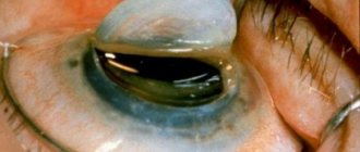
The disease usually manifests itself in early infancy. In this case, the child may notice a lack of reaction to approaching objects, a wandering gaze, without fixation on anything, a weak reaction of the pupils to light (usually in bright light the pupil narrows, and in reduced light it dilates), the eyeballs constantly move up and down or in different directions (nystagmus).
Many children develop strabismus and keratoconus (the cornea takes on a convex shape, resembling a cone). In very bright light, children experience some improvement in vision.
If the disease does not develop very rapidly, then the deterioration of vision occurs gradually, but by the age of 9-10 years it still completely disappears.
It is important that the fundus of the eye is not changed at the beginning of the disease, but later pigment begins to be deposited on it, which leads to irreversible changes in the retina.
Unfortunately, there is no special treatment for this pathology yet and vision cannot be restored. In addition to blindness, many children develop concomitant pathologies - endocrine diseases, changes in the musculoskeletal and central nervous systems.
Symptoms
The presence of a severe congenital form of the disease can be suspected already in childhood:
- The baby has no reaction even to objects located very close;
- The child is not able to fix his gaze on a specific object, and his gaze always wanders;
- There is almost no reaction of the pupil to light irritation;
- The child's eyes make constant involuntary movements in different directions.
If the disease is transient, the symptoms may differ depending on the cause, but there is always a sharp deterioration in vision:
- In case of cerebrovascular accident, in addition to loss of vision, dizziness and weakness, disorientation in space are noted;
- In case of brain injuries - loss of visual fields, nystagmus (involuntary oscillatory eye movements), inability to focus the gaze;
- In case of endocrine diseases - swelling of the orbital tissue, extraocular muscles, double vision and pain in the eyes, exophthalmos (a feeling of protrusion of the eyes);
- In case of severe poisoning - loss of visual fields, disorientation in space and confusion, blindness;
- In case of oncological diseases of the brain – headache and eye pain, nystagmus, disturbance of accommodation;
- With migraines, vision is completely absent during attacks.
Prevention
The basis of prevention is pregnancy planning, pregnancy management under the supervision of doctors, and elimination of influencing factors. You need to strictly monitor your medication intake and protect yourself from infections. After birth, it is necessary to monitor visual hygiene, protect children from eye injuries, infectious lesions, and diseases of the nervous system.
You will also learn how untimely treatment of blindness in children can be dangerous, and why it is so important to avoid the consequences. Everything about how to prevent blindness in children and prevent complications
And caring parents will find on the service pages complete information about the symptoms of blindness in children. How do the signs of the disease in children aged 1, 2 and 3 differ from the manifestations of the disease in children aged 4, 5, 6 and 7? What is the best way to treat blindness in children?
Take care of the health of your loved ones and stay in good shape!
However, essential night blindness does not develop immediately, since at least two years may pass from the onset of chronic vitamin A deficiency to the appearance of clinical symptoms. This is due to the fact that the reserves of vitamin A present in the tissues of the human body will last for about one year, provided that this compound does not come from the outside at all. However, in practice, there are no situations when vitamin A does not enter the human body at all, so reserves are depleted longer than a year and it takes at least two years for clinical manifestations of night blindness to form.
Diagnostic methods
A doctor may suspect monocular vision when external signs of strabismus are detected in adults. If necessary, specialists check the presence or absence of normal (binocular) vision in a patient using a number of tests:
- Four-point color test;
- Kalf's test;
- Bargolini test;
- Pressure test on one eye;
- Sokolov's experience.
It is necessary not only to diagnose monocular vision in a patient in a timely manner, but also to determine the cause of its occurrence. This type of visual perception can develop in connection with vascular disorders, certain types of eye injuries, and congenital abnormalities of the visual organs.
You can download the vision test chart here.
In addition to using instrumental diagnostic methods, it is important to collect information about when and how the patient’s ability to see changed.
Transient amaurosis
In a number of diseases, temporary blindness may occur, which disappears after the underlying cause is eliminated. The most common cause of amaurosis is circulatory disorders in the carotid artery system.
They can occur due to disorders or injuries in the neck and head:
- Narrowing or constriction of blood vessels;
- Neoplasms that impede blood circulation in this area;
- Trauma or mechanical compression;
- Aneurysms, arteritis, dissection, dysplasia of the vascular wall in the carotid artery basin, as well as arteriosclerotic changes in blood vessels;
- Air or fat embolism of blood vessels.
Diagnostics
A person is considered blind if he lacks visual function on both sides. In this case, there may be light perception and residual vision (from 0.01 to 0.05 diopters). In this case, corrective glasses can be selected for the better-seeing eye. A blind person cannot perceive the shape, color, size, location of objects; such patients have poor orientation in space, as they are not able to assess distance, direction of movement and other characteristics. All this leads to a decrease in sensory perception of the surrounding world. Simultaneously with the fading of visual function, in this category of patients sound perception is exacerbated. Such people find it much easier to navigate their environment based on subtle sound signals. Due to blindness, the formation of movements slows down. Some blind people experience deviations in the volitional and emotional spheres. Over time, the patient gets used to the new life, and blindness ceases to be perceived as painful. Instead of the visual analyzer, a person uses other ways of understanding the world around him (auditory, skin-sensory, motor and other types of perception)
This helps stabilize mental processes and restore voluntary attention, generalized perception, logical memory and abstract thinking. These factors help even blind people to perceive reality correctly.
When forming imaginative thinking, patients are helped by ideas that have been preserved in memory from the time of normal vision.
Total blindness is usually caused by damage to the optic nerve fibers, stroke, or other irreversible conditions. However, despite treatment, visual function, as a rule, cannot be restored. To improve the quality of life of blind patients, various devices can be used that are introduced into everyday life. These include books or manuals in Braille, special computer programs, and some devices.
With the sudden development of blindness, a person always experiences serious emotional shock. In addition to neuroses, such patients often experience depression, so not only ophthalmologists, but also psychologists or psychotherapists should treat this category of patients.
In modern ophthalmological practice, doctors are intensively looking for ways to eliminate acquired color blindness. They are mainly aimed at eliminating the causes of the disease. Sometimes vision is restored after stopping the drug that led to such consequences.
Color vision impairment
Diabetes
Atherosclerosis
1704 28 October
IMPORTANT!
The information in this section cannot be used for self-diagnosis and self-treatment.
In case of pain or other exacerbation of the disease, diagnostic tests should be prescribed only by the attending physician. To make a diagnosis and properly prescribe treatment, you should contact your doctor. Color vision impairment: causes of occurrence, what diseases it occurs with, diagnosis and treatment methods.
Description
Impaired perception of colors and their shades in medicine is called color blindness, or chromatopsia. The ability of the human eye to distinguish many colors and shades opens up enormous possibilities for orientation in space, knowledge of the surrounding world and fine art.
The conductor that helps a person perceive surrounding objects with the help of the organ of vision is light. Light is electromagnetic radiation, visible to the human eye, whose wavelengths range from 380 to 700 nm. Each object can reflect and absorb certain wavelengths of light. Those waves that are reflected pass through the transparent structures of the eye and excite special cells - cones, which are responsible for the perception of color.
The human eye can distinguish almost all color shades resulting from mixing the three basic ones - red, blue and green.
Depending on the percentage of excitation of a particular type of cone, a sensation of color arises in the brain. So, for example, when the brain perceives the color orange, then at that moment an impulse is transmitted from 99% of red cones, 42% of green and 0% of blue, and in the case when the brain perceives green, 31% of red, 67% of green and 36 % blue cones. If all cones are excited equally, then we see white color.
Color vision disorders occur due to a disruption in the excitation of cones and the transmission of impulses to the brain. If this condition is congenital, then a violation of color vision may remain unnoticed for a long time, because a person is guided by brightness and color saturation. Life experience and communication with people provide information about color: grass is green, the sky is blue, strawberries are red. Errors are detected at low brightness and object sizes.
A condition where a person has a reduced or completely absent ability to distinguish all or some colors is called color blindness.
.
Types of color vision impairment
There are congenital (most common) and acquired color blindness. Depending on the clinical manifestations, i.e. Depending on the characteristics of color perception, the following types of color blindness are distinguished:
- Achromatopsia is a complete lack of color vision, when a person perceives everything in black and white. Very rare.
- Deuteranopia is a complete loss of perception of the green part of the spectrum. Most common.
- Protanopia is a complete impairment of recognition of the red part of the spectrum. Deuteranopia and protanopia give similar color perception because... mixing red and blue, blue and green gives rise to a similar interpretation in the brain - swamp green.
- Tritanopia is a complete impairment of the perception of the blue spectrum. Rarely seen.
In addition to color blindness, there is a weakness in color sensitivity, when a person needs much more time and strong color saturation to recognize the color of an object.
Decreased perception of red is called protanomaly, green is called deuteranomaly, and blue is called tritanomaly.
Possible causes of color vision impairment
The most common cause of color vision impairment is damage to the genes involved in color perception on the X chromosome. As you know, men have a chromosome set of 46XY, and women have 46XX. If a boy received a damaged X chromosome from his mother, then there is nothing to compensate for the defective genes (there is no second X chromosome), and a violation of color perception occurs. In women, congenital color blindness is quite rare - in the case when a girl receives a damaged X chromosome from both her father and mother. The color blindness gene can be passed on through generations, manifesting itself in grandchildren and great-grandchildren. To make sure there is no genetic predisposition to color vision impairment, you can take a DNA test. This method is applicable in cases where conventional visual tests cannot be used, for example, for newborns and children in the first years of life.
Acquired types of color vision disorders are relatively rare and occur with various damage to the visual analyzer at one of the stages of receiving, transmitting or processing information.
Among their reasons are the following:
- damage to the retina by ultraviolet light if safety precautions for using UV lamps are not followed;
- Diabetes mellitus is one of the common causes of acquired color vision impairment as a result of diabetic macular degeneration. With a constant increase in blood glucose levels, damage occurs to all blood vessels, and especially to the small vessels of the fundus. Due to lack of oxygen, the retina ceases to perform its functions. Along with the symptoms of color vision impairment, distortions of straight lines and blurred vision occur;
- inflammatory diseases of the retina and optic nerve;
- cerebral atherosclerosis;
- taking certain medications (for example, cardiac glycosides can lead to seeing objects in yellow-green tones);
- cataract and glaucoma;
- retinal burns;
- chemical poisoning;
- lack of vitamin A, which results in a change in sensitivity to light and the perception of yellow-blue hues;
- mechanical damage to the retina, optic nerve, as well as traumatic brain injury;
- typhoid fever;
- neurological disorders: multiple sclerosis, previous stroke or cerebral infarction, brain tumors.
Which doctors should I contact if I have color vision problems?
If color vision is impaired, you should consult an ophthalmologist. The doctor will conduct an examination and the necessary tests to evaluate vision and color vision. If acquired color blindness is suspected, he will prescribe a set of laboratory and instrumental research methods and refer you to the necessary specialists.
Diagnosis of color vision impairment
There are several methods for diagnosing color vision disorder:
- tables E.B. Rabkin, or “polychromatic” tables, which consist of circles of different colors and brightness, which a person with color vision impairment will not be able to navigate. The circles are arranged so that some of them form a number or figure that needs to be recognized;
- the Ishihara test involves cards with spots of different colors that form numbers and shapes;
Ishihara test
- anomaloscopy;
- biochemical blood test to determine glucose levels, control cholesterol levels, very low, low, and high density lipoproteins, blood electrolytes - potassium, sodium, calcium, creatinine levels, liver enzymes - ALT and AST, total and direct bilirubin levels);
How does transient amaurosis manifest?
Visual impairment is often preceded by a transient (temporary) ischemic attack, stroke, cerebral infarction, vascular microembolism with cholesterol plaques or blood clots.
If blood circulation in the neck or head is impaired, patients may complain of a disturbance or change in sensitivity in the extremities, decreased vision, and a feeling of goosebumps crawling across the face. There may also be other phenomena - paresis of the facial muscles, difficulty swallowing or speaking, headaches, nausea, flushing of the head, sometimes convulsions, spots before the eyes and visual hallucinations.
Often, visual impairment occurs on only one side, resulting in spots appearing before the eyes, loss of visual fields, and decreased visual acuity. Occasionally, examination reveals retinal infarction, which can lead to optic nerve atrophy. However, such changes rarely develop due to the rich bypass circulatory network, which compensates for the lack of blood flow to the eye through the central vessels.
Features of the classification of ophthalmia
There are two main types of temporary or global blindness.
- A transient type of disease is called transient. The most common cause of amaurosis of this type is associated with impaired blood circulation in the head and cervical region.
- The hereditary form of the disease is called Leber amaurosis. A German ophthalmologist was the first to identify the death of light-sensitive cells in the retina without their restoration.
If, in a transient form of visual impairment, it is restored after the influence of provoking factors ceases, then the congenital pathology is associated with a bad genotype. Inheritance of Leber amaurosis is explained by a mutation in the gene that controls the development of light-sensitive cells. Both parents who do not have vision problems can suffer from a gene defect. The formation of disorders occurs at the intrauterine stage of fetal development.
Causes of transient pathology
In the case of this type of amaurosis, the blindness is temporary, vision deteriorates or disappears suddenly. The duration of the attack can be several seconds or minutes, and in rare cases it lasts an hour. The main reason for the duration of temporary loss of vision is associated with a provoking factor - impaired blood supply, blockage of the carotid artery.
We advise you to read: Retinal cancer - retinoblastoma - if diagnosed early, is not as dangerous as other types of oncology.
Other causes of transient visual disturbance:
- Failure of coherence in various parts of the optic nerve.
- Traumatic brain injuries or brain tumors.
- Atherosclerotic changes in blood vessels, their blockage.
- Endocrine pathologies, as well as infectious and toxic poisoning.
- Ischemic attacks, optic nerve atrophy, migraines.
Acquired amaurosis can be the result of pathology of the organs of vision, a complication after influenza or measles, or a consequence of meningitis. A simple example of a non-dangerous condition is a sudden change in body position.
Types of temporary visual impairment
Transient amaurosis most often occurs against the background of the following diseases.
| Name of pathology | Features of development |
| Monocular blindness | The cause of this type of blindness is changes in the blood vessels of the brain, causing problems with the arteries (ophthalmic, carotid), which leads to ischemia of the retina. The blindness is unexpected, but short-lived; recovery is surgical. |
| Ischemic oculopathy (anterior) | In the acute type of pathology, the anterior segment of the optic nerve suffers from lack of blood circulation. The result is a rapid and very sharp decrease in vision, swelling of the sclera, problems with intraocular pressure (decrease, increase), proliferation of blood vessels on the inner lining of the eye. |
| Photosensitive retinopathy | The appearance of amaurosis of this variety is associated with exposure of the eyes to light of increased brightness. The result is a slower metabolism in the retina, which results in a decrease in the rate of resynthesis of vital rhodopsin. |
Transient visual impairment is often preceded by ischemic attacks, signs of a heart attack or stroke. In patients suffering from problems with visual perception, vascular microembolism is diagnosed; the condition is associated with impaired blood circulation in the cervical area. The result of weak blood flow is not only signs of decreased vision, but also spasms of the facial muscles, difficulty swallowing, decreased sensitivity in the extremities with the appearance of headaches and nausea.
Symptoms of a congenital disorder
The cause of Leber's hereditary amaurosis lies in the impaired metabolism of the components of the complex structure of the ocular apparatus, which destroys photoreceptors. The mechanism of development of the gene disorder is associated with a failure in the production of visual pigment (rhodopsin). It provides a connection between the eye and the brain to ensure the ability to contemplate objects in different lighting angles.
The onset of pathology is caused by mutations of several types of genes located on certain chromosomes. The appearance of mutant genes leads to side metabolic disorders and disruption of rhodopsin production. In patients, ophthalmia is manifested by symptoms typical of a genetically designated type of amaurosis:
- twitching and involuntary eye movements, squint;
- difficulty focusing on nearby objects;
- absence or decreased visual reflexes;
- poor reaction of the pupil to light with a constantly wandering gaze;
- changes in the configuration of the cornea (keratoconus).
Congenital amaurosis is a systemic disease, accompanied in children by a number of severe disorders - mental retardation, hearing loss or complete loss, and there is a possibility of epilepsy. The abundance of symptoms of concomitant pathologies without obvious signs on the eyeball makes it difficult to make a correct diagnosis. Without timely diagnosis, the likelihood of pigment deposition increases, which ends in blindness.
It is important to monitor the child’s behavior, paying attention to the symptoms characteristic of Leber amaurosis. If the baby often rubs his eyes, does not react to the approach of toys, or moves awkwardly, you should quickly consult a doctor to slow down the progress of blindness and the development of other pathological conditions.
What is the difference between monocular vision and binocular vision?
Normal vision is characterized by the simultaneous operation of two eyes. A person perceives visible information with both eyes. After which the brain combines the two pictures into one and provides information to the person.
Monocular vision is a pathology in which the effect of “simultaneous” perception is absent. In this case, the visible image is distorted. Objects may not be the same height or length as they are in real life. The viewing angle of the “vision” is reduced. Double vision is common with monocular vision.
Types of transient amaurosis
Transient or transient amaurosis most often occurs with transient monocular blindness, anterior ischemic oculopathy and photosensitive retinopathy.
Transient monocular blindness
The pathology occurs in one eye, so the disease is called monocular. The duration of blindness can range from 3-5 seconds to 5 minutes. Frequently occurring episodes indicate pathology in the system of the carotid and ophthalmic arteries (spasm, thrombosis, embolism), which causes retinal ischemia.
Visual impairment most often occurs unexpectedly, against the background of relative well-being. Typically, patients have time to notice the onset of vision deterioration in the form of blurred images or spots before the eyes. In some cases, surrounding objects may acquire an unnatural color, for example, a predominance of yellow or green. Sometimes there may be a feeling that the object in question is behind a translucent screen, which makes it very difficult to clearly perceive objects.
The restoration of vision occurs as quickly as its disappearance. When examining the fundus, you can see a sharp pallor of the retina, narrowing of the arterioles, and sometimes swelling of the retina and hemorrhages in it.
Anterior ischemic oculopathy
This pathology is more dangerous than transient monocular blindness, and is evidence of blockage of the lumen in the carotid arteries.
Changes can occur in the anterior part of the eye and manifest themselves:
- Edema of the sclera;
- The appearance of cell fragments in the anterior chamber of the eye;
- Moderate dilation of the pupil and its sluggish reaction to light, and sometimes its complete absence;
- Rubeosis of the iris is a pathological growth of blood vessels on the iris and in the anterior chamber of the eye;
- Change in intraocular pressure in one direction or another.
During an ophthalmological examination, you can detect stagnation of blood in the veins (their dilation, darker color, uneven contours) of the retina and a decrease in pressure in the arteries - dilation of the arteries, a decrease in their brightness, loss of the double contour. Retinal hemorrhages are found at the ends of the capillaries. With a long course of the disease, you can notice foci of neovascularization - the formation of new pathological vessels in the anterior parts of the eye.
Photosensitive retinopathy
This type of visual impairment occurs under the influence of bright light, which causes a decrease in metabolism in the retina and, as a result, a decrease in the resynthesis of rhodopsin pigment. The problem can only be eliminated with the help of surgical techniques, for example, removal of the inner wall of the carotid arteries.
Rate this article:
(No votes yet)
Loading...
Related posts:
Causes of transient blindness
The most common cause of transient blindness is a blood clot or cholesterol plaque that can form on the walls of the eye arteries. Both blood clots and cholesterol plaque have a similar structure. Plaque is an accumulation of fats and cholesterol in the form of a solid substance, and, like a blood clot, it obstructs the flow of blood, resulting in the eye not receiving enough oxygen and nutrients.
Transient blindness can also occur as a result of the following pathologies:
- cardiovascular diseases,
- alcohol abuse, drug use, smoking;
- diabetes;
- hereditary predisposition to stroke;
- high blood pressure;
- elderly age;
- ischemic disease;
- inflammation of the optic nerve;
- migraine;
- a brain tumor;
- head injury, trauma;
- polyarthritis nodosa;
- multiple sclerosis;
- lupus erythematosus.
Treatment
Treatment is carried out in several ways. Determined individually. Depending on the type of disorder and the degree of the disease. The following types of treatment are distinguished:
- Synoptophore device. Used for research of the visual apparatus and treatment of monocular strabismus. With the help of the device, vision is corrected and allows the eye to connect two pictures together. After complete correction of vision, the results are consolidated with the help of physiotherapy.
- Diploptics is a set of techniques that are aimed at developing the fusion reflex in a person. This process helps the patient to completely overcome double vision over time.
- Prescription of corrective lenses and glasses.
- Surgical intervention. The operation does not restore binocular vision. Surgery is recommended for cosmetic effect. The “squinting” eye is “put” in its place. From this he does not acquire visual acuity. Cosmetic surgery happens quite quickly. On the day of the operation, the patient goes home (a few hours later).
Treatment is prescribed after examination by an ophthalmologist. It is important to follow the prescribed treatment. Monocular vision is not a death sentence. With proper treatment, a person can achieve binocular vision.
Notes
- https://www3.who.int/icd/currentversion/fr-icd.htm
- WHO | Magnitude and causes of visual impairment. Who.int (21 June 2012). Retrieved July 18, 2012. Archived June 16, 2013.
- Causes of blindness and visual impairment - World Health Organization.
- ↑ 123
WHO > Health issues > Blindness - WHO - 10 facts about blindness - 7
- WHO - 10 facts about blindness - 6
- WHO - 10 facts about blindness - 8
- 90% of visually impaired living in developing world. Who.int (21 June 2012). Retrieved July 18, 2012. Archived June 16, 2013.
- ↑ 1234
Blindness // Great Soviet Encyclopedia: [in 30 volumes] / ch. ed. A. M. Prokhorov. — 3rd ed. - M.: Soviet Encyclopedia, 1969-1978. - Methanol. Symptoms of Methanol Poisoning
. Canada Safety Council (2005). Archived June 23, 2013. - Methanol and Blindness. Ask A Scientist, Chemistry Archive. Retrieved June 17, 2013. Archived June 17, 2013.
- Finlay, George (1856). History of the Byzantine Empire from DCCXVI to MLVII
, 2nd Edition, Published by W. Blackwood, p. 444–445. - Eye-for-eye in Pakistan acid case, BBC News (12 December 2003). Retrieved June 30, 2008.
- International Council of Ophthalmology. "International Standards: Visual Standards - Aspects and Ranges of Vision Loss with Emphasis on Population Surveys." April 2002.
- Belote, Larry. "Low Vision Education and Training: Defining the Boundaries of Low Vision Patients." A Personal Guide to the VA Visual Impairment Services Program.
Retrieved March 31, 2006. - Living with Low Vision. American Foundation for the Blind. Retrieved July 18, 2012. Archived June 16, 2013.
- General information about the library on the official website
- Linux - use for the blind
- Gregor, P., Newell, A. F., Zajicek, M. (2002). Designing for Dynamic Diversity - interfaces for older people. Proceedings of the fifth international ACM conference on Assistive technologies. Edinburgh, Scotland. Session: Solutions for aging. Pages 151-156.
- Retinal prosthesis on IR
- Bionic eye restored sight to a blind man // Vesti.Ru. — 2009. — March 5. Archived from the original on March 4, 2020.
- Ilya Zinenko.
“Bionic eye” restored the ability to see light to a blind man // Rusbase. — 2009. — March 5. Archived March 5, 2009. - A blind man was implanted with a “bionic” eye // Komsomolskaya Pravda. - 2009. - November 27.
- Blind driver to debut new technologies at Daytona (28 January 2011). Retrieved October 27, 2012.
- The blind have a chance to see again, USA.one
. - And the blind will see. How geneticists repair eye neurons. ria.ru.
- Julius Held, Rembrandt and the Book of Tobit
, Gehenna Press, Northampton MA, 1964.
When is vision at risk?
If blindness, which occurs and goes away from time to time, occurs in youth, there is a high probability that this problem is temporary and does not pose a threat.
Also, one should not attach importance to temporary blindness that occurs after various surgical operations during which the patient loses a large amount of blood. This also applies to serious injuries, as well as childbirth - in these cases, loss of vision may appear at first, but over time the disease will disappear.
The occurrence of temporary blindness due to the use of surrogate alcohol, certain types of alcohols and their substitutes is considered as a separate problem. In this case, blindness, which manifests itself as a result of poisoning of the body on the very first day, goes away in 85% of cases. The remaining 15% accounts for complete or partial loss of vision.
In some cases, in older people, such a disorder may be a consequence of atherosclerosis and ischemia, and even if the patient does not complain of heart problems, it is worth being examined not only by an ophthalmologist and ophthalmologist, but also by a cardiologist. The older the patient, the higher the risk that frequent loss of vision for a short time is a harbinger of heart attacks and strokes.
Temporary vision loss is predominantly an age-related disease, but it can occur at any age. In such cases, you should not attribute everything to fatigue and assume that the problem will not have serious consequences. And even if this happens extremely rarely, you need to play it safe - get examined by an ophthalmologist in order to prevent more serious diseases in time.
Sudden blindness (amaurosis) can be a consequence of retinal detachment or ischemia and other eye diseases (for example, uveitis), damage to the optic nerves, or bilateral damage to the visual cortex. Patients with acutely developed visual impairment should be urgently hospitalized
At the same time, the information that the emergency doctor is able to collect about the development of the disease is important and helps to quickly establish a diagnosis at the hospital stage.
Share the article on social media. networks:
Types and classifications
The following forms of the disease are distinguished:
- Transient (or transitional) amaurosis is characterized by impaired or complete loss of vision. In this condition, the loss of visual functions is temporary, and their restoration is possible spontaneously or under the influence of medications;
- Leber amaurosis (congenital) is caused by genetic disorders. The retinal cells (rods) contain the main visual pigment rhodopsin, the formation of which can be disrupted by various types of mutations. Since the formation of visual images occurs on the basis of nerve impulses, which are impossible without a sufficient amount of rhodopsin, visual perception is impaired.
Worth test
This testing is performed only on an outpatient basis under the supervision of an ophthalmologist. Glasses with lenses of different colors (red and green) are used. The patient needs to look at five objects using such glasses: white, two green and two red. With normal vision, red and green objects will have their own colors, and white will take on a mixed color.
With monocular vision, depending on which eye is currently working, the patient will see either only green or only red objects.
conclusions
Monocular vision is the perception of the surrounding world using only one eye. Such a pathology can create two main problems for normal orientation in space and assessment of visible objects: inferior spatial vision and a significant narrowing of the visual field (by about 30 degrees). However, the limited capabilities of visual perception with monocular vision are sufficiently compensated by knowledge and ideas about the world around us: about the properties and characteristics of close and distant objects, about the ability to perceive their illumination, etc.
However, for a number of professions and life circumstances, monocular vision becomes a serious problem. In addition, the presence of such features of perception in many cases is a symptom of serious illnesses. People diagnosed with monocular vision need to take care of a high-quality medical examination and specialized treatment.
Also read about what amblyopia is and as a result of which it develops here.
Classification
Taking into account the degree of damage to the visual organs, ophthalmology distinguishes several forms of this disease:
- Full. Both eyes do not perceive visual stimuli, and the pupils do not react to light. In medical terms, complete loss of vision is called amaurosis.
- Partial (practical). Residual vision is observed, color perception and light perception are preserved.
- Subject. The pupils react to changes in the intensity of light pulses, but a person is not able to distinguish the shape of surrounding objects.
- Hysterical. Develops against the background of mental trauma, due to excessive excitability. No eye changes are observed.
- Civil. The patient cannot see his fingers at a distance of 3 meters, as a result of which he is unable to move normally and ensure a normal existence for himself. People with this form of pathology need outside help.
- Professional. Visual impairment interferes with the performance of professional duties.
- Cortical. The occipital lobes of the cerebral cortex are affected, the reaction to light is preserved.
The following specific types of pathological conditions are also distinguished, accompanied by partial loss of vision:
- Night blindness. Visibility in the dark deteriorates significantly; a person cannot see objects and cannot move independently. This form can be congenital or acquired (as a result of certain diseases). In good lighting, people with this diagnosis see very well.
- Snow blindness. This is the name for temporary blindness from bright light, which occurs as a result of powerful ultraviolet irradiation of the eyes. Temporary blindness from bright light is a consequence of swelling and growth of the cornea. With this disease, complete loss of vision never occurs; short-term blindness in one eye is often observed.
- Colorblindness. The perception of the color spectrum is impaired; a person is unable to correctly identify colors. Most often, the patient cannot distinguish only some colors; complete color blindness is quite rare. This disease is usually genetic in nature and occurs predominantly in men.
Visual impairment can be permanent or temporary. Sometimes false vision loss occurs. There is such a rare form of vision loss as onchocerciasis (river scotoma), which develops as a result of damage to the visual organs by midges that live in fast-flowing rivers.
In the presence of cognitive impairment, a cognitive form of the disease may develop.
Types of strabismus
In addition to the ability to see with only one eye, in principle, or with two, but alternately, doctors distinguish the following types of monocular vision:
Monocular strabismus - in this case, the visual function cannot be binocular, since the parallelism of the visual axes is disrupted.
The movements of the eyeballs are not coordinated with each other, so one eye begins to squint to the side.
He hardly takes part in the process of vision, and in the absence of treatment he begins to see worse and worse.
Monocular diplopia (or double vision) – a person sees with different eyes, but the pair of pictures does not combine into a common image. Diplopia develops for various reasons, which are not always associated with ophthalmological diseases or systemic diseases. It can be congenital and acquired. Provoking factors - mechanical injury, problems with the central nervous system, etc.
Transient monocular blindness - an eye disease accompanied by temporary blindness. The condition does not last long - a couple of seconds or up to several minutes. Blindness develops as a result of damage to the optic nerve, retina, and problems with the blood vessels of the brain.
With alternating strabismus, a person can alternately see with the right and left eyes.
Kalfa test
An assistant is required to conduct this test. The patient must do the following:
- The assistant and patient should sit opposite each other and hold a pencil or pen in one hand.
- Holding the pencil vertically, you need to touch its tip to the assistant’s nose.
- If there are no difficulties with this task, we can talk about binocular vision. If the patient misses or does not complete the task on the first try, this indicates monocular vision and the inability to adequately estimate the distance to the tip of the assistant’s nose, since this is impossible to do with only one active eye.