The danger of eye tumors
It all depends on what caused the formation on the eyelid. Therefore, it is important to determine the cause correctly. Depending on it, the risk of swelling should be treated and predicted:
- If the formation contains pus inside, this means that it is caused by an infection. And this process could potentially affect the inner areas of the eye and penetrate the brain. However, such situations are extremely rare, but they cannot be completely excluded;
- This or that neoplasm can develop into a cancerous tumor. This forecast also cannot be excluded. After all, any formation has the potential to become malignant;
- Often the tubercle above the eyebrow becomes a manifestation of deep processes occurring in the body. For example, damage to the liver, kidneys, and disorders of the vascular system can manifest themselves in the form of seals on the outer side of the eyelid.
| At the same time, you need to understand that not every eye seal is dangerous. It could be an ordinary wen or mole, which are absolutely harmless. |
Tumors of the eyelids
Characterized by slow progression, lack of infiltrating growth and the ability to distant metastasis. Usually the course is favorable; the reason for visiting a doctor is a cosmetic defect. More than 60% of the total number of eyelid tumors are of epithelial origin (papilloma, senile wart), about 25% come from soft tissues (lipoma, fibroma), 12% belong to the category of pigmented neoplasms (benign nevus).
Papilloma (papillary and flat acanthoepithelioma) of the eyelid is a node connected to the eyelid by a thin stalk or a wide base. Can be single or multiple. Usually located on the edge of the eyelid. In appearance, eyelid papilloma may resemble a raspberry or a cauliflower inflorescence. A long-term asymptomatic course is typical. In some cases it can become malignant. Sometimes a tumor of the eyelid, even in the absence of signs of malignancy, spreads beyond the skin to the lacrimal ducts, the mucous membrane of the eyelid and even the paranasal sinuses.
Senile wart (seborrheic keratosis, basal cell papilloma) is a tumor of the eyelids, similar in appearance to papillomatous nevus. It is an exophytic growing node of brown, grayish or yellowish color. Usually located on the lower eyelid. Painless, dense to the touch. May be oily or dry. A tumor of the eyelid develops over many years and usually does not show a tendency to malignancy. A more intense keratinization is observed compared to papilloma.
Keratoacanthoma is a dense tumor of the eyelids with a diameter of 1-3 cm with a depression in the center. Along with the eyelids, it can affect the ears, cheek area and bridge of the nose. Provoking factors include injuries, viral infections and prolonged contact with certain toxic substances. Prone to self-healing with the formation of a deforming retracted scar. The duration of the disease is 9-12 weeks. During the first 3-5 weeks, the eyelid tumor grows, then the stage of ulceration and healing begins. Usually does not recur.
Trichoepithelioma is a tumor of the eyelids arising from hair follicles. Usually develops in childhood or adolescence. It is a dense nodule measuring 1-3 mm. In some cases, it can grow up to 1 cm. It can be single or multiple, often more than 10 eyelid tumors are detected in one patient. It can become malignant, transforming into basal cell carcinoma. Treatment is cauterization or excision of nodules.
Syringoadenoma is a rare tumor of the eyelids arising from the epithelial lining of the sweat glands. It is a dense tumor-like formation consisting of many cavities. It is distinguished by very slow growth.
Benign nevus is a group of pigmented tumors of the eyelids. It appears as a flat spot, nodule, or papillomatous formation, usually located in the intramarginal space. The color of the eyelid tumor may be yellowish, light brown, deep brown, or almost black. In childhood, a benign course is observed. In adults, there is a tendency to malignant degeneration.
Fibroma is a tumor of the eyelids of mesodermal origin. It is a smooth knot on a base or narrow stalk. The diameter of the neoplasm can reach several centimeters. Characterized by very slow growth. Malignancy is extremely rare. Treatment is surgical.
Lipoma is another mesodermal tumor of the eyelids. Usually detected in young women. As a rule, it is located in the upper eyelid area. Soft, elastic, yellowish when translucent. Can be one-sided or two-sided. Sometimes it is combined with lipomas of other locations (in the chest, back, upper extremities). Treatment is surgical.
Types of neoplasms
Treatment for swelling directly depends on what type it is. Therefore, we should list and consider the main types of formations that are noted on the eyelids:
Barley
Appears as a result of infection. This could be an accidental speck or a consequence of gum suppuration. In any case, barley is not dangerous to health. It will grow and thicken due to pus.
| To make the barley ball disappear and the painful sensations to stop, it is enough to apply a hot dry compress. This can be heated sand or a boiled egg wrapped in a rag. |
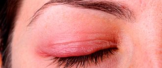
Chalazion
This manifestation is very similar in appearance to barley. Located in the inner layer of the eyelid under the skin. Externally, chalazion is expressed by pain and redness. This disease has an infectious cause and can lead to serious eye problems. Chalazion is often a consequence of damage to internal organs. And it will be possible to identify this or that pathology only as a result of blood tests, urine tests and other studies.
Wart
It is often located on the outside of the eyelid and causes purely aesthetic inconvenience. But a small, dense subcutaneous wart can increase in diameter and grow. As a result, it will hang over the eyelid and interfere with normal vision. In the absence of contraindications in the form of blood diseases, a wart can be easily removed by laser.
Lipoma or wen
It is an accumulation of tissue in the eyelid area. It will be light in color and practically no different from the rest of the skin. In this case, the appearance of redness and suppuration is not typical. Wen can appear on any side of the eye and does not pose any threat to health. It is removed through surgery.
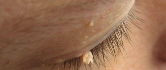
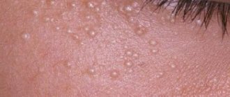
Prosyanka
It is flesh-colored and looks like a pimple. It is absolutely safe, since it is associated with the penetration of small particles of sand and dust into the subcutaneous layer. They cause irritation and local inflammation. However, it is not capable of developing into a large-scale disease. After all, the proportion of infection that gets under the skin will be negligible.
| Millet can be quickly treated with folk remedies or ointments with antibiotics. This manifestation has a purely aesthetic effect. |
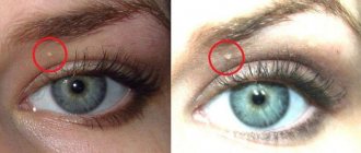
Xanthelasma
This is a benign formation that cannot lead to the formation of cancer cells. It is localized on the upper eyelid and is invisible when the eyes are open. This formation also has only an aesthetic effect. Therefore, carriers remove it using simple surgical operations. However, there is an opinion that the presence of such a formation indicates atherosclerosis and a high probability of a heart attack.
Furuncle
It is a suppuration that is completely safe. Causes redness and pain when touched. They do not affect visual function and do not transform into malignant cells.
Return to contents
Dacryocystitis
This is an inflammation of the tear duct. Manifestations include frequent fluid discharge and severe suppuration. All this is accompanied by swelling in the corner of the eye. That is, in the place where the lacrimal canal is located. This is a serious disease that reduces the quality of vision and prevents you from perceiving objects normally due to severe swelling of the eyelid. It is treated with antibiotics and other drugs that eliminate inflammation.
Human papillomavirus
It looks like a wart, has a similar structure and reasons for formation.
| This type of lump can occur in a child, but is more common in older people. This formation is harmless and can be easily removed by laser surgery. |
Melanoma
It occurs as a result of prolonged and intense exposure to the sun's rays. Such formations appear over the centuries and indicate a serious danger. The fact is that melanoma is of a malignant nature and is fraught with the development of cancer cells. In this case, the formation takes on a dark color and is very similar to an ordinary wart. But it can turn into an ulcer and bleed.
It is necessary to undergo a full medical examination to identify melanoma and separate it from the wart. But this disease has nothing to do with visual function and belongs to the field of oncology. This is why a complete examination is important rather than focusing on the ophthalmological aspect.
Non-cancerous tumors
Often in ophthalmology, diseases of the eyelids are diagnosed, in which the formation of nodules, plaques or ulcerative surfaces of a non-cancerous nature occurs. Such eyelid tumors do not pose a serious threat to human health and life and are easily treatable. The most common pathologies are:
Barley
This disease is an acute inflammation of the ciliary margin, as a result of which a painful rounded seal with purulent contents inside is formed on the eyelid. Barley (gordeolum or pisyak) most often occurs due to damage to the organs of vision by Staphylococcus aureus. The pathology manifests itself with the following symptoms:
- soreness;
- hyperemia;
- swelling;
- itching, burning;
- blurred vision.
Barley looks like a small grain with a white-yellow head, which normally goes away on its own within a few days after infection. In the event of a rupture of the pus, pus can enter the body, causing severe complications, including blood sepsis. Treatment is carried out comprehensively. Antibacterial eye drops and ointments, antiseptics and various folk remedies are prescribed.
Chalazion
A common ophthalmological disease characterized by blockage of the sebaceous gland of the eyelid. Chalazion (hailstone) usually occurs as a result of infection of the meibomian gland, seborrheic dermatitis, or rosacea. At first, the clinical picture of the disease is the same as with barley, which is why these two pathologies are often confused. But unlike hordeolum, hailstones do not burst open after a few days, but continue to grow slowly, putting pressure on the eyeball. As a result, vision becomes blurred and severe discomfort occurs.
Treatment of chalazion in the early stages is carried out by heating. If inflammation occurs, corticosteroid injections are prescribed.
In advanced cases, hailstones are excised surgically.
Xanthelasma
Xanthelasma is a noncancerous tumor of the eyelid that looks like small, flat, yellow plaques with irregular borders. Most often, this pathology occurs in women suffering from diabetes, hypertension or obesity. As the plaques grow, they merge with each other, forming a flat xanthoma. The disease is asymptomatic.
Xanthelasma is not dangerous, but creates a pronounced cosmetic effect. The only way to completely get rid of the tumor is through surgery. Usually laser surgery, electrocoagulation or cryodestruction is performed.
Prosyanka
It appears as small white formations on the skin with a diameter of 2-3 mm. These eyelid tumors, also called milia, are not accompanied by pain, itching or redness. However, they cause cosmetic discomfort. The main reason for the formation of millet is a failure in the removal of old dermal cells and stagnation of sebum.
Simply squeezing out such pimples will not work; you can only get rid of them with a needle or laser.
Furuncle
A furuncle or boil is an acute purulent necrotic inflammation of the sebaceous glands, hair follicle and connective tissue. The inflammatory process is most often caused by Staphylococcus aureus against the background of skin contamination. A boil is usually accompanied by the following symptoms:
- severe hyperemia;
- inflammation;
- swelling;
- soreness;
- hyperthermia;
- headache;
- general malaise.
Chiryak gradually increases in size and can spread to the entire half of the face. After a couple of days, a purulent tip appears on the tumor, which then opens, leaving behind a scar.
Treatment of a boil on the eyelid is carried out comprehensively, using antiseptics, anti-inflammatory and antibacterial agents.
Ptosis
Ptosis is a common eye disease that causes drooping of the upper eyelid. The causes of this pathology can be very diverse: hereditary factors, weakness of the eye muscles, diseases of the central nervous system. In addition to a pronounced cosmetic defect, the following symptoms are observed:
- eye irritation;
- headache;
- difficulty blinking;
- tilting your head back for better visibility;
- diplopia;
- strabismus.
If ptosis is neurogenic in nature, then physiotherapeutic treatment (UHF, galvanotherapy) is prescribed. In other cases, surgery is performed.
Dermatitis
This is an inflammatory skin lesion that occurs for the following reasons:
- infectious processes;
- allergic reactions;
- chemical damage;
- exposure to cold;
- dysfunction of the sebaceous glands.
Dermatitis manifests itself as a rash, the nature of which may differ depending on the origin of the pathology. In addition to small blisters or nodules, the following symptoms are observed:
- hyperemia;
- itching, burning;
- swelling;
- dry skin.
Read in a separate article: What is eye scotoma, causes and treatment
Treatment also depends on the origin of the dermatitis. The most commonly prescribed medications are antihistamines, corticosteroid ointments, anti-inflammatory drugs and hormonal therapy.
Symptoms
The causes of these neoplasms are very extensive. They appear as follows:
- The appearance of compaction. Moreover, it may have a red or white color or may not differ at all from the skin of the eyelid;
- The small, hard lump may enlarge to form a sac filled with pus. This is typical for stye, inflammation of the tear ducts and other diseases;
- Pus may leak out, and the compaction will begin to interfere with vision. The infection will cause inflammation of the inner areas of the eye if it is allowed to penetrate deep into the organs.
| These symptoms are often accompanied by severe pain, heaviness, inflammation, redness, and a feeling of constant discomfort. |
Treatment
Effective methods of therapy to eliminate tumors are:
- Cryodestruction is a new method of treating various forms of growths, which almost 100% eliminates relapses. It is inexpensive and safe, and also does not take much time for rehabilitation. Everything is quite easy; in the case of benign neoplasms, they are simply frozen with liquid nitrogen, as a result they dry out and fall off on their own.
- Surgery using a scalpel involves cutting the skin in the area of the growth. This method is used today as a last resort when other more modern methods of treatment are not suitable for the patient. Local anesthesia is used for removal. After surgery, some recovery time is required;
- The use of laser technology is the fastest and at the same time safest method of removing tumors in the eyes, which eliminates the risk of infection of the affected tissues. The duration of the procedure is no more than 15 minutes, the patient needs about 1 week to recover.
Diagnosis of eye tumors
Since neoplasms are caused by a variety of reasons, the diagnosis must be comprehensive. First of all, it is necessary to conduct a urine and blood test. This will provide information about the general state of health and the presence of diseases that can manifest themselves in the form of neoplasms.
At the same time, you need to contact an ophthalmologist and examine the organ of vision using special equipment. The doctor will determine the condition of the tear ducts, retina, and fundus of the eye. In general, diagnosis is performed by exclusion.
Microscopy
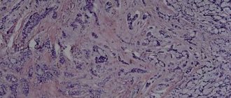
A tumor is detected growing from the cells of the basal layer of the epidermis in the form of a cell array.
Tumor complexes, including differentiated and undifferentiated cells, have different sizes. In scleroderma-like forms, malignant basal cells are found in the fibrous stroma. The diagnosis of basal cell carcinoma is established on the basis of biomicroscopy data, radioindication with radioactive phosphorus and the results of histological examination.
Differential diagnosis should include squamous cell carcinoma, melanoma, cutaneous horn, keratoacanthoma, senile wart, eczematous dermatitis and scleroderma.
Treatment of eye tumors
Treatment depends on its nature. Moreover, they can manifest themselves in infants and adults. However, the main types of treatment for such manifestations should be indicated:
Drug treatment
It will be effective if we are talking about an infectious lesion. In these cases, it is necessary to take measures to destroy the infection. Accordingly, antibiotics are taken. For example, this is how inflammation of the tear duct is treated. To prevent recurrence of symptoms, you should complete the full course of treatment. But first you need to find out exactly the type of tumor.
Operation
Often it is of an aesthetic nature. For example, papilloma and warts are removed using laser intervention. Typically, surgery is not required. If the tumor is temporary, it should be treated with antibiotics, ointments or folk remedies.
Return to contents
Laser removal
It is the most popular method of getting rid of formations. Timely intervention will help protect yourself from a malignant tumor. Moreover, such removal occurs quickly, in a clinical setting, and does not require long-term recovery or hospitalization.
Treatment with folk remedies
The most effective folk remedies for removing barley or millet. It is quite enough to warm up the affected area. This will neutralize the effect of the infection and resolve the resulting abscess.
Forecast
The prognosis for vision depends on the stage of development of the process and the treatment used.
Radiation therapy (brachytherapy and external irradiation) can lead to radiation cataracts, ulceration and xerosis of the cornea. The prognosis for life is usually favorable: the tumor rarely metastasizes, mainly to regional lymph nodes. However, basal cell carcinoma can grow into deep, vital structures of the eye. We describe a case of basal cell carcinoma invasion into the brain 25 years after the start of treatment for a tumor of the eyelid.
A.F. Brovkina, V.V. Valsky, G.A. Gusev
Published by Konstantin Mokanov
What not to do if a lump appears on the eyelid
To establish a diagnosis, you must consult a doctor and undergo an examination. It will consist of tests and examination, including the use of ophthalmological equipment.
| However, there are some things you shouldn't do when you have lumps on your eyelid. This will protect the person from dangerous consequences and prevent the infection from penetrating further. |
So, it is unacceptable to try to squeeze the pus out of the sac on your own. This will only lead to a wider spread of infection. Under no circumstances should the formation be treated with vinegar or other potent agents. They will cause necrosis of the affected tissues. But the infection will not go away and will appear again even more intensely.
Diagnosis of basal cell carcinoma of the eyelid
If any skin disorders appear in the eye area, you need to go to the hospital. For help, you can contact a therapist, dermatologist, ophthalmologist or oncologist. Diagnostics includes:
- taking an anamnesis;
- inspection of the lesion site;
- dermatoscopy and biopsy.
A biopsy is a mandatory procedure that allows you to determine the nature of the tumor (benign or malignant) and the depth of the lesion. A scraping or puncture is taken from the tumor site and the tissue is examined under a microscope.
After diagnosing and establishing an accurate diagnosis, the doctor prescribes a treatment method.
The neoplasm must be examined with a dermatoscope
Prevention
It is impossible to prevent the appearance of such formations. This will not succeed due to the differences between the reasons that cause the formation of seals. After all, it is impossible to exclude the possibility of dust particles, grains of sand getting into the eyelid, or the development of pathologies of the arterial system. At the same time, the immune system should be strengthened to prevent the development of the disease.
| General strengthening procedures will be effective means of prevention. For example, taking vitamin complexes, giving up bad habits such as smoking or drinking alcohol. A proper diet is also important, avoiding fatty and toxin-laden foods. |
In addition, you need to protect yourself from the sun's rays by using safety glasses and caps. This will prevent the development of melanoma, which is a malignant tumor.
Treatment methods
A growth has appeared on the eyelid, a photo and description of which can be found in reference books and on the Internet, and it often hurts and causes great discomfort to any patient. At the initial stage of any eyelid change, any sensations from the neoplasm may be absent.
However, as the tissue grows and the growth increases, painful sensations become more common, so you should immediately consult a doctor for examination and treatment.
Treatment of a growth on the upper or lower eyelid depends on the disease and comes in different types:
So for the treatment of infectious lesions and barley the following is prescribed:
Albucid
An effective antiseptic drug that is active against infectious and purulent lesions of the eye and eyelid.
The medicine suppresses the proliferation of pathogenic bacteria and viruses and helps cleanse the eye of their vital activity. The drug is prescribed 1-2 drops up to 6 times a day for a week.
Levomycetin
Levomycetin eye drops contain a broad-spectrum antibiotic that can quickly relieve inflammation from the eyelid and have an antibacterial and healing effect. The drug is prescribed 2-3 drops 3 times a day for 5-7 days.
Oftalmoferon
The drug ophthalmoferon is considered an effective antiviral agent for the eyes, which actively helps to suppress the pathogenic environment and restore the normal condition of the eye and eyelid. The drug is instilled 2-3 drops 3 times a day for 5 days.
Oksolin
Oxolinic ointment as an antiviral and antibacterial agent is effectively used to treat growths on the eyelids. The drug is applied to the affected area 2-3 times a day in a thin layer for a week.
To treat growths with reduced immunity, the following is additionally used:
Arbidol
The latest generation antiviral drug has an active effect on viruses and pathogenic bacteria and is an immunomodulator.
The medicine is taken 1 capsule 4 times a day for 5 days.
Alphabet
The alphabet preparation contains a complex of vitamins and minerals that are necessary for the body and helps to quickly restore the vitamin balance in the body. The drug is taken 1 tablet. 3 times a day for 20-30 days.
Laser removal
Laser removal is the excision of a neoplasm on the eye by pinpoint cauterization with a directed laser beam. This method is considered an effective way to combat warts, papillomas and moles.
The painless procedure is carried out within a few minutes, after which the skin on the eyelids is quickly restored without scars. This method is considered gentle and effective, since after removal, new tumors do not form on the eyelid.
Cryotherapy
Cauterization with liquid nitrogen allows you to precisely freeze a new growth on the skin, which dies off on its own within a few days. The skin at the treatment site heals easily and quickly.
This procedure is widespread in cosmetology and effectively combats papillomas and warts. However, nitrogen does not allow one to accurately determine the depth of the effect, so a similar procedure after healing is repeated 2 more times.
Surgical intervention
Malignant and benign formations that cause discomfort are subject to mandatory surgical removal. Eyelid surgery is performed by excision of the growth with a scalpel and joining of intact tissues. This procedure is carried out under local, less often general, anesthesia and requires a highly qualified specialist.
So, when excising a cyst on the upper eyelid, deep layers of tissue must be removed, so any awkward movement can cause serious consequences for the patient’s health. Surgical removal of growths on the eyelids is carried out only in specialized hospitals using the latest technologies.
Celandine
Celandine herb juice is widely used to treat warts. Purified juice extract can be purchased at the pharmacy. The liquid product is applied pointwise to the wart 2-3 times a day for 7-10 days.
The top layer of damaged skin is gradually burned off, forming a thin crust at the application site, which falls off on its own. This procedure must be carried out carefully on the eyelids. After cauterization, rinse the eyelids thoroughly with clean water to avoid getting the composition into the eye.
Castor oil
Castor oil has an effective effect on growths.
- To prepare the composition, castor oil (1 tsp) is mixed with tea tree oil (0.5 tsp).
- The composition is mixed and poured into a dark container and stored in the refrigerator.
- The mixture is applied to the site of the tumor with a cotton swab for 5-7 days. Then, after a week's break, the procedure is repeated.
This remedy actively helps reduce tumor growth. It has antiseptic and anti-inflammatory effects.
Walnut
A tincture of walnut leaves is used effectively to combat the human papillomavirus.
- Fresh walnut leaves (50 grams) are chopped finely and pour 100 ml of boiling water.
- Infuse the composition in a water bath for 15 minutes. and cool to room temperature.
- Then the infusion must be filtered and poured into a glass container.
- The product is applied to cotton pads and applied as a compress on the eyelid to the growth for 15-20 minutes. 3-4 times a day for 10 days. This decoction, together with prescribed drug treatment, can stop the growth of papillomas and gradually helps to reduce them.
Basalioma of the eye, cancer of the eyelid: causes, types, treatment, types of operations
.
There are three types of skin cancer - basal cell carcinoma, squamous cell carcinoma and melanoma. The least aggressive form is basal cell carcinoma or basal cell carcinoma. It rarely metastasizes, so in most cases it has a favorable prognosis. A tumor develops in exposed areas of the skin, including the eyelids. Most often, basal cell carcinoma of the eyelid affects older people.
Basalioma of the eye often develops in old age
Reasons for the development of basal cell carcinoma of the eyelid
The main risk group for developing basal cell carcinoma on the eye is people over 40 years of age. Skin cells at this age are already aging, mutations occur more often, and the general condition of the body contributes to the development of oncology. Most often the tumor affects the lower eyelid.
Basalioma of the lower eyelid also appears in young people under the influence of external factors and disorders within the body. Tumor development is promoted by:
- Genetic predisposition. The risk of the disease increases if a close relative suffered from basal cell carcinoma on any area of the skin.
- Ultraviolet irradiation.
- Xeroderma pigmentosum. This is a congenital disease characterized by intolerance to ultraviolet radiation.
- Chemical burns to the eyes.
- Chronically reduced immunity. This condition is caused by diseases such as AIDS and diabetes.
Basal cell carcinoma of the eyelid rarely appears in children and adolescents, but there are cases of congenital Gorlin-Goltz syndrome, a form of basal cell carcinoma.
Types and symptoms of basalioma on the eye
Basalioma does not metastasize and grows slowly, over several months and even years. But it cannot be called completely safe. Basal cell carcinoma can affect different areas of the eye:
- lower eyelid;
- medial commissure of the eyelids;
- upper eyelid;
- external commissure of the eyelids.
Tumors of the medial commissure spread to the sinuses and orbit. This type of basal cell carcinoma is difficult to diagnose and treat, and has a high tendency to recur.
Basaliomas can be located on different parts of the eye
Nodular or sclerosing form - definition
There are nodular ulcerated and very rare sclerosing forms of basalioma.
Nodular form
The nodular form in the early stages appears as a small tubercle. It has a developed vascular network and is painless when pressed. Over time, ulcers appear on the surface of the tumor and it becomes covered with a crust.
The tumor grows in width over several months. In later stages, the tumor takes the form of a plaque with raised, fibrous edges or a large mushroom-shaped nodule.
The blood vessels on the sides of the tumor are dilated.
For the first six months, the tumor grows slowly, then growth accelerates, and ulceration appears in the center of the node or plaque.
Sclerosing
A sclerosing tumor grows from under the skin and causes deformation of the eyelid. Its edges are not clearly defined. When palpated, the size of the tumor turns out to be larger than it appears during visual inspection.
Diagnosis of basal cell carcinoma of the eyelid
If any skin disorders appear in the eye area, you need to go to the hospital. For help, you can contact a therapist, dermatologist, ophthalmologist or oncologist. Diagnostics includes:
- taking an anamnesis;
- inspection of the lesion site;
- dermatoscopy and biopsy.
A biopsy is a mandatory procedure that allows you to determine the nature of the tumor (benign or malignant) and the depth of the lesion. A scraping or puncture is taken from the tumor site and the tissue is examined under a microscope.
After diagnosing and establishing an accurate diagnosis, the doctor prescribes a treatment method.
The neoplasm must be examined with a dermatoscope
Treatment methods for ocular basal cell carcinoma
It is very difficult to treat a tumor on the eye. It is necessary to make sure that the tumor cells are completely removed, while preserving as much healthy tissue as possible.
Microsurgery
Micrographic surgery is often used. To completely remove the cancer, the sections are removed with serial freezing. The sections are color coded so that the remaining areas of the cancer lesion can be easily identified using the histological method.
Microsurgery with freezing
Micrographic surgery with tissue freezing and analysis improves surgical success. It is used when the tumor is located on the commissures of the eyelids, the boundaries of the tumor are poorly defined and are not identified when there are processes.
If histology does not reveal cancer cells, the treatment is considered complete and restoration of the eyelid begins.
Reconstruction of the century
Reconstruction of the century is a very important stage. The operation is needed to restore the integrity of the skin and restore normal eyelid mobility. Reconstruction of the century plates is a very complex process. In case of severe damage to the plate, it is replaced with tissue with a similar structure.
Reconstruction techniques, depending on the size of the defect:
Partial
Violations that occupy less than a third of the century are sutured. This is done if the tissue around the wound is elastic.
Percentage
Violations that occupy an area of less than half a century are sutured with a semicircular skin flap.
Minimum
Large violations are reconstructed:
- using a flap of skin taken from the cheek (closure of the lower eyelid);
- using cartilage and mucous membrane of the nasal septum (restoration of the posterior plate);
- division of the century;
- glabellar flap (correction of defects in the medial corners of the eye fissure).
The reconstruction is carried out very carefully in order to fully preserve the functions of the upper and lower eyelids, their tone and mobility.
Eyelid reconstruction allows minimizing the consequences of basal cell carcinoma removal
Prevention of recurrence of ocular basal cell carcinoma
Basalioma of the eye, depending on the location and shape, has a different tendency to relapse. Prevention will help avoid recurrence of the disease after treatment.
- Regular examinations with your doctor (1-2 times a year).
- Reducing sun exposure.
- Compliance with the rules of tanning in a solarium.
- Balanced diet enriched with vitamins.
- Reducing the impact of stress.
- Timely and complete treatment of viral infections.
- Eye hygiene. Do not use expired or untested cosmetics, or rub your eyes with dirty hands, scarves or towels.
People over 40 years of age, regardless of whether they have had the disease before, need to undergo preventive medical examinations.
Basalioma on the tissues of the eyelid is a treatable disease and has a favorable prognosis if detected in a timely manner. If you notice any noticeable growths or pain in the eyelid area, you should consult a doctor. The therapy will preserve the beauty of the eyes and comfortable sensations.
Source: //kozhmed.ru/zabolevaniya/bazalioma-glaza.html
Prevention and prognosis
To prevent the development of tumors, adhere to the following recommendations:
- maintain visual hygiene: wash with cool water, carefully blot your eyes with a clean towel;
- To remove makeup, use mild, hypoallergenic detergents without aggressive detergent components;
- diseases of the visual organs are promptly treated under the supervision of an ophthalmologist;
- adhere to recommendations regarding the care and storage of contact lenses;
- monitor the quality of nutrition so as not to cause a deficiency of fat-soluble substances;
- take dietary supplements to support the functional state of the visual organs: vitamins A and E, lutein, zeaxanthin.
The prognosis depends on the underlying factor that provoked the growth of the tumor, the thickness and type of tumor. For benign neoplasms, the prognosis is favorable in most cases. For malignant lesions, it depends on how timely the disorder was detected.
Are blisters on the eyelids dangerous and how to deal with them?
30.10.2018
Blistering rashes can occur in people of all ages. A bleb on the eyelid is not only a cosmetic defect, but sometimes also a sign of illness.
Why do blisters appear on the eyelids?
Low-quality cosmetics, poor hygiene, improper care of lenses, burns, infectious and allergic factors can cause bubbles in the eyes. May be affected: the lower or upper eyelid, the skin above and below the eyes, the membranes of the eye.
Adenoviral conjunctivitis
The classic triad of symptoms of adenovirus infection: conjunctivitis, pharyngitis, fever. The follicular form of adenoviral conjunctivitis is manifested by the formation of blisters on the inside of the eyelid. A person feels a foreign body in the eye, the eye turns red and swells.
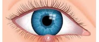
Conjunctival cyst
A round, watery formation on the conjunctiva is called a conjunctival cyst. A transparent bubble forms on the mucous membrane of the eye, which gradually increases, creating a feeling of discomfort. Conjunctival cysts can be congenital or acquired (after injury, inflammation of the eye).
Diagnostics
The effectiveness of treatment depends on a correctly established diagnosis. The doctor conducts a detailed survey and clarifies complaints. The person must tell why the bubble appeared in the eye or on the eyelid, how quickly it grows, and what sensations it is accompanied by.
Then the doctor examines the eyelids, eyes, and assesses the condition of the fundus at the slit lamp. An ophthalmologist may prescribe laboratory and instrumental examinations:
- CBC to assess the general condition of the body.
- ELISA for detecting viruses.
- Cytological examination of scrapings to evaluate cell characteristics.
- Histological examination of removed elements to exclude oncology.
If the cause of blistering rashes is allergic, it is necessary to identify and exclude the allergen. For this, the person is referred to an allergist.
Treatment of blistering rashes
It is advisable to show a transparent pimple on the eye or eyelids to a specialist. Some diseases (herpetic infection, burn) can irreversibly damage vision. The ophthalmologist will determine the cause and tell you what to do when a bubble pops up on your eyelid:
- Antiviral drugs: drops “Ophthalmoferon”, “Aktipol”; ointments "Acyclovir", "Zovirax"; tablets "Acyclovir", "Valacyclovir".
- Antihistamines to relieve allergic manifestations: drops “Cromohexal”, “Allergodil”, “Opatanol”; tablets "Suprastin", "Cetrin". It is imperative to exclude the allergen.
- Surgical treatment. Removal of papillomas, conjunctival and Moll cysts is carried out using a laser, cryodestruction, or classical surgery. After removal of the wart or cyst, the material is sent to the laboratory for histological examination.
- Treatment for burns depends on the extent of the injury. In mild cases, wound-healing ointments for external use “Bepanten”, “Solcoseryl”, painkillers “Pentalgin”, “Sedalgin”, moisturizing drops “Vizin”, “Systane” are prescribed. Severe burns are treated in a hospital.
Watery pimples on the eyelids should not be scratched. This can damage them and cause bacterial infection. Do not heat the bubbles, as this may provoke the growth of the element.
Treatment of a swollen eyelid
Therapy begins with determining an accurate diagnosis. All treatment steps are controlled by an ophthalmologist or other specialist if the cause lies in diseases of the internal organs.
Pharmacy drugs
Treatment with medications is carried out to get rid of the primary disease. Eliminating the underlying cause will solve the problem with a swollen upper eyelid.
We recommend reading: Anti-edema tablets
The following pharmaceutical medications are used:
- Antihistamines - for an allergic reaction. Medicines for the treatment of allergies: Suprastin, Tavegil, Akrivastine, Ebastine, Olopatadine, Telfast, Allegra. Many antihistamines are powerful anti-inflammatory agents. The effect of modern medications begins 30 minutes after ingestion and continues throughout the day. The latest generation of antihistamines have the lowest risk of drowsiness. The dosage of any medications for an allergic reaction is calculated depending on the patient’s age and weight.
- Diuretic medications and teas - for kidney dysfunction. Furosemide is considered the most powerful diuretic. The medicine acts quickly and is often used for fluid accumulation in the upper eyelid. Furosemide leaches beneficial substances from the body; improper use leads to dehydration. Therefore, the use of the medicine is possible only in emergency cases, after consultation with a doctor.
- Hormonal medications - for disrupted cycles. Menstrual irregularities with insufficient production of progesterone and excess estrogen are treated with the hormone of the second phase of the cycle - progesterone. Birth control pills or oral contraceptives are often prescribed.
If the cause is organ disease, the doctor will prescribe the optimal treatment directly for the internal organ.
In case of swelling due to poor lymph drainage, treatment is based on lymphatic drainage. To do this, they use the method of electrical stimulation, which restores lymph exchange, restores microcirculation and metabolism.
Folk remedies
Isolated cases are treated with home remedies. Traditional methods of treatment will help if it is allergies, cold swelling or lack of sleep.
Home Recipes:
- If your eyelid is swollen, baking soda will help. Dissolve 1 tbsp in 200 ml of water. l. facilities. Compresses are made from the prepared solution. Baking soda will reduce swelling and disinfect the eyelids.
- Brewing tea will quickly eliminate the problem, relieve tension, and make dark circles less obvious. Place tea bags on your eyes or brew a strong drink, soak a cotton pad in it, and apply for 15 minutes.
- Mix beeswax or honey with water in a 1:2 ratio. Drop the medicine into the eyes 2 times a day.
- Wipe your eyes with a decoction of chamomile or chamomile daily. A frozen decoction will help better.
- Chilled spoons, cucumber slices, and ice help quickly get rid of swelling.
If the eyelid swells due to an internal disease, traditional methods will not help. They stop the symptom, but do not fight the cause.
How does eye basal cell carcinoma manifest, causes and treatment methods?
Among the various forms of skin oncology, ocular basal cell carcinoma is quite common. This disease affects older people, those with snow-white skin, blond hair and blue eyes, and those who like a bronze tan.
What are the signs by which ocular basal cell carcinoma can be identified?
As a rule, basal cell carcinoma is localized on the lower eyelid, less often on the upper eyelid, and also affects the area of the internal (medial) junction of the upper and lower eyelids. Symptoms of basal cell carcinoma of the eyelid depend on the location and form of the tumor.
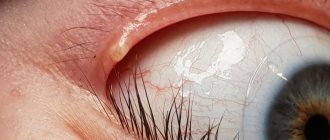
A nodular-ulcerative tumor manifests itself in the form of a hardened pearlescent nodule rising above the surface of the eyelid with visible blood vessels, which can be clearly seen in the photo. Over time, pus or a bloody mass may be released from the neoplasm.
A sclerosing tumor appears as a plaque growing from the epidermal layer of the skin, severely deforming the eyelid.
The sclerosing form of basal cell carcinoma of the eyelid is often confused with chronic blepharitis, since the neoplasm itself does not have clear boundaries upon visual inspection, and when diagnosed, compactions are noted by touch.
Causes of eye cancer, what diseases can trigger the development of basal cell carcinoma
There are a number of factors that provoke the development of ocular oncology. These include:
- intense exposure to ultraviolet radiation on the eyes, including procedures in a solarium;
- injury to the eyelids;
- cosmetic procedures involving injury;
- low-quality decorative cosmetics intended for the eyes;
- genetic predisposition;
- bad habits;
- weakened immune system.
Diseases preceding ocular oncology
Basalioma can develop as a result of diseases such as xeroderma pigmentosum and Gorlin-Goltz syndrome.
Xeroderma pigmentosum is expressed in skin hypersensitivity to UV rays.
Gorlin-Goltz syndrome - affects the skin, bone and nervous systems, and organs of vision.
Basalioma of the eye, existing treatment methods
A timely diagnosis and correctly selected treatment method for ocular basal cell carcinoma determine how successful the treatment will be, as well as the risk of relapse.
There are certain difficulties in treating a tumor: the oncologist must not only excise the tumor, but also leave as much healthy tissue as possible unaffected.
Removal of basal cell carcinoma is done in several ways.
Cryodestruction
Microsurgery with partial freezing is effective for non-aggressive forms of ocular tumors. For large tumors, a different method is used.
Surgical method
Basalioma of the lower eyelid is successfully removed with a surgical scalpel. When a significant portion of healthy tissue, more than 4 mm, is excised, reconstructive surgery is performed.
Microsurgery with frozen sections
Modern technique for removing basal cell carcinomas using frozen sections. At the same time, a histological examination of the samples is carried out to determine the presence of cancer cells.
Radiation therapy
If the tumor is located in the medial angles, surgery is contraindicated. Treatment is carried out with radiation. Radiation therapy may be prescribed if the patient himself refuses surgery.
The Center for Modern Surgery successfully performs operations to remove basal cell carcinoma on the face. Consultation, examination and surgery are carried out by oncologist M. A. Popovtsev.
Find out more about the symptoms of basal cell carcinoma and its stages.
Question:
What is the survival prognosis for treatment of basal cell carcinoma under the eye?
Answer:
Ocular basal cell carcinoma responds well to treatment; the prognosis for five-year survival is over 90%, provided timely contact with an oncologist.
Question:
Is it possible to treat an eye tumor using traditional methods?
Answer:
It still won’t be possible to avoid surgery using different grandmother’s recipes. Treatment with traditional methods will only worsen the situation.
Question:
Is it possible to remove basal cell carcinoma on the eyelid with a laser?
Answer:
Laser treatment is effective for small tumors; with the help of a laser, tumors localized in hard-to-reach places are removed.
Source: //modernsurgeon.ru/napravleniya/dermatoonkologiya/bazalioma/bazalioma_glaza/