Symptoms and photos of atheroma of the eyelids and lacrimal caruncle
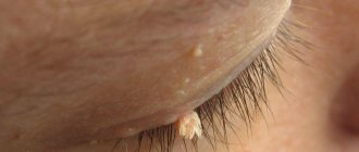
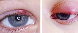
Atheromas on the eyelid are benign cystic formations, characterized by a round shape, dense structure, clear outlines and mobility. Single or multiple cysts do not cause pain, grow slowly and can reach sizes of more than three centimeters. In the central part of the wen there is a dark point - a clogged duct, which, when pressed, can release a thick mass.
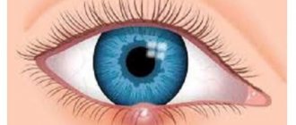
With atheroma of the lacrimal caruncle, the component of the eye enlarges and turns red, the person feels the presence of a foreign body and a burning sensation.
Removing a cyst on the eye
A tumor on the eye must be removed using various methods. Intensive growth of a tumor is an indicator for surgical intervention.
Laser removal
The cyst is removed in a gentle manner, leaving healthy tissue unaffected. The laser acts in a targeted manner, producing a bactericidal effect. Characterized by rapid rehabilitation and a short postoperative period. There are no defects in vision or appearance. It is practiced only for small formations that do not show a growth trend.
Dissolution of tissue occurs when a laser beam is applied.
The whole process consists of three important stages:
- preparing the eye for laser exposure (the tissue around it is treated with a solution, a micro-hole is made);
- dissolution of neoplasm tissue;
- aspiration (pulling out contents from the eye).
Tissue dissolution occurs when a laser beam is passed through a thin tube whose walls protect healthy tissue. The laser burns the neoplasm tissue, and at the same time the damaged vessels are fused. The liquefied contents are removed using a vacuum tool - it is pulled out of the cavity.
Surgical removal
Indicators for surgery are intensive growth, or an advanced state, and if drug therapy has proven to be ineffective. Surgery is often the only solution, since the eye area is dangerous to be affected by long-term drug treatment. This will also lead to relapse or the development of cysts. Surgery is required if a dermoid cyst of the eye appears.
The site for excision of the tumor is marked.
Excision of the cyst does not take more than an hour; the operation is performed under anesthesia. Afterwards, a bandage with antibiotic ointment is applied, and the eye is completely covered with a protective bandage. The patient is observed for a week, or comes for examination on his own within 5 days. Dressings are only done in the hospital to avoid infection. The bandages are removed after the operated area has completely healed. Medicines are prescribed to support the immune system, weakened due to the intervention.
After the operation, a bandage with antibiotic ointment is applied, and the eye is completely covered with a protective bandage.
After complete removal of the cyst, the eye organ quickly recovers. There are no complications. It is recommended to avoid physical and psychological stress for six months. It is also necessary to avoid stressful situations. Intense sports, swimming and baths or saunas are completely contraindicated. Exposure to high temperatures and steam combined with humidity can cause the cyst to recur.
The only noticeable drawback is that after the operation, vision is fully restored within 1-2 months. A complication may also arise in the form of decreased vision, after which maintenance therapy or even prophylactic wearing of glasses will be required.
At-risk groups
The appearance of a wen in the facial area is provoked by several factors:
- hormonal imbalance in the body (adolescence or menopause);
- increased testosterone production or infrequent shaving in men;
- excessive exposure of maternal hormones to the fetus during pregnancy (subcutaneous congenital atheroma is formed);
- metabolic disorders caused by exacerbation of diseases or decreased immunity;
Diseases of the gastrointestinal tract, nervous system, adrenal glands.
- diseases of the gastrointestinal tract, nervous system, adrenal glands;
- using large quantities of low-quality decorative cosmetics;
- failure to comply with hygiene standards;
- excess hair at the location of the excretory ducts;
- atheroma occurs in the seborrheic areas of the face due to a hereditary factor.
Sometimes the provoking factors are increased temperature indoors or outdoors, excessive sweating, and acne. A cyst on the face can form due to injury.
When pinched or cut, an epidermal cyst appears in the epidermis. Superficial cells are transported to deeper tissues.
What is eye atheroma: causes and mechanism of formation
Atheroma on the eyelid appears due to an increase in the thickness and viscosity of the secretion of the Zeiss glands. Due to its high density, the fatty secretion cannot be evacuated to its destination, so it remains in the gland ducts. From excess sebum they stretch, the inflammatory process begins, and as a result the eyelid swells. Swelling completely blocks the ductal outlets of the gland. This is how cysts form on the eyelid.
Doctors associate the reasons for the thickening of the secretion of the Zeiss glands with diseases of the gastrointestinal tract (peptic ulcer, gastritis), as well as with pathology of the pancreas.
What is the danger of wen
Lipomas themselves are not dangerous, since they are benign formations and degenerate into cancerous tumors extremely rarely. But the appearance of a capsule and the isolation of adipose tissue inside it is a favorable condition for the development of harmful microorganisms that can cause liposarcoma and tumors.
When trying to remove a wen on the eyelid or near the eye on your own, it may begin to grow. In addition, when squeezing or piercing there is a risk of infection. Therefore, you cannot try to remove a lipoma at home.
https://www.youtube.com/watch?v=XftxYCVYPdM
This situation leads to the fact that it becomes a breeding ground for pathogenic bacteria, which can, in turn, push towards the development of liposarcoma - a malignant formation.
| Attempts to squeeze out the wen provoke its further growth, which entails the creation of favorable conditions for other ailments. Entropion, otherwise known as entropion, may develop. |
Opening the pathology yourself by squeezing or using a needle opens access to all kinds of infections. And its piercing itself is very dangerous! There is a huge risk of injury.
An attempt to remove a wen on your own hides huge risks associated with a high probability of infection and damage to the organs of vision. Do not try to perform surgical procedures yourself; entrust your health to specialists.
How to get rid of a cyst on the face?
All doctors studying the problem say that the cyst cannot resolve on its own. It cannot be left unattended, otherwise it will increase in size and may become inflamed. It is forbidden to try to squeeze out the contents yourself. This will help for a short time, and soon the secretion will begin to accumulate in the capsule again.
Local treatment
A festering wen will not be able to resolve on its own.
The experience of specialists suggests that festering atheroma cannot resolve on its own. Today there are no medications that can be used to treat compaction. According to doctors, all possible methods only help slow down the growth of the tumor. Surgical intervention is considered the only effective method.
Removal in the clinic
Removal of any atheroma on the face is carried out in a medical facility. You cannot open the thickening yourself. Cases when it ruptures spontaneously also do not bring the desired result. Penetration of pus into internal tissues and infection cannot be ruled out. The patient will experience recurrence of the cyst.
The most effective way to combat atheroma is the complete removal of the capsule along with its contents. Only in this case will re-appearance be excluded. The earlier the surgery is performed, the lower the risk of forming a noticeable scar on the face. The most common methods are the following:
Laser removal.
- Laser removal of atheroma. Performed with local anesthesia. Laser cauterization, excision or evaporation is used. The method depends on the size of the tumor. If it is more than 5 mm, sutures will be required.
- Radio wave excision of atheroma is used for small cysts on the face or body. The waves burn out the capsule along with the secretion. A small depression forms at this place. The entire procedure takes no more than 20 minutes and is performed under local anesthesia.
- Surgery is a more traditional method. During surgery, the capsule may be removed or left in place.
After removal of the atheroma, the inflammation site heals within a few days. The duration of the recovery period depends on the size of the atheroma and what method was used. At this time, antibiotics are additionally used to prevent possible complications. No side effects from the procedure were noticed. Surgery is considered a simple way to correct a cosmetic defect.
Treatment
To get rid of atheroma forever, you will have to remove it. Other treatments are not effective. Radiosurgical devices are used more often. This method has a number of advantages over the traditional surgical method using a scalpel:
- Minimum damage to the skin.
- There is practically no blood loss observed.
- Doesn't hurt.
- The scars after the operation are faint.
- Wound healing is quick and painless.
If you remove atheroma using a laser method, local anesthesia is first applied and the procedure lasts an average of 30 minutes. If inflammation begins, then the atheroma is not temporarily removed. If pus accumulates, it must be opened and the contents cleared of liquid. After the pus is removed, the wen is treated with a local antiseptic and drainage is installed. When the inflammation subsides, the capsule can be excised.
It happens that the cyst bursts on its own. You should not immediately run to the doctor with an open purulent wound.
First, use a sterile cotton swab to clean the contents of the capsule from pus, treat everything with an antiseptic and cover the atheroma with a band-aid.
After this, go to the doctor, since you will not be able to completely get rid of the pus on your own; only a specialist will clean out the cyst so that there are no complications in the form of an abscess.
Unfortunately this is not possible. Even if you manage to open this cyst, after some time it will appear again. The fact is that the subcutaneous capsule will remain and will produce secretion that fills it. The process of formation of atheroma on the back will begin again.
Today, the following treatment methods for sebaceous cysts are possible:
- Removal using laser.
- Radio wave therapy.
- Surgical excision.
Laser treatment is recommended for mild forms of atheroma, when the size of the formation is small. The method is bloodless, there are no scar formations after its use. After laser therapy, relapses are extremely rare.
Laser and radio wave treatment are conservative treatment methods. When they don't help, the only option is surgery.
The operation is performed under local anesthesia. It does not last long and includes the following stages:
- a small incision above the surface of the cyst;
- opening the capsule;
- removal of the capsule with its contents.
After surgery, the doctor applies stitches and bandages until the skin heals. All procedures are carried out only under sterile conditions.
After surgical excision, antibiotic therapy is often prescribed. Local drugs that have anti-inflammatory, antimicrobial and healing effects are also used - for example, Levomekol or Vishnevsky ointment.
A few months after treatment of the inflammatory process, the atheroma can be removed.
ICD10 code
ICD10 is an international classification of diseases that collects coded information about diseases. The list is updated at regular intervals; today’s version of classified diseases is already the 10th.
The ICD was created to facilitate the work of medical specialists; it includes all diseases, including skin diseases, which include atheroma.
This disease is included in the section on neoplasms of the skin and appendages (benign). In the ICD-10 encoding it is included in the interval from L72 to L72.9.
Skin atheroma is treated by a surgeon. He will tell you in detail what it is, show photos and other visual materials. After diagnosis, appropriate therapy is prescribed. Usually the atheroma is removed surgically. It is also possible to remove atheroma using laser and radio wave methods. The method is selected by the doctor individually, depending on the location.
Surgery is performed under local anesthesia. The atheroma is peeled off along with the capsule or completely excised. The operation is performed on an outpatient basis; the total time, including preparation and suturing, can take about an hour.
Atheroma is a disease in which a relapse may occur if the capsule is not completely removed. Therefore, you should carefully consider the choice of doctor and method of treatment. Sometimes traditional methods can be used, but one should not forget that in case of suppuration, one should consult a doctor as soon as possible.
Is it possible to remove eyelid atheroma on your own?
Atheroma of the eyelid is a benign skin tumor that forms in the hair follicles. The causes of the disease are not fully known, but effective treatments exist.
Not all atheromas of the century pose a threat to human life, but some can be fatal. Knowing certain symptoms helps to identify a dangerous pathology in time and consult a doctor.
Diagnosis and types of atheromas
Atheroma is a collective name for epidermal cysts in the area of hair follicles with various etiologies and histologies. These include epidermal cyst, trichymemal cyst and steatocystoma. The British Association of Dermatologists distinguishes “true” from “false” atheromas.
Epidermal cysts are usually traumatic due to the breakdown of squamous epithelium into subepithelial tissue. The most common complication is rupture with secondary sepsis. Healing with atypical scarring occurs if the cyst is not surgically removed.
Epidermal cysts make up about 70% of all benign skin cysts and are most often seen on the hairless scalp, face, torso, feet and hands. Men suffer more than women.
The defeat occurs mainly in the 2-4 decades of life. Epidermal cysts are sliding subcutaneous nodules with a sticky consistency ranging in size from a few millimeters to 2-3 centimeters.
Doctors often use the term "atheroma" very inconsistently. Strictly speaking, the following variants of atheroma can be distinguished. “True” atheromas (epidermal cysts) form on the hair follicles - in the upper part where the hair emerges from the skin. They arise from skin cells and subcutaneous tissue.
The nodes appear spherical, convex and elastic. Epidermal cysts usually do not have an optically recognizable excretory duct. Horny cells and hair accumulate in the cyst; the contents smell unpleasant when the doctor opens the atheroma. True atheromas often occur in families and can be inherited.
“False” atheroma forms in the lower part of the hair follicles. The knots also feel rough, hard, and rubbery. False atheromas occur when one or more sebaceous glands become clogged, such as loose skin cells or dried sebum.
Examination is one of the methods for diagnosing atheroma of the eyelid
Fat does not come out and gradually accumulates in the tissues. The location of a blocked exit passage is sometimes recognized as a black dot. The cyst grows slowly because the sebaceous glands produce only a small amount of oil each day.
Atheroma can occur almost anywhere on the body because hair follicles and sebaceous glands extend throughout the skin. Atheroma on the head, neck, ear, chest, back, neck, face or genital area is not uncommon. However, most dermatologists do not always distinguish between “true” and “false”. Therefore, both forms are often called simply “atheroma.”
Therapy methods
Excision of atheroma without infection is relatively simple. It is important to remove the sebaceous duct and capsule. If the capsule is open or part of the duct remains in the skin, recurrence of atheroma is very high. Careful attention must be paid to complete removal, as this is often very difficult, especially if the capsule is damaged during removal.
Conservative treatment
Sometimes an abscess forms and pus accumulates in the atheroma. In this case, it is important not to touch the tumor. If a pustule bursts, bacteria can enter the bloodstream and cause blood poisoning. This is especially dangerous when the atheroma is on the head - microbes can enter the brain.
In the case of inflamed atheroma, doctors first suppress the inflammation with antibiotics. Medicines spread infection. In addition, the doctor tries to ripen the atheroma with ointment. The ointment softens the skin, soothes pain and slows down inflammation. This also slows down sebum production and ensures that the abscess opens and drains faster.
Conservative treatment of eyelid atheroma includes ointment
For non-inflammatory atheroma, the ointment is of no benefit: although it compresses the atheroma, because the secretion is emptied, the capsule does not dissolve the neoplasm. It can only be removed surgically.
The surgeon then carefully opens the atheroma, removes the pus, and rinses with a disinfectant solution to remove as many germs as possible.
Only after the inflammation has passed after some time, the doctor removes the entire atheroma surgically. As with any surgery, wound pain may occur.
The larger the atheroma, the more pronounced they can be. Pain medications effectively control the symptoms of the disease.
Atheroma has no medical value. This means that the cyst may be unpleasant and cosmetically disturbing, but not a health hazard. In any case, atheromas have nothing to do with cancer. Health insurance companies usually do not cover atheroma removal for aesthetic reasons.
Removing a formation
The atheroma must be removed by a surgical specialist. He cuts the tumor out of the skin with a scalpel. The doctor not only removes the contents of the cyst, but at the same time removes the sebaceous glands from the capsule. There should be no remnants of coating or ducts on the skin, otherwise there is a high probability that the atheroma will grow again.
Small and moderate atheromas are removed under local or regional anesthesia. For large or inflamed atheromas, removal is a little more difficult and takes longer. Sometimes general anesthesia is needed.
Self-removal
Even if the atheroma is irritating or looks like a pimple at first, it is forbidden to remove it or pierce it with a sharp object. Anyone who tinkers with a cyst runs the risk of inflammation. Broken skin can allow bacteria and other germs to enter. The skin then swells, turns red and can cause quite a lot of pain.
You should always consult a doctor for atheroma.
Treatment of atheroma on the eyelid should be carried out by a specialist, because he works in sterile conditions. This avoids the risk of infection, but also that the atheroma grows again.
Possible complications
Atheromas are harmless in most cases. However, an inflamed tumor can also break through. As a result, bacteria are distributed throughout the body. Therefore, in case of signs of inflammation, there is a risk of delayed septic shock.
Relapse prevention and prognosis
There are no preventive measures in the strict sense of the word. However, regular skin care is recommended. Hairy skin areas deserve special attention. The prognosis of non-inflammatory atheroma is relatively favorable.
Source: https://oonkologii.ru/ateroma-veka-01/
Treatment of pathological formation
Often patients try to treat the so-called pimple in the eyes on their own. But treatment of atheroma of the lower or upper eyelid must be performed by a qualified specialist in order to avoid serious complications. Conservative therapy consists of treating the tumor with antiseptic solutions and anti-inflammatory drugs in ointment dosage forms. But if signs of suppuration have already appeared, immediate surgical intervention is required. When the operation is performed as planned, there is a chance to avoid the formation of a rough keloid scar. If pus breaks out from the atheroma, the wound heals by secondary intention, after which a keloid is formed. Surgical intervention consists of excision of the epithelial surface of the formation, drainage of pus and suturing of the wound defect. After this procedure, the patient is given a patch with an antiseptic and an antibacterial drug is placed in the wound.
Preventive recommendations
In order to prevent pathology, it is necessary to regularly consult with your family doctor. If the doctor suspects the onset of the disease, you need to promptly contact a dermatovenerological or surgical hospital. The patient can prevent complications by leading a healthy lifestyle, eating low-calorie foods, doing moderate exercise, and regularly taking blood tests for biochemical tests. All these measures will help prevent serious complications for the visual organs.
Atheroma on the forehead
A sebaceous gland cyst “selects” a specific place for formation; it requires either a hair follicle, which includes the excretory duct glandulae sebacea, or an area rich in many alveolar glands. Atheroma on the forehead most often develops in the hair growth zone, that is, closer to the scalp itself; such neoplasms are considered benign, retentional, formed due to the accumulation of sebaceous secretions and blockage of the duct outlet.
Atheroma on the forehead can be provoked by the following factors:
- Disruption of the sebaceous glands as a result of age-related hormonal changes (adolescence, menopause, old age).
- Improper care of the skin of the forehead, blockage of the excretory ducts of the glands, skin pores with cosmetics.
- Endocrine pathologies (diseases of the ovaries, adrenal glands).
- Taking medications (glucocorticosteroids).
- Indigestion, diseases of the gastrointestinal tract.
- Chronic acne, acne.
- Demodicosis is a microscopic mite that parasitizes hair follicles and sebaceous glands.
- Hypotrophic scars after injury, post-acne.
Atheroma on the forehead may be similar in clinical manifestations to lipoma, fibroma, epithelioma, and therefore requires precise differentiation. In addition, a specific neoplasm related to sexually transmitted diseases can develop in the forehead area - syphilitic gumma, which also represents a painless, dense subcutaneous node that is not fused to the skin.
Treatment of sebaceous gland cysts is always surgical; atheroma can be removed at any stage of its development, and differential diagnosis is carried out in parallel, when tissue is collected for histology during enucleation. Removal of atheroma on the forehead can be carried out in various ways; their choice depends on the size and condition of the tumor. Small cysts can be easily removed using a laser; purulent atheromas of the forehead are first opened, treated, and drained; total excision of the capsule and its contents is possible only after the symptoms of inflammation have been neutralized. One of the most effective and safest methods is the radio wave method, in which there is practically no scar left on the skin. It should be noted that proposals for removing atheroma on the face without stitches and incisions are incorrect. It is impossible to remove the cyst without a minimal incision in the skin, since its capsule must be completely removed, otherwise the atheroma will recur, so the operation will have to be repeated more than once. The radio wave method involves dissecting the skin within 1.5-2 millimeters, evaporating the contents of the tumor, its capsule and tissue coagulation. From an aesthetic point of view, this method is the most gentle; thus, forehead atheroma can be removed forever.
Take care of your eyes, do not allow atheroma to appear on the eyelid
Atheroma does not belong to the category of tumors. This is a cyst that forms due to blockage of the sebaceous gland. It forms where there are sebaceous passages, and the eyelid area is no exception.
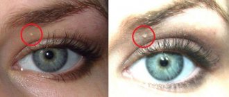
Neoplasms of the eye
On the eyelid, atheromas are not large, reaching a diameter of 5-10 mm, but sometimes they can be the size of a walnut (up to 2 cm). If the formation is small, then it does not cause inconvenience to a person, but a large atheroma, and even in a visible place, brings many problems to its owner.
But not only in aesthetic terms, a wen can cause harm. If it has grown to an impressive size, then such a formation begins to compress neighboring tissues, blood vessels and nerve endings, causing headaches and dysfunction of those systems and organs that the atheroma has compressed. The person may also feel soreness in this area.
When removing atheroma on the eyelid, the doctor must refer the patient to a consultation with an ophthalmologist .
Causes
The following factors can influence the occurrence of atheroma of the upper and lower eyelids:
- Hormonal disorders in the body, often they occur during adolescence or during menopause in a woman.
- A congenital cyst can occur due to the effect of maternal hormones on the child.
- Due to diseases of the central nervous system or the autonomic nervous system, lipid metabolism may be disrupted.
- Violation of metabolic processes.
- Problems with the adrenal glands.
- Gastrointestinal diseases.
- Seborrheic dermatitis.
Why is a lacrimal caruncle cyst dangerous?
The task of the lacrimal apparatus is to protect the eyes from environmental influences and protect the cornea and conjunctiva in order to maintain normal humidity levels.
A cyst in the area of the lacrimal caruncle is formed extremely rarely and only in those people whose lacrimal canaliculi are covered with thin hairs. This organ is not a working one in the human body.
Fortunately, atheroma of the lacrimal caruncle does not become malignant and does not affect vision . But still, a person experiences the following unpleasant symptoms:
- burning sensation in the eye;
- it feels like there is a foreign body in the area of the lacrimal caruncle;
- dry eye;
- pain is usually absent;
- The lacrimal caruncle may become enlarged and reddened.
For what reason atheroma forms in this part of the eye, scientists will not say with certainty, but the following circumstances can presumably influence it:
- when fallen eyelashes penetrate the eye;
- foreign objects entering the eye;
- microtrauma of the eye and penetration of infection through it.
The following changes in the eye are complications of atheroma of the lacrimal caruncle:
- suppuration;
- inflammation;
- infectious damage to other structures of the organ of vision.
Doctors recommend not to wait for complications and to remove such wen; the operation is performed under local anesthesia; children under 7 years of age receive general anesthesia.
Important! If the purulent contents of the atheroma penetrate the skin, it will enter the bloodstream, and this is fraught with blood poisoning, which most often leads to the death of a person.
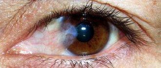
Photo
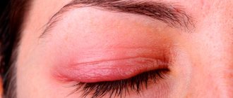
In the photo below, you can find out what atheroma of the eyelid mucosa looks like:
Treatment
Due to the peculiarity of its structure, the sebaceous gland cyst on the face, including the eyelid, does not resolve on its own . It is useless to use medication or folk treatment; they still won’t help.
The only correct solution is to completely remove the atheroma along with the capsule in which it is located.
It is not recommended to open a cyst on your own , especially if it is located in the eyelid area; in addition to the risk of loss of vision, there are other complications of such a rash step:
- abscess;
- phlegmon;
- sepsis;
- and even blood poisoning.
Thanks to modern technologies, atheroma can be removed without visible scars, pain and subsequent complications.
The doctor chooses the surgical removal tactics based on many circumstances. This is also due to the fact that atheroma of the eyelid can often become inflamed and suppurate . Therefore, it is better to remove it immediately after a small formation appears, and not wait for it to grow.
Inflamed atheromas, even after complete removal, can recur, this is due to the fact that sometimes access to them is limited due to the structure of the eye. And the capsule has no clear boundaries. Therefore, precise removal of the wen is almost impossible.
With a purulent cyst, it is necessary to wait for complete remission. And only then carry out its removal. The recovery period lasts on average 1.5 months, the seam on the eyelid is of minimal size, and therefore is not considered a cosmetic defect.
Regarding the newest techniques, namely laser coagulation or radio wave destruction, the patient must first
consult with an ophthalmologist .
Some doctors combine treatment methods. For example, the atheroma is first removed surgically by cutting it out, and then the removal site is treated with a laser.
Reference . Conservative and alternative treatment is not able to rid a person of a cyst on the eyelid, since even if the atheroma is cleared of pus, it will appear again. It is necessary to get rid of the wen along with the capsule, and this is achieved surgically.
Why it occurs and how it is diagnosed in children and adults
The neoplasm is usually benign. It does not cause any trouble because it practically does not hurt. If an infection occurs, the wen begins to fester, become inflamed, the affected area turns red, and pain appears when palpated. In this case, it is recommended to open the cyst surgically.
Based on their origin, benign neoplasms in the form of atheromas are divided into 2 types:
- primary (congenital);
- secondary formations.
Congenital atheroma is characterized by the following features:
- located on the head or scrotum;
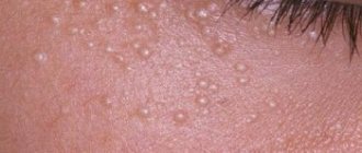
- consists of multiple clusters of cysts the size of a grain of lentil;
- no painful sensations are observed when palpated;
- the formation is soft, mobile.
People suffering from acne and seborrhea are most susceptible to the disease. With secondary atheroma, a person feels some pain when palpating the affected area, the cyst becomes dense, and the skin becomes pale.
The increase in size is explained by the abundance of connective tissue around the lesion.
Based on the structure of the tissue, atheroma is divided into 4 types:
- trichodermal;
- epidermal;
- follicular;
- Steacystoma.
In some cases, atheroma matures and opens to the outside, releasing a sebaceous secretion.
Atheroma is a disease of the sebaceous gland of the skin, which implies a benign tumor and is a consequence of its blockage.
The formation of cystic growths is associated with blockage of the sebaceous ducts. This phenomenon occurs mainly as a result of excessive production of sebum by the body. Various factors can provoke this. Most often, they are not associated with disturbances in the functioning of internal organs.
The cause of atheroma on the face can be:
- Disturbance of metabolic processes in the body. As a result, the properties of the secreted sebaceous secretion change. A blockage occurs, and as a result, a new formation appears.
- Hormonal imbalance provokes excessive secretion of sebum. There is increased sweating. Skin diseases often progress - seborrhea, acne. These diseases contribute to the development of subcutaneous tumors.
- Mechanical tissue damage. Due to trauma to a certain area of the facial skin, its structure may be disrupted. As a result, the natural excretion process is disrupted.
- Insufficient personal hygiene. If a person ignores basic hygiene procedures, pores may become clogged with dead epidermal cells.
- Excess body fat. Obesity is a problem throughout the body. With excess body weight, all processes are disrupted, including the production of sebum.
Causes
The main reasons for the development of sebaceous gland cysts are:
- swelling of the follicle caused by injury or hair removal, leading to blockage of the exit;
- injury or rupture of the sebaceous gland, leading to the formation of a subcutaneous cyst;
- acne;
- increased sweating;
- heredity;
- increased testosterone levels;
- seborrhea;
- puberty, accompanied by increased activity of the sebaceous glands.
Cyst on the eyelid: treatment with folk remedies
Good day! My name is Khalisat Suleymanova - I am a herbalist. At the age of 28, I cured myself of uterine cancer with herbs (read more about my experience of recovery and why I became a herbalist here: My story).
Before being treated using traditional methods described on the Internet, please consult with a specialist and your doctor! This will save your time and money, since the diseases are different, the herbs and treatment methods are different, and there are also concomitant diseases, contraindications, complications, and so on.
There is nothing to add yet, but if you need help in selecting herbs and treatment methods, you can find me at my contacts:
Instagram page: instagram.com/fitoterapevt1
Telephone
I consult for free.
A healthy person perceives the beauty of the world around him through his eyes. But sometimes our vision can fail us due to the occurrence of an unpleasant disease. This may be a growth on the surface of the eyeball. It may be benign. Don't get upset right away. There are several folk remedies for treating cysts on the eye.
Symptoms and factors that provoke their appearance
In itself, this is a benign neoplasm, but without the necessary assistance measures it can develop into malignant. Among them, there are several types that must be taken into account when selecting treatment:
- traumatic,
- congenital,
- spontaneous,
- post-inflammatory,
- conjunctiva,
- pigment,
- serous,
- pearl,
- exudative,
- degenerative.
The main reasons that provoked the occurrence of such an illness may be: the strong effect of certain medications,
- inflammation, inflammation
- genetic disorders
- dissection of the iris leaf,
- ophthalmological diseases,
- injuries and so on.
Ophthalmologists recommend treating eye cysts with folk remedies in combination with medications that will help achieve a positive effect in a shorter period of time. The most common symptoms of this disease may be:
- blurred visual fields,
- a small formation on the surface of the sclera,
- disturbance of visual perception,
- redness of the eyes,
- dull bursting pain.
INTERESTING fact: Lavender for colds, coughs and sinusitis
Folk remedies for illness
Although most doctors say that independent folk treatment for eye cysts will not help eliminate the problem, no one refuses to admit that it will give a chance to increase the recovery rate when combined with traditional approaches. Here are the most commonly used tools.
Seaweed infusion
Buy algae called "fucus" at the pharmacy. Pour 3 tablespoons of them into a thermos and fill with water. Leave it overnight. After this, pour the resulting infusion into ice cube trays. Before going to bed, take out the cube and wipe the area around your eyes and eyelids with it. This will also help relieve pain.
Simple ways to treat complex diseases:
Flatbread with clay
Take some clay and put it in a container. Fill with water until it slightly covers. Leave to brew and then knead everything well. Place a piece of fabric and form the cakes a little larger than necessary. The thickness should be 3 cm. Place it on your eye for 3 hours. It will dry out a little. Remove and rinse skin well.
Prefabricated infusion
Pour a tablespoon of cumin into a container, a teaspoon of cornflower petals and plantain leaves. Grind all the herbs well. Pour a glass of boiling water. Don't forget to strain afterwards. Apply 3 drops 5 times a day.
INTERESTING fact: Bowel cancer after surgery treatment with folk remedies
Acacia decoction
Boil acacia leaves in water. Soak a piece of cotton cloth in the liquid and apply it to the affected eyelid for 10 minutes 3 times a day.
Chamomile tea
Buy chamomile tea in filter bags at the pharmacy. Brew it according to the instructions. Then remove and cool slightly to a tolerable temperature. Apply to the sore spot. And tea can be used for rinsing.
Guava decoction
Make a decoction of guava leaves. Soak a piece of gauze or a cotton pad in the prepared liquid. Apply to the surface of the new growth for a quarter of an hour. Such treatment with a folk remedy for a cyst on the eyelid will help eliminate the formation, relieve redness and pain.
Remember that self-administration of any medications can worsen the course of the disease. To avoid harm, you should consult your doctor first.
Health to you!
Treatment options
When an eyelid cyst ruptures on its own and all its contents come out, the disease still returns after a few weeks. For this reason, in ophthalmology clinics, atheroma of the upper or lower eyelid is treated only by planned removal. If it becomes inflamed and then begins to fester, then surgery is prescribed immediately. Additionally, such a patient, in order to relieve inflammation and increase the body's defenses, is prescribed antibiotics, as well as immunostrengthening drugs.
Conservative
Atheroma of the century is curable, but such a result can be achieved not by folk or traditional treatment, but by surgical medicine. Local medications help stop the growth of the epidermal lump and relieve inflammation. Medicines can draw out stagnant sebaceous secretions from it, heal the wound, but do not exclude relapse.
The list of safe folk remedies often used for atheroma of the century includes:
- Silver item. A ring, spoon or coin is applied 3 times a day for 30 minutes to the inflamed eyelid. Treatment lasts until the epidermal ball is reabsorbed (about a month).
- Chicken egg shell film - apply the wet side to the atheroma overnight. The procedure is repeated until recovery.
- Lamb fat - rub the sore eyelid at night, stop using it after the contents of the cyst are released.
- Watercress herb juice - under the influence of the components of the juice, the resorption and healing of the opened formations occurs. You need to take the juice daily for at least 2 months in a row.
Effective ointments for atheroma - Levomekol, Ichthyol, Vishnevsky. But ophthalmologists do not recommend using them on eyelids, since the products can damage the mucous membrane.
Radical
Complete removal of atheroma on the eyelid is usually performed surgically:
- The skin over the tumor is opened with a scalpel.
- Its capsule is cleaned and dehusked.
- Self-absorbing sutures are applied.
The patient needs to be prepared for the appearance of a scar at the site of the cyst after its removal.
Surgical treatment of atheromas is not used for inflamed tumors. There is a high probability that the surgeon will not be able to completely remove the cyst capsule. Subsequently, this will lead to the growth of the pathological fragment remaining under the skin.
If the inflamed lump has purulent contents, the surgeon opens it, cleans it of pus and drains the wound. As soon as the inflammation goes away, the patient is scheduled for surgery to remove the cyst shell.
Self-treatment of an epidermal lump in the eye area (steaming, heating, squeezing) can lead to infection and decay, and can also cause decreased vision.
Laser therapy is often used for atheroma of the eyelids. Removal is carried out using one of the following methods:
- Photocoagulation - used for tumors no more than 5 mm in diameter. The laser beam evaporates even cysts containing pus.
- Excision with membrane - prescribed for epidermal bumps no larger than 20 mm in size. During the operation, in addition to the laser beam, a scalpel and forceps are used, as in conventional surgery.
- Capsule evaporation - used for atheromas larger than 20 mm. First, the tumor capsule is cut, its contents are cleared with gauze swabs, then the shell is evaporated with a laser.
Another relapse-free method for removing sebaceous cysts is radio wave therapy. But it is used only for small epidermal bumps, without inflammation or suppuration. Radio waves affect the pathological lump in such a way that its tissues die and dissolve. At the site of the operation, only a crust remains, under which new skin is formed.
Removal of atheroma from the upper and lower eyelids
In addition to the methods described above, atheroma on the lower or upper eyelid can be evaporated by exposing the capsule to a laser. During the procedure, the cyst is opened with a scalpel, and it is cleaned of exudate.
Then the skin edges are moved apart, and the capsule shell is evaporated from the inside using a laser. At the end of the operation, the wound is sutured, and the sutures are removed on the 10th day.
However, laser evaporation of a cystic formation on the eyelid has some contraindications. You cannot carry out such an operation if you have:
- diabetes mellitus,
- immunodeficiency states,
- pregnancy,
- malignant tumors, etc.
Radio wave therapy makes it possible to prevent the re-development of pathology after surgery; the wound is not sutured when using this technique, it heals very quickly, and only a slight scar remains.