How to treat eye staphyloma?
Staphyloma is a pathological process that results in pathological protrusion of the posterior surface of the sclera.
The development of such a disease leads to the fact that the patient’s quality of vision significantly decreases and the visual fields narrow. It should be noted that this pathology can occur as an independent disease extremely rarely. Most often, the development of this pathological process is caused by myopia. That is why there is a high risk of complete loss of vision.
Myopic staphyloma is diagnosed most often at the age of 20-40 years and is quite often complicated by other ophthalmological diseases. This course of the clinical picture leads to disability in working age.
The clinical picture is characterized by a decrease in visual acuity, a general deterioration in well-being and other nonspecific symptoms, therefore, an accurate diagnosis requires a number of instrumental studies. Diagnosis and treatment are carried out by an ophthalmologist .
The prognosis in this case is only individual, since everything will depend on the form of the disease, the stage at which treatment was started and on the general clinical indicators of the patient, including his age.
Etiology
The development of this clinical picture is based on the development of myopia. With the development of such a disease, the collagen fibers of the sclera become loosened, which causes an increase in the number of proteases in the posterior pole.
Excessive amounts of proteases lead to atrophy of adhesive bonds in elastic fibers, which is the main reason for the formation of staphyloma.
However, it should be noted that the clinical manifestation of this ophthalmological disease occurs only in patients with high myopia.
Predisposing factors for the development of such a pathological process include:
- the presence of chronic ophthalmological diseases.
- genetic predisposition.
- long work that requires eye strain.
- astigmatism.
- strabismus.
- sedentary lifestyle.
- non-compliance with doctor's recommendations regarding the use of glasses or contact lenses.
In more rare cases, it is not possible to establish the cause of staphyloma of the eyeball.
Classification
In this case, there are only two forms of development of this pathology - false and true. Only a qualified ophthalmologist can determine which form occurs in a particular clinical case.
True staphyloma
Almost always, true corneal staphyloma is a consequence of a false form of this disease. Its clinical picture is characterized as follows:
- there is a strong stretching of the sclera near the optic nerve;
- The entire circumference of the optic disc is affected, which leads to the development of optic disc staphyloma.
This form of the disease is characterized by extremely negative prognosis. Even if specific treatment is started in a timely manner, there is no guarantee that vision will be fully preserved.
False staphyloma
In this case, the development of the pathological process is slow, so in the early stages it is practically not diagnosed. The exception is those cases when a person is systematically, on a regular basis, examined by an ophthalmologist .
The clinical picture of the development of this form of pathological process will be characterized as follows:
- Due to retinal degeneration, tissue gradually begins to break down.
- dystrophy of the vascular surface begins with the involvement of the optic nerve head in the pathological process.
- the last stage leads to the development of the true form of the disease.
Most often, the full clinical picture with significant deterioration of vision appears already in old age. It is because of this factor that treatment will not be as effective as in younger people.
Symptoms
As already mentioned, retinal staphyloma can occur without any symptoms for a long time. As the pathological process worsens, the following symptomatic complex may appear:
- significant visual impairment;
- increased fatigue even after long and prolonged rest;
- feeling of heaviness in the eyes;
- narrowing of the field of view in one visual organ;
- albino fundus color;
- symptoms of hemorrhagic retinal detachment.
It should be noted that in this case, both unilateral and bilateral damage to the visual organ . Due to the fact that the symptoms are nonspecific, only an ophthalmologist can establish an accurate diagnosis by carrying out the necessary diagnostic measures.
Diagnostics
The first step is to conduct a physical examination of the patient, during which the clinician should determine the following factors:
- How long ago did the first clinical signs begin to appear?
- features of the clinical picture, with what intensity symptoms appear.
- whether there were any typical diseases in the family history.
- Do you have chronic ophthalmological diseases?
- whether the patient is currently taking any medications without a doctor’s prescription, and if so, which ones.
In addition, the following diagnostic measures are carried out:
- tests to determine visual acuity;
- tonometry;
- biomicroscopy using a slit lamp;
- eye refraction test;
- Ultrasound of the eye with measurement of the anterior and posterior axis;
- computer perimetry;
- optical coherence tomography;
- electroretinography.
According to the results of the study and taking into account the data that was collected during the initial examination, the doctor can determine the form of the pathological process and, therefore, prescribe the most effective treatment.
Treatment
In this case, only an integrated approach is used, since therapeutic measures should be aimed, first of all, at eliminating the root cause, that is, myopia.
The medicinal part of treatment may include the following drugs:
- to strengthen the scleral membrane.
- to improve hemodynamics of the eye.
- to stabilize metabolic processes in the retina and choroid.
- hemostatic.
- absorbable.
- desensitizing.
In addition, physiotherapeutic procedures are prescribed, namely:
- magnetophoresis;
- electrophoresis;
- laser stimulation.
In order to reduce the rate of progression of the pathological process, the doctor may prescribe orthokeratology lenses .
As for surgical intervention, in this case all actions will be aimed at preventing even greater stretching of the scleral membrane. The surgical procedure is determined on an individual basis.
The insidiousness of this pathological process is that even with the timely initiation of therapeutic measures, the prognosis is unlikely to be positive, since progression of myopia is observed.
Possible complications
Without treatment, the risk of developing the following complications increases significantly:
- cataract.
- open angle glaucoma.
- detachment of the mesh surface.
- complete loss of vision.
The latter, as a rule, leads to disability.
Prevention
If we talk about the congenital form of the disease, then there are no methods of prevention. In general, regarding this pathological process, only general recommendations can be followed:
- compliance with visual hygiene rules;
- prevention of ophthalmological diseases;
- strengthening the immune system;
- the workplace must have high-quality and proper lighting;
- when working at a computer for a long time, you should use special protective equipment - drops or glasses;
- For preventive purposes, you should visit an ophthalmologist at least once every few months, and if you have a genetic predisposition to such diseases, it is advisable even more often.
In addition, it should be remembered that self-medication or ignoring symptoms can only aggravate the course of the pathological process and lead to the fact that the disease can no longer be eliminated. Therefore, you need to promptly seek qualified medical help.
Source: https://brulant.ru/health/stafiloma/
Etiology
The development of this clinical picture is based on the development of myopia. With the development of such a disease, the collagen fibers of the sclera become loosened, which causes an increase in the number of proteases in the posterior pole. Excessive amounts of proteases lead to atrophy of adhesive bonds in elastic fibers, which is the main reason for the formation of staphyloma. However, it should be noted that the clinical manifestation of this ophthalmological disease occurs only in patients with high myopia.
Predisposing factors for the development of such a pathological process include:
- the presence of chronic ophthalmological diseases.
- genetic predisposition.
- long work that requires eye strain.
- astigmatism.
- strabismus.
- sedentary lifestyle.
- non-compliance with doctor's recommendations regarding the use of glasses or contact lenses.
In more rare cases, it is not possible to establish the cause of staphyloma of the eyeball.
Staphyloma
Staphyloma
is a pathological protrusion of the posterior surface of the sclera. Clinically manifested by decreased visual acuity and narrowing of the visual field. The fundus reveals diffuse atrophy of the pigment epithelium of the retina, and the presence of peripheral vitreochorioretinal dystrophy or retinal traction is possible.
For diagnosis, an external examination is used, visual acuity and the nature of vision are examined, tonometry and biomicroscopy are performed. Additional methods include ultrasound of the eyeball, computer perimetry, and electroretinography.
Treatment of staphyloma is conservative (drugs and physical procedures to improve blood supply to the retina, strengthen the sclera and relax the accommodation of the eye) and surgical (aimed at strengthening the posterior surface of the sclera).
Staphyloma (staphyloma; Greek staphylē – bunch of grapes + -ōma) – severe deformation of the sclera with pathological prolapse and elongation of the eye axis. Scleral staphyloma occurs with high myopia.
Myopia is the most common disease and the leading cause of blindness in developed countries. In Russia, 15% of the population suffers from refractive errors, 3% of them have a complicated form with pronounced changes in the fundus.
Myopic staphyloma develops between the ages of 20 and 40 years. It is often accompanied by other pathological changes in the structure of the eye and is a cause of disability in young working age.
Rehabilitation of patients with high myopia and prevention of complications still remain important problems in modern ophthalmology.
Causes and symptoms of staphyloma
The scleral membrane is the outer opaque capsule of the eyeball and contains in its structure cellular elements that are immersed in a ground substance consisting of glycosaminoglycans, protein, and polysaccharide complexes. 70% of the sclera consists of collagen protein, its bundles - fibrils - form a special plexus with elastic fibers.
Thanks to this structure, the scleral membrane performs its main functions - maintaining the strength and elasticity of the eyeball. With the development of high myopia, loosening of the collagen fibers of the sclera occurs.
In the posterior pole, the number of proteases increases, which destroy adhesive bonds in elastic fibers and lead to the formation of staphyloma.
Clinically, staphyloma manifests itself when complications develop in a patient with high myopia. Most often, there is a significant decrease in visual acuity, fatigue, and a feeling of heaviness in the eyes. There may be a narrowing of the field of vision in one eye.
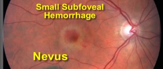
During examination, an ophthalmologist may detect extensive staphyloma in the fundus (a white focus of atrophy in the form of a ring at the posterior pole), diffuse atrophy of the pigment epithelium, “albinotic” color of the fundus, the presence of peripheral vitreochorioretinal dystrophy or traction. The lesion is often bilateral.
Complications of myopic staphyloma include the occurrence of retinal dystrophy, the formation of hemorrhagic retinal detachment, destruction of the vitreous body, the development of cataracts, and open-angle glaucoma.
Diagnosis and treatment of staphyloma
Diagnosis of staphyloma begins with the collection of anamnestic information. Then the ophthalmologist performs an external examination, examines visual acuity and the nature of vision, and conducts tonometry. Biomicroscopy using a slit lamp is the main method for diagnosing staphyloma. A refractive examination using cycloplegia is required.
Additional methods include ultrasound of the eye with measurement of the anterior-posterior axis and computer perimetry (to identify paracentral visual field defects). Optical coherence tomography is used to diagnose the condition of the macular area. Electroretinography helps to identify functional disorders in the retina and choroid of the eyeball.
Treatment of myopic staphyloma is complex and includes both surgical and conservative techniques. The primary goal of therapeutic measures is to reduce the progression of myopia.
Conservative therapy for staphyloma includes the use of drugs that affect the relaxation of accommodation, help strengthen the scleral membrane, improve the hemodynamics of the eye, metabolic processes in the retina and choroid and increase visual functions.
With the development of hemorrhages in the retina, the use of hemostatic, absorbable and desensitizing agents is necessary. Physiotherapeutic treatment is also indicated. Electrophoresis, laser stimulation or magnetophoresis are prescribed.
Rigid orthokeratology lenses can be used to reduce the rate of myopia progression. Surgical treatment of staphyloma is aimed at preventing further stretching of the sclera.
Various techniques are used to strengthen the posterior pole of the eyeball.
Prognosis and prevention of staphyloma
The prognosis is most often doubtful. Prevention of myopic staphyloma is aimed at reducing the progression of myopia.
Includes activities to promote health and physical development in childhood and adolescence, teaching children and adults the rules of visual hygiene.
It is necessary to organize high-quality lighting in schools and workplaces, monitor compliance with sleep and rest schedules, limit the use of tablets and phones by children, and regularly visit an ophthalmologist for preventive examinations.
Source: https://MyMedNews.ru/stafiloma/
Therapy and prevention of myopic staphyloma
Modern ophthalmology uses conservative, surgical and combined methods of treating the disease. At the initial stage, the goal of therapeutic intervention is to stop the progression of myopia. For this purpose, medications are used that cause:
- relaxation of accommodation;
- strengthening the surface of the sclera;
- improvement of hemodynamics of the organ of vision;
- optimization of metabolic reactions in the retina and blood vessels;
- enhancing visual capabilities.
To eliminate hemorrhages, hemostatic, absorbable and desensitizing materials are used. Physiotherapeutic procedures have a beneficial effect on the healing process: electrophoresis, laser activation or magnetophoresis.
A rigid orthokeratology lens is used to reduce the rate of myopia development. Surgery is resorted to when it is necessary to prevent ongoing protrusion of the sclera.
To date, no effective means or special methods for combating staphyloma have been developed. Doctors recommend treating laser coagulation with a certain degree of caution. Prevention should begin in childhood.
First of all, it is necessary to follow all hygiene rules, strengthen the immune system, conduct regular physical exercise, and eat well. At home and at school, conditions should be created for normal lighting of the workplace. You especially need to limit watching TV, sitting at a computer, tablet or smartphone.
Treatment of staphyloma of the eye sclera, its types and diagnosis
Staphyloma is one of the most common ophthalmological diseases leading to disability in developed European countries. More often, the pathology occurs in people with a high degree of myopia, but it often occurs at an early age in the presence of keratitis and keratomalacia or trauma.
The pathology is difficult to treat, and in the absence of timely treatment can cause complete blindness. So, approximately 10% of Russian residents suffer from visual impairments, of which about 3% suffer from severe degrees of myopia.
What is staphyloma
The occurrence of the disease is associated with disruption of the functioning of the sclera - the outer white layer of the eye. The sclera consists of cellular elements, polysaccharides and a large number of collagen fibers (2/3 of the composition), which intertwine with each other and form a dense capsule. It is the white shell that gives the eyeball stability and shape.
In a number of cases, scleral collagen loses its elasticity, as a result of which characteristic disturbances, a kind of protrusion, appear at the site of weakening of tension.
Causes of staphyloma in the eyes
Among the factors causing the occurrence of staphyloma, there are several. However, the main condition for the appearance of the disease will be a violation of the elasticity of the sclera, which can arise as a result of the following.
- High degree of myopia, in which there is an elongation of the axis of the visual path, which causes keratoconus and provokes posterior myopic staphyloma. The myopic form of the disease affects young people aged 20 to 40 years. In this case, a false posterior staphyloma is more often formed, which is located at the posterior pole of the eye and surrounds the optic nerve. It causes a number of negative phenomena on the retina: ring-shaped or cluster-shaped atrophy (staphyloma translated as cluster), hemorrhages in the retina as a result of stretching and weakening of blood vessels, retinal detachment and ruptures.
- Keratitis or keratomalacia. In this case, the development of staphyloma occurs as a result of perforation of the anterior part of the sclera - the cornea. The eye fluid flows out through the resulting hole, and the iris, carried away by the fluid, is soldered to the edges of the perforated hole. As a result, the anterior chamber disappears and an obstacle to the movement of ocular fluid is formed. Intraocular pressure increases, a protrusion forms - this is an anterior staphyloma. It is characterized by the formation of walls covered with scar tissue and overgrown pathological vessels; inside there is fluid from the anterior chamber. When a staphyloma is perforated, some of the fluid flows out of it, and after closing it is collected again; in some cases, the hole does not close and a fistula is formed. When staphyloma persists for a long time, the corneal cells degenerate and tissue similar in appearance to skin is formed.
- Eye injuries. This type of staphyloma occurs as a result of traumatic penetration of foreign objects into the eye.
Types of staphyloma
Regardless of the location of scleral staphyloma, it is divided into the following types.
- Complete. With this form, the entire sclera is involved in the process, and the protrusion can be so large that it prevents the eyelid from closing. The occurrence of complete staphyloma causes complete blindness or vision is fixed at the level of light perception.
- Partial. As a rule, the partial form is characterized by less visual impairment and can be either posterior or anterior.
- Lobed (lumpy). This type is characterized by the appearance of several scleral staphylomas, which have the shape of a cluster or tubercles, which gave rise to the name of the disease.
Posterior myopic staphyloma, in turn, is divided into the following.
- False myopic staphyloma, which is formed as a result of lengthening of the visual path with strong degrees of myopia. In this case, the myopic cone, which initially forms a white crescent on the optic nerve head, lengthens, gradually forming a ring on the retina (false staphyloma); in difficult cases, it can also capture most of the macula. Characteristic disturbances occur, which the ophthalmologist sees as white or gray spots in the fundus. This causes dystrophies and pathologies on the part of the retina, which is less elastic than the sclera and cannot stretch as well as it. Foci of peripheral dystrophy, hemorrhages (as a result of vascular fragility), and local detachments appear in the fundus.
- True posterior staphyloma (protrusion of the sclera in the posterior pole of the eye) is rare; it worsens depigmentation and diffuse dystrophy of the epithelium, which promotes the formation of pathological retinal vessels, causes multiple diffuse hemorrhages and provokes extensive retinal detachments. A true scleral staphyloma forms.
Symptoms and diagnosis of staphyloma of the sclera of the eyes
Many signs of staphyloma are similar to other signs of myopia, the most characteristic are:
- Rapid, sharp decrease in the quality of vision;
- Feelings of heaviness in the eyes;
- Fast fatiguability.
When examined by an ophthalmologist, the doctor will detect characteristic “albino” (white) lesions, epithelial dystrophy, and the presence of retinal traction in the fundus.
https://www.youtube.com/watch?v=4WhiKQOURO8
To clarify the diagnosis, visual acuity testing and fundus examination using a slit lamp are performed. The following will be very informative: fluorescein angiography, eye ultrasound, computer perimetry.
The doctor may prescribe a number of other examinations of the eye structures.
Treatment method for staphyloma
Treatment of staphyloma can be either conservative or surgical. The choice of treatment always depends on the degree of scleral protrusion.
Initially, when myopia occurs and throughout the course of treatment, myopia therapy is necessarily used; drugs are used here for:
- relaxation of accommodation;
- stimulation of metabolic processes in the retina;
- improving blood supply to the retina and sclera;
- strengthening the sclera.
- strengthening the immune system.
Often, therapeutic measures include physiotherapeutic procedures: laser, UHF, magnetophoresis. One of the accessible and easy-to-use techniques is the use of hard special lenses that correct keratoconus and prevent the occurrence of staphyloma of the posterior pole of the eyeball.
If retinal detachment is detected, surgical treatment and laser coagulation are performed.
When diagnosing anterior staphylomas of the sclera and cornea, especially with a fistula, more drastic treatment will be required:
- corneal transplant;
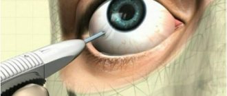
- in the most difficult cases, enucleation (removal) of the eyeball is indicated.
For uncomplicated forms, penetrating or partial cone keratoplasty is performed and rigid contact lenses are used.
Possible complications and prevention of the disease
Among the complications that corneal staphyloma causes is complete loss of vision and the eyeball.
Among the complications of posterior myopic staphylomas the following are observed:
- retinal detachment;
- destructive changes in the vitreous body;
- open-angle glaucoma;
- cataract.
It is very important to carry out preventive measures aimed at preserving vision in early childhood and adolescence. Among the mandatory requirements will be sufficient illumination of places for games and activities, common areas for children to stay, as well as limiting the time that children spend watching television programs, playing on tablets and phones.
Tips and tricks
Source: https://MoeOko.ru/zabolevaniya/stafiloma.html
Diagnostics
The first step is to conduct a physical examination of the patient, during which the clinician should determine the following factors:
- How long ago did the first clinical signs begin to appear?
- features of the clinical picture, with what intensity symptoms appear.
- whether there were any typical diseases in the family history.
- Do you have chronic ophthalmological diseases?
- whether the patient is currently taking any medications without a doctor’s prescription, and if so, which ones.
In addition, the following diagnostic measures are carried out:
- tests to determine visual acuity;
- tonometry;
- biomicroscopy using a slit lamp;
- eye refraction test;
- Ultrasound of the eye with measurement of the anterior and posterior axis;
- computer perimetry;
- optical coherence tomography;
- electroretinography.
According to the results of the study and taking into account the data that was collected during the initial examination, the doctor can determine the form of the pathological process and, therefore, prescribe the most effective treatment.
Retina for myopia: myopic cone and staphyloma
In patients with myopia, in most cases there are changes in the fundus of the eye (around the optic nerve head).
There are several types of such pathologies:
- Myopic cone;
- Peridiscal light reflexes, presented in the form of an arc;
- True staphylomas.
Even in patients with the initial form of myopia, parallel reflexes are detected near the optic nerve head, which are located on the border of the disc. They can be single or double. In the case of detection, we are often talking about initial anomalies in the structure of the eye wall in the region of the posterior pole.
Myopic cone
Myopic cones look like crescent-shaped white formations with clear boundaries. They are located in the temporal region of the disc.
At the border of such pathological formations there is often pigmentation, the severity of which can vary (up to complete coverage with dark pigment).
Sometimes pigment formations in the form of lumps are located around the cone. In some cases, it is possible to identify embryos of choroidal vessels in the cone itself.
In the case of a small diameter, less than 20% of the diameter of the optic nerve head, the cone is called a falx. If the diameter of the formation exceeds these values, then it is called a cone proper. Sometimes such circular cones are also called staphylomas, but their structure differs from the structure of true staphylomas.
Sickles are formed as a result of the fact that the optic nerve canal is not perpendicular to the plane of the scleral membrane. In this case, the wall of such a channel looks like a sickle. The color of this neoplasm is associated with the translucency of the white sclera.
The formation of cones is associated with atrophic processes in the pigment epithelial layer near the disc and with stretching of the sclera itself. This leads to improved transparency of the choroid, which is also subject to degenerative processes. The sclera shines through this membrane.
The formation of cones most often does not affect visual acuity.
In patients with high myopia, a ring-shaped cone covers the area around the optic disc.
Treatment
In this case, only an integrated approach is used, since therapeutic measures should be aimed, first of all, at eliminating the root cause, that is, myopia.
The medicinal part of treatment may include the following drugs:
- to strengthen the scleral membrane.
- to improve hemodynamics of the eye.
- to stabilize metabolic processes in the retina and choroid.
- hemostatic.
- absorbable.
- desensitizing.
In addition, physiotherapeutic procedures are prescribed, namely:
- magnetophoresis;
- electrophoresis;
- laser stimulation.
In order to reduce the rate of progression of the pathological process, the doctor may prescribe orthokeratology lenses .
As for surgical intervention, in this case all actions will be aimed at preventing even greater stretching of the scleral membrane. The surgical procedure is determined on an individual basis.
The insidiousness of this pathological process is that even with the timely initiation of therapeutic measures, the prognosis is unlikely to be positive, since progression of myopia is observed.