More about pathology
Exposure to unfavorable factors during intrauterine development leads to failure. A defect in the iris is formed in the form of additional round or irregularly shaped pupils. Acquired forms of polycoria resulting from mechanical damage are possible.
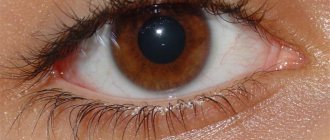
Polycoria most often occurs in one eye (unilateral), less often in both (bilateral). It is more common to have 2 or 3 pupils located closer to the center. One pupil is usually larger in size than the others.
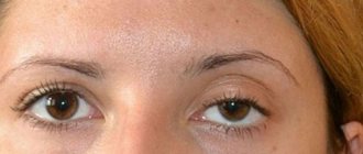
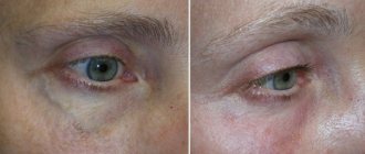
The photo shows classic examples of eyes with polycoria.
There are 2 types of polycoria of the eye:
- True form. This defect is characterized by several pupils, each of which has its own sphincter. The sphincter is a circular muscle that is responsible for the contraction of the pupils. Therefore, they react normally to light.
- False form (pseudopolycoria). With pseudopolycoria, the pupils do not react to light because they do not have contractile muscles.
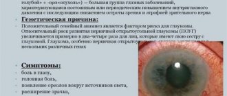
Ocular herpes
Ocular or ocular herpes is often caused by the chickenpox virus or is herpes simplex type 1. Unlike genital herpes, ocular herpes is not sexually transmitted. It is worth noting that most people are exposed to some form of herpes throughout their lives without even noticing its symptoms. Ocular herpes is felt as pain or throbbing in the eyelid. The illness usually resolves within a week and is characterized by red eyes, migraines and light sensitivity.
In some cases, the virus can affect the cornea, leading to more complex symptoms. Very rarely, herpes can be transferred to the eyeball, which will lead to temporary loss of vision.
6
Causes of the disease
There are no statistics on people with polycoria, but there are reported cases of Axenfeld-Rieger syndrome. There are 35 thousand people in the world with this syndrome. From these figures one can indirectly judge the prevalence of polycoria.
Causes of the defect:
- Teratogenic effects on the fetus. A pregnant woman's intake of alcohol, drugs, medications, as well as being in the zone of ionizing radiation lead to the formation of polycoria.
- Genetic abnormalities.
- Partial closure of iris coloboma.
- TORCH infections (rubella, herpes, cytomegalovirus, toxoplasma). Infections suffered by the mother during pregnancy lead to disruption of the intrauterine formation of organs and systems of the fetus.
- Iridocorneal endothelial syndrome. Polycoria occurs due to dystrophic processes. A triad of symptoms is characteristic: corneal edema, polycoria, secondary glaucoma.
- Iatrogenic damage. A false form occurs due to defects in iridoplasty, when the doctor accidentally mechanically damages the iris or incorrectly sets the laser power.
- Eye injury. Pseudopolycoria can form after traumatic damage to the visual organ or burn.
- Axenfeld-Rieger syndrome. With this syndrome, congenital underdevelopment of the iris and displacement of the pupil, abnormal development of teeth and abdominal organs are noted.
Cat's eye syndrome
Cat's eye syndrome is an extremely rare chromosomal disorder. It is congenital and cannot be treated. The syndrome was named due to its symptom when some tissues in the eye are missing, which causes the pupil to become constricted and located inside the iris. It is worth noting that this symptom is not observed in all people with cat eye syndrome.
This syndrome also affects the bones, heart and kidneys. In addition, people suffering from cat eye syndrome often have mild mental retardation or hyperactivity. The disease is easily detected at an early stage, accompanied by noticeable growth retardation. It is impossible to completely get rid of the syndrome, however, thanks to individual treatment, the severity of symptoms can be significantly reduced and vision improved.
2
Symptoms of the disease
The diameter of the pupils in polycoria affects how a person sees. A hole diameter of less than 1.5 mm significantly impairs the quality of vision: clarity decreases, the boundaries of the visual field are limited, and vision becomes blurred. With increased visual load, rapid fatigue of the organ of vision and photophobia occurs.
If the diameter is more than 1.5 mm, then vision is not impaired. A person is only concerned about the external defect.
Polycoria is often complicated by amblyopia, less often by ocular migraine and secondary glaucoma. People with polycoria are more susceptible to inflammatory processes in the eye organ. Structural features and frequent wearing of lenses predispose to the frequent development of degenerative phenomena in the iris.
How does a person with polycoria see?
Typically, with polycoria, the pupils are unequal in size, and their shape is noticeably different from the standard one. As a result, the holes in the iris are unable to function normally and have an insufficient response to light.
Most often, polycoria is accompanied by a significant deterioration in vision, a noticeable decrease in its acuity, and problems with recognizing color shades. In the presence of unfavorable circumstances, the pathology may be accompanied by the development of amblyopia (lazy eye syndrome).
There is a belief that with this disorder a person sees all objects in two. Sometimes diplopia does occur, leading to a significant deterioration in the patient's quality of life. With polycoria, the inability may appear:
- read any texts;
- work with small parts;
- navigate in space.
Sometimes people with polycoria suffer from more or less severe photophobia. With this condition, a painful reaction of the eyes to light sources develops.
In some cases, patients with polycoria experience an exacerbation of visual perception. It is believed that people with such an anomaly can acquire the ability to better focus on surrounding objects and distinguish small objects located at large distances.
Diagnostics
Congenital polycoria is determined in the neonatal period. Defects that appear as a result of injury are determined when contacting an ophthalmologist.
To establish a diagnosis, the following is carried out:
- external inspection;
- assessment of pupil reaction to light;
- test with mydriatics.
During the examination, the doctor evaluates the characteristics and location of the pupils and checks the pupillary reaction.
To do this, cover the eyes one by one with your palms, then remove your palms. Normally, the pupils constrict in light and dilate in dark conditions. If they react normally to light, then they speak of the true form. If they are motionless, then it is pseudopolycoria.
The mydriatic test helps evaluate the pupillary response. Mydriatics are drugs that dilate the pupil. For the test, drops from the group of M-anticholinergic blockers are used. The normal reaction to a mydriatic is expansion (mydriasis). This is characteristic of true polycoria. If the pupils do not respond to drops with M-anticholinergic blockers, then they speak of a false form of the disease.
Additional examinations to clarify visual impairments:
- Assessment of visual acuity (visometry).
- Determination of the boundaries of the visual fields (perimetry).
- Examination of the fundus (biomicroscopy).
- Visualization of eye structures (ultrasound of eyeballs).
The video shows the reaction of the pupils to light in the true form of the disease.
Hemolacria
Supposedly one of the most surprising medical conditions, hemolacria is better known as "crying blood." About 10 years ago, teenager Calvino Inman discovered he was bleeding from his eyes. Reasonably assuming he was dying, Calvino went to the hospital, where he was subjected to a CT scan, MRI and ultrasound. None of the procedures revealed any abnormalities in him. Until today, doctors have not fully understood the cause of Calvino Inman's bleeding eyes while crying.
Another patient, Michael Spann, experienced a severe headache followed by bleeding from his eyes, mouth and nose. Since then, he has had “attacks” of hemolacria at least once a week. This greatly influenced Spann's future life. According to the man, he was fired from many jobs due to bleeding from his eyes. What's interesting is that Spann, like Inman, is from Tennessee.
In a 2004 study, doctors looked at four children with hemolacria who were eventually cleared of the diagnosis without any treatment. The cause of the development of the disease in Spann and Inman is unknown, but Spann is already recovering - at the beginning of the discovered condition, he cried blood at least 3 times a day.
Treatment methods
The iris defect can only be radically corrected surgically. The task of the operating surgeon is to restore the correct shape of the iris.
Correction of an iris defect is possible in the following ways:
- Iridoplasty – restoration of the anatomical structure of the iris. A large number of defects require iridoplasty with the installation of an iridolenticular diaphragm.
- Surgery with suturing. Surgical correction is carried out by applying sutures that form one correct pupil. Performed in cases where the diameter is more than 2 mm.
- Optics for the eyes. When operations are contraindicated or a person does not want to undergo surgery, contact lenses matched to the color of the iris will help hide a cosmetic defect. Refractive errors can be corrected using optics for vision correction (glasses, lenses).
- Symptomatic therapy. Symptoms of rapid eye fatigue will help reduce drops based on natural tears: “Artificial tears”, “Visine”.
Methods for treating double pupil
If the pupil is slightly divided, then special cosmetic lenses are suitable. If polycoria is true and the two pupils are functioning correctly, surgery is required. Usually, iris surgery is performed without opening the eyeball.
If an anomaly is detected in a newborn and the pupil diameter is more than 2 mm, an open radical correction is performed. Sutures are placed on the eye and a single pupillary opening is formed. For adults, such correction is practically not used - a positive result can only be achieved on a growing eye.
Congenital polycoria is not dangerous and can be corrected surgically. But acquired ones can lead to negative consequences and loss of vision if the pathology is not corrected. Cosmetic lenses usually cope with this successfully.
Preventive measures
There are no measures to prevent polycoria. The likelihood of fetal development abnormalities can be reduced if a pregnant woman gives up bad habits. Before pregnancy, it is recommended to undergo screening for infectious diseases. If detected, undergo treatment together with your spouse.
To avoid traumatic polycoria, you need to protect the organ of vision from injury and burns.
The prognosis is favorable. Timely surgical correction allows you to forget about visual discomfort and cosmetic defects. Even without surgery, the prognosis is relatively favorable: the external defect and visual impairment are easily corrected with contact lenses.
Tell us in the comments, have you met people with iris defects? Share the article with your friends on social networks. Be healthy. All the best.