Causes of nevus formation on the eye
A nevus on the eye is an accumulation on a very small area of the tissue of the eyeball of a special pigment - melanin, which is responsible for the color of hair, skin and eyes. The reasons for this can be a variety of factors, but most often it is caused by changes in hormonal levels.
Often, a nevus on the eye occurs during pregnancy and lactation (breastfeeding), against the background of severe stress, infectious diseases, taking oral contraceptives, inflammatory skin diseases, natural aging of the body and as a result of ultraviolet and/or ionizing radiation.
It has been proven that there is also a genetic predisposition to the formation of a nevus on the eyeball. It is the hereditary factor that usually explains the occurrence of this pathology in children.
Conjunctival nevus: features of the mole, its types
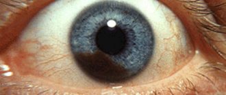
The conjunctiva is the thin, transparent lining of the eye. The nevus on it, as a rule, is located in the inner corner of the eye or the tear month, so it is not very noticeable. However, sometimes you can find a mole on the iris of the eye, which looks like a small dark spot.
Interestingly, this feature on the iris does not affect vision. This growth can look different. As mentioned above, it all depends on the amount of “coloring” substance.
The mole may be on the retina of the eye, faded, or black or red in color. Very often it is completely colorless and therefore invisible.
Conjunctival nevus can be divided into three types depending on the formation factor:
- Vascular moles. They have a reddish tint, since they are based on the capillaries of the conjunctiva;
- Melanin moles. Appear accordingly due to the accumulation of melanin;
- Cyst-shaped nevus. The reason for this formation is the combination of lymphatic vessels.
Clinical manifestations
A nevus on the eye can be located on the outer or inner part of the tunica albuginea, the lacrimal caruncle, the limbus, and sometimes on the retina. True, in the latter case it is not visible to the naked eye and is detected only during ophthalmoscopy (examination of the fundus). An ocular nevus can have different colors (pinkish, black, brown, yellow), shape and size.
As statistics show, the more melanin a person has in his skin, the lower the likelihood of developing a nevus on his eye. That is why pathology is more often observed in fair-skinned and blue-eyed people, in particular residents of Scandinavian countries.
Types and forms of pathology
Choroidal nevus occurs:
Typical nevi:
Progressive nevi:
Atypical nevi
Why is a nevus on the eye dangerous?
Regardless of its location, a nevus is dangerous due to its ability to degenerate into a malignant tumor - melanoma. A mole that is benign in nature usually practically does not change its color and size, while melanoma is characterized by fairly rapid growth.
But it is practically impossible to determine the malignancy or benignity of a birthmark on the eyeball by this sign, because Nevus of the eye is characterized by changes in both size and color, regardless of the characteristics of the histological structure.
Reasons for appearance
The etiology of the formation of a mole on the pupil or eyeball is always of the same origin. A mole in the eye means that the cells of the organ are oversaturated with a large amount of pigment - melanin. There are provoking reasons for the formation of a benign spot in the form of a nevus:
- Labor activity. Daily exposure to environmental factors during professional activities affects the pigmentation of any part of the organ of vision. Typically, an acquired benign nevus is found in hot shop workers - welders and metallurgists. In this case, the mucous membrane of the eye comes into contact with heated air and a bright source.
- Ultra-violet rays. Tanners who spend a lot of time in direct sunlight and do not wear sunglasses may develop excess pigmentation in the eyeball. This is how the body reacts to ultraviolet radiation, which has a negative effect on vision.
- Hereditary predisposition. Birthmarks are passed from parents to children with information in the DNA. In most cases, the dermatological defect passes through the male line, so the female half of the population almost does not suffer from this pathology.
- Frequent visits to the solarium. Artificial ultraviolet rays have a negative effect not only on the pupil, but also on the entire visual organ. Although protective glasses are used, the procedures may result in the formation of a benign mole.
- Chronic inflammatory processes caused by fungal or viral microorganisms can also lead to the appearance of a nevus. This provoking factor is the most dangerous, since a mole that arises due to chronic inflammation can transform into a malignant formation.
- Adolescence. During adolescence, the child's hormonal system is restructured. Increased secretion of hormones leads to excess accumulation of melanin both throughout the body and in the cells of the eye.
What to do with an ocular nevus?
If you suddenly notice a nevus appearing on your eye or your loved ones, then under no circumstances try to get rid of it using home methods. This can cause irreparable damage to your visual function!
You should contact an ophthalmologist and be registered with him at the dispensary. If the doctor has doubts about the benign quality of the formation or if it suddenly begins to change its color and size, the doctor will most likely recommend removing it, as this will prevent the development of melanoma.
Treatment
If the doctor has confirmed the presence of a typical type, then drug therapy is not necessary. Regular monitoring and frequent visits to the ophthalmologist will be required. It is enough to carry out an inspection several times a year. If the nevus begins to grow and its structure changes, then surgical intervention is necessary to remove it.
At each examination, the ophthalmologist must record the condition of the nevus and keep a photo report. This will help monitor any changes. Therapeutic treatment can be prescribed taking into account the location of the tumor, its size and the development of the process.
Each patient must be registered with a doctor.
It is important to control the dynamics and intensity of growth of such neoplasms. If necessary, they are simply excised. This procedure is safe and is performed exclusively in a hospital setting. To do this, use the following methods:
- Electroexcision. This excision consists of removing the pigment spot. This method helps to carry out the removal as accurately as possible. In addition, the patient undergoes plastic correction of the defect of the conjunctiva and cornea.
- Laser therapy. This technique is most often used to remove nevi. This carries a low risk of injury. This will also help remove moles that are located in hard-to-reach places.
- Surgery. Prescribed in case of emergency. This usually occurs with benign tumors. Emergency help is needed when large nevi develop.
- Surgical intervention. Used when severe symptoms of a malignant nature develop, especially when visual acuity decreases. The operations are not dangerous or painful for the patient.
It is impossible to get rid of such unusual moles on your own. The patient needs a full examination and diagnosis by an ophthalmologist.
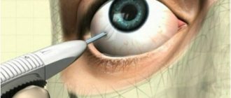
How to remove a nevus on the eye?
Until recently, the only safe treatment for nevus on the eye was its surgical removal. The operation was performed under a special microscope using a radioscalpel or microscalpel.
The use of a laser beam was considered dangerous by many experts, since it could damage neighboring eye tissues.
Currently, the development of modern laser technologies has made it possible to develop new methods of surgical treatment of nevus. And now it is increasingly being removed from the eye using laser radiation.
The main advantages of using laser technologies to remove a nevus are that they are painless, bloodless, and most importantly, the absence of a rough postoperative scar.
Progressive nevus
A progressive nevus is characterized by an increase in size and a change in its color. The surface of the nevus becomes variegated: along with non-pigmented or weakly pigmented areas, zones of intense pigmentation appear, the boundaries of the tumor become less clear due to the spraying of pigment. Accumulation of pigment can also be observed at a distance from the tumor. The nevus's own vessels expand sharply, and their number increases (Fig. 4.8).
Rice. 4.8. Progressive nevus. a - general view. b — histological specimen
The presence of a triad of signs: increased pigmentation, vascularization of the nevus and blurred boundaries makes it possible to differentiate the true progression of the tumor from its increase due to reactive epithelial hyperplasia. The appearance of limited displacement of the nevus in relation to the sclera is a late symptom indicating the development of melanoma.
Morphology
Nevi are represented by proliferating melanocytes that form cell nests in the basal layer of the conjunctival epithelium (the so-called border nevus).
During growth, nevus cells can penetrate the subepithelial layer, forming a mixed nevus. Borderline nevi are more often diagnosed in children; mixed nevi, especially in the area of the lacrimal caruncle, are usually detected in adults. If a border nevus is characterized by a flat shape, then a mixed nevus acquires a slight prominence.
The diagnosis of nevus is made on the basis of biomicroscopy.
Differential diagnosis is especially difficult for non-pigmented nevi. It is necessary to differentiate from lymphoid hyperplasia, sarcoidosis, papilloma, juvenile xanthogranuloma.
Treatment is indicated when signs of growth appear and consists of excision of the nevus. If the nevus is not completely removed, a relapse with transformation into malignant melanoma is possible. Information about the incidence of melanoma is contradictory.
According to Z.L. Stenko (1971), just over 40% of conjunctival melanomas develop from pre-existing nevi. F. Jacobiec et al. (1989) believe that about 20% of nevi become malignant. According to the latest information, the incidence of malignancy of conjunctival nevi reaches only 2.7%.
It should be remembered that malignancy of a conjunctival nevus can occur not only in the first years of its existence, but also decades later. F. Daxecker (1988) described a case of the development of conjunctival melanoma 58 years after the discovery of a nevus. In this regard, the operation should be performed using not only microsurgical, but also laser technology.
With adequate treatment, the prognosis is usually good for both vision and life.
Symptoms and diagnosis
The neoplasm has no external symptoms; a person does not feel it in the eye. Only when the nevus enlarges is observed:
The main diagnostic method is ophthalmoscopy. The doctor may also use:
Removal
Before removing a nevus in the eye, the doctor determines the nature of the neoplasm. Interfering birthmarks can be removed in several ways. The ophthalmologist chooses the method of excision of a dangerous tumor. Patients are given:
- electroexcision followed by plastic surgery of affected tissues;
- microsurgical operation;
- laser excision.
With progressive choroidal nevus, the individual characteristics of the patient are taken into account. Treatment tactics are influenced by:
- localization of the birthmark;
- tumor growth rate;
- age and condition of the patient;
- accompanying pathologies.
Progressive growths are removed using traditional microsurgery techniques or laser coagulation. Hard-to-reach moles are removed with laser. Removing problematic moles allows you to avoid the development of a cancerous tumor, the transformation of a nevus into melanoma.
In a child, a nevus is treated as a last resort when the eye mole grows rapidly.
Progressive medical technologies prevent the degeneration of eye nevi into cancerous tumors. Thanks to them, it is possible to preserve vision and health. The main thing is to seek medical help in a timely manner and undergo regular preventive examinations with an ophthalmologist.
Every person has nevi The number and location of their localization are different. It is believed that on average there are nine to fifteen moles on the body of an adult.
Sometimes a nevus can be located in the most seemingly inappropriate place, for example on the eyeball! In this case, nevus in the eyes occurs at any age. It can occur in both infants and the elderly.
What is the danger
Eye moles do not manifest themselves for many years and do not cause a person any discomfort. However, when certain factors come together, they begin to undergo transformation. Progressive nevus is the most dangerous type of eye mole. Changing pigment spots cause:
- deterioration and loss of vision;
- degenerate into a cancerous tumor - melanoma.
After discovering a birthmark, you should regularly visit an ophthalmologist. This will prevent the development of melanoma. In case of negative dynamics, the doctor will draw up a treatment regimen or decide to remove the nevus. You should consult a doctor immediately if the following complications occur:
- a mole makes it difficult to see;
- the quality of vision decreases;
- a foreign body is felt in the eye;
- the size and color of the spot changes.
With timely consultation with a doctor and proper treatment, the prognosis of the disease is favorable. A nevus in the eye transforms into cancer in 1 patient out of 500. Close attention must be paid to the spots:
- the thickening of which has reached 2 mm;
- with subretinal exudate;
- with orange pigmentation;
- located on the posterior disc of the eyeball.
How to treat
Typical nevi do not require intervention, only observation.
Typical nevi do not require treatment, only dynamic observation.
If growth and change in the nevus is observed over 6-12 months, surgical treatment is necessary. The same as when diagnosing an atypical nevus.
The operation performed is laser coagulation of the retina or photocoagulation.
Stationary and progressive nevi have a favorable prognosis. Atypical ones are considered as a malignant formation and the prognosis is made according to the behavior of the tumor.
Most Caucasians have moles (nevi). Pigment spots form on the body, face and even the eyeball. Nevi differ in shape, color, size, and location. A mole in the eye occurs in people belonging to different age groups. Nevus tumor is found in adults, elderly patients, adolescents and young children. Eye moles are benign.
Forecast and prevention of the disease
. The prognosis of the disease and the effectiveness of treatment directly depend on the following factors:
- Regular observation by an ophthalmologist and strict compliance with all his instructions.
- The type of disease and its severity.
- Correct placement of the eyeball.
- Age at which amblyopia was first identified.
- Initial visual acuity.
- Age of start of treatment.
- The therapeutic methods and regimens used.
The sooner the disease is detected and intensive treatment is started, the greater the chances of achieving success and restoring normal functions of the eyeball.
https://youtube.com/watch?v=qCUs7TRzoy8
Preventative measures include:
- Compliance with a rational regimen, regular good rest and dosed physical activity.
- Complete balanced nutrition. The diet should include foods containing B vitamins, ascorbic acid, and retinol. They help maintain good visual acuity.
- Walks in the fresh air and dosed exposure to a computer monitor or TV screen.
- Regularly visit an ophthalmologist and undergo preventive medical examinations 2 times a year.
- For children under 1 year of age, routine medical examination is required.
If a child under the age of 5 years is diagnosed with strabismus, constant medical observation by an ophthalmologist and intensive treatment are necessary. It is necessary to begin treatment as soon as amblyopia is identified.
Amblyopia disease
Originally posted 2018-02-07 12:16:06.