Therapy according to Dashevsky
Indications: correction and prevention of myopia, relieving spasm of accommodation. It is not performed for glaucoma, divergent strabismus, if the patient has only one eye. Performed in courses of ten sessions each. Combines with magnetotherapy, generation of color pulses and laser stimulation.
The patient sits down, the ophthalmologist places a diverging lens in front of him. The first setting is 0.5 dpt. The doctor waits until visual acuity begins to increase. The optical power is then increased by another 0.5 dpt. The doctor again expects an increase in visual acuity. Visual acuity after Dashevsky procedures increases significantly.
With regular 10-minute procedures, children's visual acuity increases. The duration of the course is three weeks. A good effect is enhanced by walks in the fresh air and physical exercise.
Violation of eye accommodation
- Spasm of accommodation is an acute pathological excessive tone of accommodation, causing a change in the existing refraction of the eye towards myopia (negative refraction) and reducing the maximum corrected (corrected) visual acuity.
- Habitually excessive tension of accommodation (HATS) is a long-term accommodative tone that causes myopization of the existing refraction and does not reduce the maximum corrected visual acuity. This condition is increasingly common among teenagers who are not limited in their communication with a computer, telephone, smartphone, or tablet.
- Paresis or paralysis of accommodation is an acute disorder of accommodation, in which a change in the optical system of the eye to any distance, due to a change in refraction, becomes temporarily impossible.
- Weakness of accommodation is a long-term state of insufficient or unstable accommodation.
- Accommodative asthenopia is a condition of a disorder of the visual system in which the performance of visual work becomes difficult or impossible due to a violation of the refractive and accommodation system.
- Presbyopia is a weakening of accommodation, which physiologically occurs after 40 years.
- Violation of accommodation after refractive surgery. Most often this is a decrease in near vision, rapid fatigue, difficulty in focusing and moving the gaze to a close distance.
What contributes to the disturbance of accommodation?
- Long-term work on a computer, tablet, phone, smartphone, TV.
- Doing homework in the evening.
- Poor workplace lighting.
- Computer games.
- Incorrect seating position at a workstation at home or school.
- Weakness of the neck and back muscles.
- Neglect of sports, exercise, physical education.
- Lack of proper daily routine.
- Abuse of any work at close range.
- Insufficient physical activity.
- Presence of somatic concomitant diseases.
Any work at close range involves constant tension of the accommodative apparatus. In children and young people, a disorder of the accommodative apparatus leads to false myopia. Violation of accommodation in children can aggravate existing visual disorders: myopia, farsightedness, astigmatism and amblyopia.
Violation of eye accommodation, symptoms
- Rapid fatigue when working close.
- The appearance of unpleasant sensations, burning, stinging, redness of the eyes, sometimes watery eyes or, on the contrary, a feeling of dryness.
- The image up close becomes less clear, ripples, pulsates, when you move your gaze into the distance, the image blurs, double vision may appear, and you cannot focus your vision on distant objects.
- Frequent headaches.
- Children get tired faster in school, which contributes to a decline in academic performance.
What is drug-induced cycloplegia in children and adults, principles of treatment
21.01.2019
In ophthalmology, there are 2 concepts of cycloplegia. The first involves drug therapy that dilates the pupil and creates paralysis of the ciliary muscle in the eye. The procedure involves instillation of mydriatics and cycloplegics. This helps to identify changes in the peripheral part of the fundus.
The second term means a disease in which paralysis of the ciliary muscle occurs. Therefore, it is necessary to distinguish what it is: drug-induced or conventional cycloplegia.
What is cycloplegia
Unlike drug-induced cycloplegia, the usual disease paralyzes the eyelash muscles. Therefore, it is difficult for a person to focus on a specific object. In this case, the sphincter of the pupil does not work.
Cycloplegia is uncommon in ophthalmology. It is important to notice changes in time. In the absence of proper treatment, a person’s nerve impulses are disrupted. Paralysis of accommodation appears. A person can no longer see an object, no matter how far or close it is.
The disease develops due to:
- Damage, severe trauma to the visual organs.
- Taking medications. These include “Atropine”.
In the following video, an ophthalmologist will talk about cycloplegia:
Symptoms
When pathology appears, a person experiences symptoms:
- decreased visual acuity when focusing into the distance;
- pain in the eyes, temples, forehead;
- as the disease progresses, the sharpness of near vision decreases, it is difficult for a person to read;
- rapid fatigue when working if objects are located close to the eyes.
The disease is otherwise called false myopia or tired eyes syndrome. Cycloplegia occurs more often in adolescent children. According to statistics, every 6th child is diagnosed with this pathology.
The child does not like to constantly keep a distance when working close to an object. As the distance changes, the clarity of vision deteriorates.
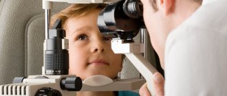
How to diagnose
Diagnosis of cycloplegia includes examining the medical history and interviewing the patient. The examination includes:
- Visometry. Visual acuity is determined.
- Vision test using a phoropter.
- Refractometry. The refraction of the eye and changes in the direction of vision are determined.
- Medication-induced cycloplegia. The condition of the peripheral areas of the fundus is studied. A solution of atropine sulfate is dripped into children and adults.
- Ophthalmoscopy. Prescribed if it is necessary to identify additional eye pathologies. The entire fundus of the eye is examined.
Treatment
In most cases, when prescribing treatment, it does not matter what cause contributed to the development of cycloplegia. Usually, various methods of treating the disease are used. But, if cycloplegia is diagnosed due to refractive error, vision correction is performed with glasses or surgery.
When selecting spectacle vision correction, the main thing is to take into account the individual development of cycloplegia. In cases where the cause of the disease is underdevelopment of the ciliary muscle, treatment methods are prescribed:
- performing eye exercises using the Avetisov method;
- use of Dashkovsky diverging lenses.
Dashevsky's method
The treatment is supervised by an ophthalmologist. Let's consider the sequence of actions of Dashevsky's method:
- The man sits down. A diverging lens is placed in front of the eyes. The first stage involves using a lens with a diopter of 0.5.
- The muscles contract reflexively. Because of this, visual acuity increases. Then the diopter increases by 0.5.
So the sharpness of the diopters gradually increases. Systematic exercises help improve visual acuity.
Exercises are carried out daily for 10 minutes per procedure. The duration of the course of treatment is prescribed individually. This usually takes 2–3 weeks. After rest, another course is carried out. It is important to maintain the result with restorative therapy. A person is recommended to engage in physical activity outdoors.
Also see another method of improving vision according to Dashevsky:
Medical therapy according to Avetisov
Treatment of cycloplegia according to Avetisov involves performing the following exercises:
- A round shaped mark with a diameter of 5 mm is glued to the window.
- The person is seated 30 cm from the mark.
- The gaze is directed through the mark to an object that is located in the distance.
- A person should focus his gaze on the mark, and then on an object in the distance. This needs to be done several times.
The exercise is done every day, 10 minutes is enough. The duration of the procedures does not exceed 3 weeks. The course is repeated after 15 days of rest.
In addition to viewing, eye exercises using the Avetisov method:
Cycloplegia in ophthalmology is considered a disease that is difficult to treat. The main thing is to promptly treat the disease in school-age children, since the vision situation may worsen in the future.
Source: https://ozrenieglaz.ru/bolezni/drugie/tsikloplegiya
Pathogenesis
The oculomotor, trochlear and abducens nerves located in the eye muscles are affected. The ability to transmit nerve impulses is impaired, and the tone of the external muscle fibers is lost. The eyes do not respond to the body's commands and stop moving. There is no pupillary reaction, since the function of the fibers innervating the dilator and sphincter of the pupil is impaired.
Causes
The main causes of cycloplegia are damage to the ciliary muscle, any trauma, blows and ruptures. There is also a risk of developing cycloplegia when taking atropine.
The patient begins to have trouble seeing into the distance. Near vision remains normal for some time. As the disease progresses, near vision also deteriorates. The patient notes severe eye fatigue, and pain in the temples may occur.
Therapy according to Dashevsky
Indications: correction and prevention of myopia, relieving spasm of accommodation. It is not performed for glaucoma, divergent strabismus, if the patient has only one eye. Performed in courses of ten sessions each. Combines with magnetotherapy, generation of color pulses and laser stimulation.
The patient sits down, the ophthalmologist places a diverging lens in front of him. The first setting is 0.5 dpt. The doctor waits until visual acuity begins to increase. The optical power is then increased by another 0.5 dpt. The doctor again expects an increase in visual acuity. Visual acuity after Dashevsky procedures increases significantly.
With regular 10-minute procedures, children's visual acuity increases. The duration of the course is three weeks. A good effect is enhanced by walks in the fresh air and physical exercise.
Avetisov's technique
A round mark with a diameter of 5 mm is glued to the window. The patient sits from the window at a distance of 0.3 meters.
The ophthalmologist determines a distant object based on the direction of view. The patient needs to focus on the mark, looking far and near.
The exercise is done every day. Duration 10 minutes.
The course lasts three weeks, then after a break of 15 days, treatment of the disease continues.
Phones
Drug cycloplegia is used in ophthalmology to dilate the pupil and create paralysis of the ciliary muscle. Such measures are required to examine the periphery of the fundus. However, cycloplegia is also a disease that manifests itself in the form of paralysis of the ciliary muscle. The disease provokes the inability to focus on objects.
Why does pathology develop?
The disease can occur together with other ailments. The following factors mainly cause the development of cycloplegia:
- damage to the visual organs due to a strong impact;
- use of medications, for example, Atropine.
What symptoms indicate illness?
The first sign that indicates the occurrence of cycloplegia is deterioration in distance vision. The patient sees normally up close.
However, if the pathology is not treated in a timely manner, over time, patients lose the ability to read at close range. That is why this disease is also called tired eyes syndrome or false myopia.
The pathology is often diagnosed in children and adolescents.
In addition, cycloplegia causes increased fatigue when interacting with objects that are at close range. Patients complain of difficulty focusing their gaze. Pain syndrome in the temporal region is often observed. Pain can also occur in the forehead area, as well as in the eyes.
Diagnostic measures
If a person's vision deteriorates and there is a suspicion that cycloplegia has occurred, it is important to visit a medical facility as soon as possible. At the appointment, the doctor will conduct a survey of the patient, during which he will find out how long ago visual functions have deteriorated and what symptoms are troubling the patient.
To confirm the preliminary diagnosis, the doctor evaluates visual acuity and performs ophthalmoscopy. Sometimes additional diagnostic measures are required.
How is the treatment carried out?
Treatment methods are related to the cause that contributed to the development of the pathology. If age-related changes are to blame for the disease, they resort to wearing glasses or surgical intervention.
When the disease is caused by underdevelopment of the ciliary muscle, patients are prescribed to wear special glasses with holes and lenses, as well as exercises to train the muscles.
How is the disease treated?
Treatment of paralysis of accommodation is prescribed only after confirmation of the diagnosis by a medical specialist and depends on the diagnosis of the primary disease.
In case of poisoning, detoxification therapy is carried out. It is necessary to treat diseases that lead to general intoxication of the body.
When it is difficult to immediately determine the cause, the doctor creates conditions to relieve tension from the damaged eye. Suitable, sometimes specialized, lenses or glasses are selected. Miotics, drugs for constricting the pupils, are prescribed to be instilled into the conjunctival sac.
Paralysis of accommodation of traumatic origin does not require special treatment. Over time, its effects disappear in whole or in part. This depends on the location and severity of the injury. Treatment can only slightly reduce inflammation and relieve pain if the patient complains of it. If the cause is neurological, therapy is prescribed aimed at getting rid of or remission of this disease. As a rule, after recovery from the underlying disease, paralysis of accommodation also heals on its own.
If necessary, laser and surgical intervention is used.
When should mydriatics not be used?
They are contraindicated in the presence of the following pathologies:
- glaucoma (as drops increase intraocular pressure);
- diabetes mellitus, which leads to diabetic nephropathy and chronic renal failure (medicines will be slowly eliminated from the body);
- arrhythmia, angina (mydriatics increase heart rate);
- thyrotoxicosis (with this disease the level of heart rate is also increased).
Mydriatics are contraindicated for pregnant women and women during breastfeeding due to the fact that toxic substances can get into the milk. Vasodilator drugs are rarely prescribed to children.
If necessary, the doctor resorts to instilling a very small dose of drops into the child's eyes.