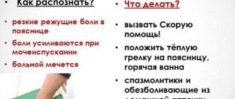
Renal colic according to the “pain scale”
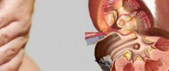
Stones in the ureter are always a problem. The movement of a stone along the ureter can cause severe pain attacks. Patients figuratively describe their feelings, saying that sometimes they “climb the wall.” This is not a figure of speech, but a real description of the strength of the colic that occurs. According to the “pain scale”, which doctors compiled when comparing the pain symptoms of diseases, renal colic ranks first.
Pain from stones passing through the ureter or stopping at places of anatomical narrowing is highly intense
In addition to severe pain, renal colic often causes painful urination, increased blood pressure, nausea, vomiting, impaired bowel function and other negative consequences.
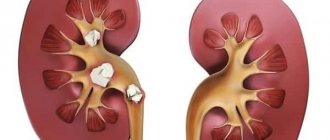
Symptoms of coral lithiasis
There are several stages of development of coral stones (coral lithiasis):
- Hidden
- there are no manifestations of the disease. There may be minor pain in the lower back. - Initial
- periodic increases in temperature and pressure, thirst, weakness, and minor swelling of the face are added. - Clinical
– constant pain in the lower back, loss of strength, and urinary problems appear. There are traces of blood and small pebbles in the urine - broken fragments of coral. - Hyperazotemic
- renal failure increases, edema appears, temperature rises, and blood pressure surges are observed. When pyelonephritis occurs, pus is detected in the urine, and leukocytes in the blood increase and ESR accelerates. The expansion of the pelvis leads to hydronephrosis - the accumulation of urine inside the kidney. Acute urinary retention occurs, requiring urgent hospitalization.
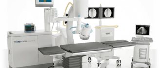
Techniques for crushing kidney stones
The solution to the problem of radically getting rid of stones was the procedure of crushing them. Indications for use:
- Large stones, ranging in size from 6 mm in diameter.
- Crystalline stones, the sharp edges of which cause spasm of the urinary tract and pain.
- An inflammatory process in the kidneys or ureter, which is supported by the presence of a stone.
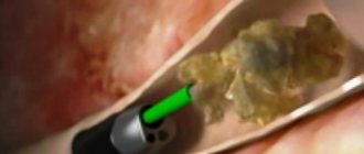
Contact crushing is the most effective method, which often does not require opening the abdominal cavity. The stones are destroyed as a result of wave action. In modern medicine, various wave sources are used:
- electricity;
- pneumatics;
- ultrasound;
- laser generator.
Uclinica has chosen the two most effective methods of crushing stones: ultrasonic and laser.
Contact crushing of stones in the ureter is low-traumatic; in many cases, one day is enough to prepare, carry out the procedure and primary rehabilitation
The main contraindications for all types of contact lithotripsy are systemic problems of the body: tumors, exacerbations of a number of chronic diseases, and others. Before prescribing the procedure, a general examination of the patient is carried out.
What you need to know about the postoperative period
The cost of surgery to remove kidney stones in our clinic includes the patient’s hospital stay for at least 3 days. After the operation is completed, the patient is under the supervision of a doctor in a separate room. A catheter is placed in the patient's bladder - it is important that it remains there for at least 3 days. At this time, the medical staff performs all necessary procedures and injections, monitors the condition - it is important that the patient remains in the hospital for the entire required period. If a stent was installed during surgery, it will be removed after some time after completion of therapy.
After the procedure, the patient may experience pain during urination and blood in the urine. This feeling is normal for this period.
However, there are a number of symptoms that indicate that you need to seek medical help.
These include:
- temperature increase;
- vomit;
- severe pain in the side or pubic area;
- problems with urination - drip or complete absence;
- severe bleeding from the urethra.
In any of these cases, you should report your symptoms to your nurse and get help from a doctor as soon as possible.
Ultrasound: crushing and cleaning
The ultrasonic crushing technique not only destroys the stone, but also gets rid of its fragments. After the foreign body is destroyed by ultrasonic waves, liquid is supplied to the kidney or ureter, which is immediately removed using suction from the manipulator. The escaping liquid captures all the destroyed stones, completely removing them from the body. The kidney or ureter is cleared of the foreign body and all its remains, fragments and sand.
Advantages of the technique:
- Effective crushing of stones.
- Quickly cleans the canals from foreign bodies and their traces.
- Minimal risk of leaving small fragments of stone in the kidney.
- Less traumatic.
- Reasonable price.
Disadvantages of the technique:
- The size of the instrumentation does not allow the use of contact ultrasound lithotripsy for all parts of the ureter.
Contraindications:
- Position of the stone in the ureter. As a rule, its upper (or middle) third is inaccessible for contact ultrasonic crushing.
- Anatomical indicators. In all other respects, ultrasonic crushing can be safely called an ideal method for removing stones, completely eliminating foreign formation.
Pros and cons of the procedure
Removing kidney stones with a laser, the price of which is quite affordable, is a low-traumatic procedure. After the doctor prepares patients for it by answering all the questions, most of them do not feel fear.
The advantages of laser treatment are that:
- there is no need to use anesthesia - the patient does not experience pain;
- therapy is effective even for super-strong stones;
- To remove all kidney stones, one session is enough;
- the likelihood of re-formation of stones is small;
- there are no scars after the procedure;
- immediately after completion, the patient experiences relief - the process of urination is normalized.
In addition to the advantages, there are also disadvantages, which we honestly warn our patients about. The laser is not able to crush large stones - more than 20 mm. It is possible that a piece of stone may get stuck in the urinary tract upon exiting - the risk is small, but nevertheless it exists.
If we compare the ratio of advantages and disadvantages, then the laser procedure has a good effect for most patients. However, the final decision on its prescription is made by the doctor based on the results of tests and examination using ultrasound.
Laser: new generation technologies
Laser is the most powerful source. With its help, it is possible to crush stones of any density in all parts of the ureter. Nothing is impossible for a laser; it can handle all hard-to-reach places, difficult locations, stones of all characteristics, shapes and sizes.
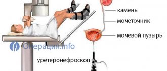
Thanks to the flexible urethroscope, you can crush stones even in the most difficult to reach places
Advantages of the technique:
- Possibility of removing all stones of any location.
- Less traumatic.
- No contraindications (except for general ones).
Disadvantages of the technique:
- High price.
Not only the laser equipment itself is expensive, but also the consumables for each operation. Therefore, not every medical institution can purchase and use a laser lithotripsy device.
At Uclinica there is such an opportunity, so our patients have access to a laser procedure for crushing stones in the ureter.
The clinic uses the most effective and safe techniques: laser and ultrasound lithotripsy. Specialists will remove even a large stone in any location and return patients the joy of a full life without pain.
Endoscopic treatment of ureteral stones
ERSHOV A.V.
Krasnodar State Medical University, Department of Urology
Krasnoyarsk
Currently, there is a worldwide trend towards an increase in the incidence of urolithiasis (UCD). In recent years, this disease has occurred in at least 1–3% of the world's population. According to the Krasnoyarsk Regional Medical Information and Analytical Center, up to 8% of the region’s population suffers from urological diseases, the growth rate is 13.9%. It should be noted that in the region since 2003, the number of patients with urolithiasis has increased by 29.6% and currently amounts to 7.4 cases per 1000 people, more than 16 thousand patients with urolithiasis are registered annually.
Among the various forms of manifestation of urolithiasis, the most common are ureteral stones, which now (due to the introduction of radiotherapy) account for more than 35% of clinical cases. It is also worth noting that renal colic caused by a ureteral stone in 89.5% of cases is the diagnosis of hospitalization of patients for emergency reasons. Despite their apparent simplicity, it is ureteral stones that cause the most pronounced pain, suffering and fear in patients, and most often they can lead to the development of severe complications.
Ureteroscopy is widely used in clinical practice, especially in the treatment of urolithiasis, in particular ureteral stones. Even after the introduction of extracorporeal shock wave lithotripsy (ESWL), it has not lost its significance. This is explained by the fact that not all stones can be crushed using ESWL due to the peculiarities of localization, duration of stay in the ureter and other reasons, and the “stone path” formed after ESWL also requires the use of endoscopic intervention. On this slide you can see the effectiveness of KUL in the treatment of ureteral stones according to various urological societies: the Russian Society of Urology, the European Association of Urology and the American Urological Association.
Endoscopic Contact ureterolithotripsy and lithoextraction allow:
Relatively quickly relieve the patient from a ureteral stone (from long-term physical and psychological suffering)
Reduce treatment costs and recovery period, the duration of which is reduced by 3-5 times
In this connection, the goal was set: To identify diagnostic criteria that would improve the effectiveness of endoscopic treatment of patients with ureteral stones
Material from 153 operations for ureteral stones in patients undergoing treatment in 2011 was analyzed. on the basis of the Road Clinical Hospital at Krasnoyarsk station of JSC Russian Railways
Thirty-two patients were operated on using traditional open ureterolithotomy, and CLT ureteroscopy was performed in 121 cases. Among the patients there were more men - 85 people, women - 63 people. It should also be noted that patients of working age (from 21 to 50 years) were more than 56.7% (87 people), which indicates the socio-economic significance of the problem.
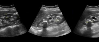
To establish a diagnosis, determine indications and draw up an operation plan, a comprehensive urological examination was carried out, including the study of complaints and anamnesis, physical examination, clinical blood and urine tests, bacteriological analysis of urine with identification of the pathogen and determination of sensitivity to antibacterial drugs, as well as: standard ultrasound a study during which the size, location of the stone and changes in the kidney parenchyma were determined.
Peristalsis of the ureter during its obstruction was studied using color Doppler ultrasound. Using this method, the nature of obstruction (complete, incomplete), the number of emissions per unit of time on both sides and separate diuresis of each kidney were determined.
Survey and excretory urography. Using this technique, the presence or absence of urinary bladder obstruction was determined, the function of the affected and contralateral kidney was assessed, as well as the severity of the degree of expansion of the collecting system
An imaging method that allows you to choose the optimal method for surgical removal of ureteral stones is MSCT.
Diagnostic criteria for which:
Changes in the renal parenchyma
Anatomical and functional structure of the VMP
Nature and severity of changes
Dimensions, location, configuration, density of stones
Changes in the wall of the ureter at the site of contact with the stone
According to dynamic nephroscintigraphy, a secretion deficiency of 51 to 70% was determined in 8 (25.0%) patients.
In case of pronounced pathological changes according to ultrasound, R-studies and dynamic scintigraphy, traditional open ureterolithotomy with revision of the affected kidney is indicated
The main criteria for choosing the type of endoscopic intervention are the size, density and shape of the stone, as well as the duration of its presence in the ureter. Ureterolithoextraction was used to remove small mobile stones or after contact lithotripsy of a large stone.
Ureteroscopy and contact lithotripsy were performed in cases where the size and shape of the stone did not allow lithoextraction, as well as, regardless of size, for occlusive stones that had been in the ureter for a long time. It was carried out using pneumatic, electrokinetic and laser lithotripters. For stones located in the upper third of the ureter, preference was given to traditional ureterolithotomy, while stones in the middle third and lower third were removed using the endourological method.
Stones in the upper third of the ureter were present in 24 patients; in the middle third – 37; in the lower third – in 92 patients; Of these, 6 patients had 2 stones in the ureter, 3 had 3, and 14 were diagnosed with a “stone path” formed after DLT. X-ray negative stones were present in 13 patients.
In 48 cases, contact lithotripsy of the stone was performed, in 73 cases, ureterolithoextraction was performed, and in 2 (2.0%) cases, the stone of the lower third of the ureter migrated into the pyelocaliceal system during lithotripsy. These patients underwent fibroureteropyeloscopy with contact laser lithotripsy and lithoextraction.
In 80 cases, postoperative drainage of the upper urinary tract was performed with a ureteral catheter, in 7 cases - with a stent. The ureteral catheter was installed after complete disintegration of the stone in the absence of migration of large fragments into the cavity of the renal collecting system.
In 22 patients, the presence of a stone was combined with a ureteral stricture (in these patients, ureterolithotripsy and ureterolithoextraction were performed after preliminary dissection of the stricture using a laser).
COMPLICATIONS:
Exacerbation of chronic pyelonephritis 3
Perforation of the ureteral wall 1
In the open ureterolithotomy group, urodynamic disturbance on day 5 was 27.3%, while in the endourological surgery group, urodynamic disturbance on day 5 was only 7.6% (9 people)
Thus, ureteroscopy was effective in 98.3% of cases. In 2 (1.7%) cases, ureteroscopy was not possible due to severe deformation of the ureter in the middle third.
Considering the high efficiency, low morbidity, short period of postoperative clinical and labor rehabilitation of patients, the CULT method is the operation of choice in the treatment of patients with urolithiasis with stones in the lower third of the ureter with a density of over 1000 HU in the absence of severe concomitant pathology.
Topics and tags
Young scientists
Comments
To post comments you must log in or register