Basic methods of treating urolithiasis or how to treat urolithiasis
Small stones
In the event that the stone is small (that is, up to 4-5 mm - although some of the patients, I’m sure, will argue with me!), the pain is easily relieved with the help of drugs and there are no complications (fever, complete kidney block), you can carry out active drug treatment of urolithiasis, aimed at spontaneous passage of the stone and prevention of complications.
Large stones
Treatment of large stones is no longer possible without surgical intervention, especially in the case of pronounced pain. Modern treatment of urolithiasis involves the following types of operations:
External beam lithotripsy or EBRT
External lithotripsy of ureteral stones (EBLT) is the crushing of kidney stones using special equipment. Allows you to destroy the stone into small fragments without the need for surgical intervention. Fragments of the stone pass away on their own during urination, while the kidney itself and nearby tissues and organs are not affected.
Modern medical devices for crushing kidney stones allow the patient to be discharged immediately after DLT without the need for hospitalization.
Lithotripsy machine with dual guidance system
In addition, my clinical base allows us to crush stones on an emergency basis, on the day of treatment.
By ordering the service of emergency crushing of kidney stones on the day of your visit, you will receive:
- the ability to complete the procedure without unnecessary formalities
- lowest possible prices for crushing kidney stones in Moscow
- safety and guarantees of positive results.
Contact lithotripsy
Fibrourethroscopy is a radical surgical removal of stones. This treatment of urolithiasis is carried out when complications occur (for example, acute pyelonephritis).
The essence of the operation: a special thin instrument with an optical system and a video camera is inserted into the urethra, and under the supervision of a surgeon, it is carried into the ureter and brought to the stone. Next, the stone is destroyed by contact method using a pneumatic probe or laser.
Duration of hospitalization: 24 hours.
Fiber urethroscope Richard Wolf Cobra
Percutaneous lithotripsy
It is performed when the stone is large, when even when destroyed, the fragments cannot pass through the ureter.
The essence of the operation: the instrument is inserted directly into the kidney through a puncture in the skin, the stone is destroyed and removed.
Duration of hospitalization: 3 - 5 days.
Endoscopic removal of stones from the ureter (contact lithotripsy)
The standard access for this operation involves the formation of a canal with a diameter of up to 10 mm, but nowadays mini-access (with a diameter of up to 5-6 mm) is increasingly used. Percutaneous nephrolithotripsy is contraindicated in patients receiving anticoagulant therapy. Anticoagulants should be stopped before surgery (if possible).
Other contraindications for percutaneous crushing of kidney stones include untreated urinary tract infections, tumors in the surgical canal area, potentially malignant kidney tumors, and pregnancy. Careful detailed preoperative visualization of the stone and elements of the kidney and upper urinary tract is important for the safe and successful performance of percutaneous nephrolithotripsy. The standard and most informative method is multispiral computed tomography of the kidneys with intravenous enhancement.
Percutaneous nephrolithotripsy is usually performed under general anesthesia and can be performed with the patient in either the supine or prone position. In the supine position, the surgeon has the opportunity, if necessary, to carry out transurethral access to the kidney and ureter. Most urological surgeons perform the operation under X-ray guidance, although ultrasound (or a combination of these methods) can also be used. Traditionally, percutaneous nephrolithotripsy ends with the installation of a nephrostomy, although with the use of a mini-access, a small volume of surgery and the absence of complications, drainage-free completion of the intervention is also possible. Complication rate of percutaneous nephrolithotripsy (based on observation of about 12 thousand patients): fever 10.8%, blood loss with the need for blood transfusion 7%, pleural damage, pneumothorax 1.5%, sepsis 0.5%, damage to adjacent organs 0.4% , fatal outcome 0.05%.
Treatment methods
For small kidney stones (no more than 5 mm), the prognosis is favorable: the stone can pass out on its own naturally in the urine, without causing discomfort to the patient.
For large stones, both conservative and surgical treatment can be used*.
A conservative method of treating ICD is litholytic therapy. It is effective for urate stones. Since they are formed in an acidic environment, alkalizing the urine with special medications (citrate mixtures) leads to their gradual dissolution.
To combat stones that cannot be dissolved with medications, three surgical methods are used:
- 1. Remote lithotripsy.
The most modern and most gentle method for the patient. People call it “crushing kidney stones.” The main advantage is the absence of direct penetration into the body. A shock wave is sent to the stone, under the influence of which, in about 40 minutes, it collapses into dust and is excreted in urine. - The process is painless for the patient and is carried out even in a clinic. After the session you can go home straight away. However, this method is not suitable for stones with a density of 1000 Hounsfield units.
- 2. Percutaneous surgery.
The same crushing of stones, but using the contact method, i.e. with violation of the integrity of organs. To do this, special instruments are inserted into the ureter, the surgeon sees the stone on the monitor and crushes it with a laser, and then, using special gripping devices, removes its fragments. This operation refers to the so-called one-day surgery: after it, the patient spends one night in the hospital under the supervision of a doctor. - 3. Laparoscopy.
When the stone is removed entirely, without crushing, by capturing it with special surgical instruments. But if earlier open surgery was used for this (i.e., an incision was made with a scalpel in the peritoneum, and then the ureter or renal pelvis), now the operation is performed in a minimally invasive way - through several punctures on the abdomen or in the lumbar region, which significantly speeds up the rehabilitation process. Until now, in some cases this method is the most effective.
*Please note that the decision to choose one or another treatment method is made by doctors, depending on the individual indications and contraindications of a particular patient.
Recommendations of the European Association of Urology for the treatment of urolithiasis
For a patient with an acute attack of renal colic, the first step is to relieve pain. Non-steroidal anti-inflammatory drugs are recommended:
- Metamizole,
- Paracetamol,
- Diclofenac,
- Indomethacin,
- Ibuprofen
The latter two drugs are also used to reduce the frequency of pain relapses after an attack. Long-acting alpha-blockers (for example, Tamsulosin) can be prescribed for the same purpose. When using drugs, you need to remember about contraindications and side effects.
In case of recurrent attacks of renal colic and low effectiveness of drug treatment, it is recommended to immediately drain the kidney using an internal stent or puncture nephrostomy, as well as removal (crushing) of the ureteral stone.
Dissolving urinary stones (or their fragments) can only be effective if they are composed of uric acid. Dissolution is achieved by alkalinizing the urine using chemicals (potassium citrate or sodium bicarbonate). Stone-dissolving therapy is carried out under mandatory monitoring of urine pH, the target value is 7.0 - 7.2. Excessive alkalization can lead to the formation of phosphate stones. In the process of treating urolithiasis, it is necessary to observe a urologist, monitor urine tests and ultrasound of the kidneys (or MSCT).
The effectiveness of extracorporeal lithotripsy depends on the lithotripter used, crushing modes, size, location and density of the stone, as well as on the characteristics of the patient (for example, obesity). When crushing large stones, pre-installation of an internal stent may reduce the risk of stone walk formation.
Modern aspects of drug treatment of patients with urolithiasis
The article analyzes modern trends in drug treatment (antispasmodic, lithokinetic, citrate therapy) for patients suffering from urolithiasis. Despite the development of modern treatment methods, there remains a need for the use of pharmacological drugs, since their use reduces the risk of recurrent stone formation by correcting biochemical changes in the blood and urine, and also promotes the passage of stones.
Table 1. Medicines used in the treatment of patients with urolithiasis
Table 2. Drug treatment of patients with urolithiasis and specific urinary disorders
Urolithiasis (UCD) occupies one of the leading places in the structure of urological diseases, which is due to its prevalence and tendency to recur. According to official data from the Russian Ministry of Health, for the period 2002–2012. the absolute number of patients with urolithiasis increased by 25.1% and amounted to at least 787,555 people. In approximately 65–70% of cases, the disease is diagnosed in people aged 20–25 years, that is, in the most productive period of life. According to global data, the prevalence of urolithiasis varies from 1 to 20%. In countries with a high standard of living, such as Sweden, Canada or the USA, the incidence of urolithiasis exceeds 10% [1–4]. It is known that the most common are calcium stones, which account for at least 80% (85-90% - calcium oxalate, 1-10% - calcium phosphate, 5% - oxalate and calcium phosphate in combination with uric acid), Uric acid (5–10%), struvite (5–15%) and cystine (1–3%) stones are less common [5, 6].
Modern technologies of remote lithotripsy and X-ray endoscopic surgery (percutaneous nephrolithotripsy, ureteroscopy) make it possible to most effectively rid the patient of stones. However, none of the existing methods for removing urinary stones solves the problem of stone formation, which requires complex methods of treatment and metaphylaxis in the postoperative period [7, 8]. Various metabolic disorders and types of stone formation determine the use of certain medications and their combinations in the complex treatment of patients with urolithiasis (Tables 1 and 2) [9].
An important step in the treatment of patients with urolithiasis is the relief of renal colic using combinations of various drugs: metamizole, diclofenac, indomethacin, ibuprofen, tramadol. According to the recommendations of the European Association of Urology 2021, when choosing a first-line treatment drug, preference should be given to non-steroidal anti-inflammatory drugs. They effectively relieve pain in patients with renal colic and are superior to opiates in their analgesic effect. However, it must be taken into account that diclofenac and ibuprofen increase the risk of cardiovascular complications. Diclofenac is contraindicated in patients with heart failure, coronary artery disease, peripheral arterial disease and cerebrovascular pathology. If there is a high risk of cardiovascular complications, diclofenac is used only in emergency situations. Due to the fact that increasing the dose and duration of therapy increases the risk of complications, drugs should be prescribed in the lowest doses and for the shortest possible period of time [10–12].
To relieve an attack of renal colic, antispasmodics are also used, which promote the passage of small stones and also reduce tissue swelling in the area where the stone is located. The myotropic antispasmodic drotaverine (No-shpa) has found the greatest application in urological practice. The drug selectively blocks phosphodiesterase 4 contained in smooth muscle cells of the urinary tract, which leads to an increase in the concentration of cyclic adenosine monophosphate. The latter is associated with muscle relaxation, reduction of edema and inflammation, in the pathogenesis of which phosphodiesterase 4 is involved [13].
An important aspect of the management of patients with nephrolithiasis is the correct selection of drug expulsive (lithokinetic) therapy that promotes the passage of stones or their fragments. Lithokinetic therapy includes two main groups of drugs: alpha-blockers and herbal diuretics.
Tamsulosin is one of the most commonly used alpha-1 blockers in urology. The pharmacological effect of the drug in urolithiasis is due to a decrease in the tone of the smooth muscles of the urinary tract, which leads to the relief of an attack of renal colic, facilitates and accelerates the passage of ureteral stones [14, 15]. The clinical effectiveness of this group of drugs is also confirmed by studies demonstrating an increase in the incidence of stone passage when taking doxazosin, terazosin, alfuzosin and silodosin. The administration of tamsulosin and nifedipine is safe and effective for patients with renal colic and stones localized in the distal ureter. However, tamsulosin is much better than nifedipine in stopping an attack of renal colic, facilitating and accelerating the passage of ureteral stones [16, 17].
To this day, such a direction of conservative treatment as the use of herbal preparations remains relevant. They apply:
- to prevent relapses of stone formation;
- improving the spontaneous passage of stones, including their fragments after extracorporeal lithotripsy;
- prevention of exacerbations of chronic inflammatory diseases of the genitourinary system, mainly chronic pyelonephritis [18].
The advantage of herbal medicines is the minimal number of side reactions and unwanted drug interactions, as well as the possibility of long-term use, which is very important in the metaphylaxis of recurrent urolithiasis.
Phytotherapy with essential oils (terpenes) has been used effectively in the treatment of patients with urolithiasis for a long time (more than 60 years). The lithokinetic effect of this group of drugs is due to a combination of diuretic, anti-inflammatory and antispasmodic properties. The main pharmacological effect of terpenes is to relieve spasm of the smooth muscles of the collecting system and ureter, as well as to increase renal blood flow, which leads to increased diuresis. In addition, terpenes in high concentrations have a bacteriostatic effect.
One of the drugs, which is based on a special combination of plant terpenes, is Rowatinex (Medintorg). The drug contains anethole 4 mg, borneol 10 mg, camphene 15 mg, pinene (alpha and beta) 31 mg, fenchone 4 mg, cineole 3 mg. Anethole, borneol and camphene have a diuretic, anti-inflammatory (antibacterial) effect, and also increase renal blood flow. Pinene (alpha and beta) and fenchone are responsible for the antispasmodic and anti-inflammatory (antibacterial) effect, cineole - for the anti-inflammatory (antibacterial) effect. It has been established that Rovatinex has an antispasmodic effect, promotes the passage of stones and their fragments, reduces the severity of pain, increases renal blood flow, improving renal function and increasing diuresis. Combining anti-inflammatory and antimicrobial properties against gram-positive and gram-negative microorganisms, Rowatinex enhances the pharmacological effect of antimicrobial drugs. Prescribing the drug increases the percentage of stone fragments passing after extracorporeal lithotripsy while reducing the intensity of the pain syndrome. The drug Rovatinex helps reduce leukocyturia, increase daily diuresis and stabilize urine pH. Taking the drug is not accompanied by the development of complications or side effects, which makes it possible to take it for a long time as part of complex lithokinetic therapy and for the metaphylaxis of recurrent stone formation [19].
The herbal combination drug Canephron N (Bionorica) has proven itself as a component of lithokinetic therapy and metaphylaxis of urolithiasis. The essential oils and phenolcarbolic acids contained in Canephron N have an antiseptic, antispasmodic, anti-inflammatory effect on the organs of the urinary system, have a diuretic effect, and potentiate the effect of antibacterial therapy [20].
The group of herbal medicines also includes the dietary supplement NefraDoz (Stada), a phytocomplex of seven plant compounds that have a lithokinetic effect. It has been established that NefraDoz statistically significantly reduces the density of urine due to the diuretic effect, reduces bacteriuria and leukocyturia, reduces the concentration of urates, oxalates and phosphates, and also increases the frequency of passage of stone fragments after extracorporeal lithotripsy while reducing the intensity of pain [21]. In addition, NefraDose contains the plant polyphenol resveratrol. Resveratrol is characterized by antitumor, neuroprotective, cardioprotective, anti-inflammatory, antidiabetic and antibacterial effects.
If relief of renal colic and restoration of impaired urine passage cannot be achieved with medications and there is no effect from conservative therapy, they resort to drainage of the upper urinary tract or emergency disintegration (lithoextraction) of the stone using remote lithotripsy, ureteroscopy, etc.
Particular attention should be paid to patients with urate nephrolithiasis, since they may have chemolytic dissolution (citrate litholysis) of stones. This process is based on alkalinization of urine through oral administration of citrate mixtures or sodium bicarbonate. The main indicator of a properly occurring process of chemolysis (litholysis) is the dynamics of urine pH. It is necessary to maintain urine pH in the range of 6.2–6.8, and citrates should be prescribed and dosed not according to a single urine pH test, but according to the average daily pH fluctuations (morning, lunch, evening). Within these pH values, chemolysis is most effective, and at higher pH values, the risk of calcium phosphate stone formation increases [22]. Alkalinization of urine with parallel use of tamsulosin allows one to achieve a higher rate of complete clearance of stones located in the distal ureter [23].
Drugs for citrate therapy include Blemaren (Esparma). The composition of the drug is dominated by citric acid, and a significant part of the buffering function is performed by potassium hydrogen carbonate. The reduced sodium content in the drug promotes accelerated dissolution of uric acid in the renal tubules and prevents their further crystallization. The above properties provide a dose-dependent shift in urine pH from acidic to neutral or alkaline, while the acid-base state of the blood does not change. Citrate binds calcium ions from the gastrointestinal tract, where it reduces calcium absorption, to the urinary tract, where this effect is most active due to the highest concentration of citrate. In addition, by stabilizing solutions, citrate prevents crystallization processes in the urine. The complex effect of citrate on the physicochemical state of urine leads to an increase in the solubility of urates, calcifications and, first of all, oxalates, complex magnesium-ammonium phosphates and some other salts, helping to inhibit stone formation and dissolve already formed stones [24].
The drug Uralit-U (Madaus) has also proven itself as one of the drugs for litholysis. Uralit-U is a mixture of alkaline salts that combine with weak acids. The pharmacological effect of the drug is based on regulating the pH of urine and long-term maintenance of the urine reaction within the alkaline range (pH 6.2–7.5), at which uric acid salts are in solution and do not form stones. The salt of alkali metals and a weak acid, excreted in the urine, shifts the urine pH to the alkaline side (to 6.2–7.5), which determines an increase in the degree of dissociation and solubility of uric acid. The bioavailability of Uralit-U is about 100%. The drug is quickly and almost completely absorbed from the digestive tract. It is excreted from the body along with urine; during treatment, the electrolyte balance of the blood does not change [25].
Thus, despite the development of modern treatment methods (external lithotripsy, percutaneous nephrolithotripsy, ureteroscopy), the need for the use of pharmacological drugs remains, since complex drug therapy reduces the risk of recurrent stone formation by correcting biochemical changes in the blood and urine, and also promotes the passage of stones .
External shock wave lithotripsy
The number of shock wave pulses depends on the type of lithotripter and the power used. The gradual (rather than sudden) increase in power during the procedure prevents damage to the kidney tissue. In many cases, repeated crushing sessions are necessary, the intervals between which can range from 1 to 7-10 days or more. External lithotripsy of kidney stones is performed under close X-ray or ultrasound guidance and is an operator-dependent method, that is, the effectiveness and safety of the procedure depends on the experience of the doctor performing it.
In patients with “infectious” stones (usually struvite and carbonate apatite), in the presence of bacteria in the urine or a foreign body in the urinary tract (internal stent, nephrostomy), it is advisable to carry out antibacterial prophylaxis (taking into account the results of bacteriological examination of urine).
Complications in the treatment of kidney stones
Complications with extracorporeal lithotripsy are much less common than with other methods of surgical removal of kidney stones (ureteroscopy, percutaneous nephrolithotripsy). Complications of lithotripsy may be associated with the process of removal of fragments (recurrent renal colic, the formation of a “stone path”), with the inflammatory process (pyelonephritis, sepsis) and with damage to the renal tissue (hematoma formation). Recurrent renal colic can be observed in 2-4% of patients after crushing, the formation of a “stone path” - in 4-7% of patients. According to statistics, severe infectious and inflammatory complications (sepsis) can develop in 1.0-2.7% of patients. Asymptomatic hematomas are diagnosed in 4-19% of cases, and symptomatic hematomas are diagnosed in no more than 1%. To date, there is no convincing evidence of persistent long-term complications of extracorporeal lithotripsy.
Technical improvements, including miniaturization of endoscopes, flexible and disposable instruments, and improved optics, have led to increased use of transurethral endoscopic procedures (ureterorenoscopy) for both ureteral and kidney stones. The greatest progress has been made in retrograde intrarenal surgery. Recent clinical trials for the treatment of large (> 2 cm) kidney stones have demonstrated the high effectiveness of transurethral endoscopic stone removal (91% stone-free rate in 1.45 procedures per patient) with an acceptable risk of complications.
In most cases, ureterorenoscopy is performed under general anesthesia, although spinal anesthesia is also possible.
In order to achieve maximum efficiency and safety, ureterorenoscopy is performed under mandatory x-ray control. The use of modern disposable instruments, such as flexible guidewires, ureteral dilators, ureteral sheaths, baskets and extractors for removing stones or fragments, makes the procedure even more effective.
What is urolithiasis?
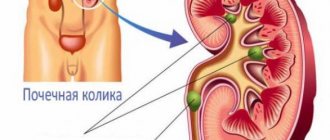
Urolithiasis (in medicine it is also called urolithiasis) is a chronic pathology caused by the formation of stones (calculi), which differ in their chemical composition.
The basis for the formation of kidney stones is a metabolic disorder in the body. It has been proven that people of any age can suffer from urolithiasis. If previously it was believed that kidney stones were more common in men than in women, recent studies on this topic indicate that the gender difference has been smoothed out.
The main danger of urolithiasis is that the stones can become large and block the ureteral duct. In this case, the kidney may completely stop performing its function.
“Who is at risk?”
The likelihood of developing urolithiasis increases if there is a hereditary predisposition, congenital kidney abnormalities (horseshoe kidney, hydronephrosis, duplication of the ureter), hyperfunction of the parathyroid glands, osteoporosis, metabolic syndrome. Also at risk are patients who were forced to lie motionless for a long time due to some kind of injury (car accident, etc.), which made the normal outflow of urine from the kidney difficult.
Vigen Andreevich Malkhasyan
Head of the Urology Department No. 4 of the City Clinical Hospital named after. S.I. Spasokukotsky, Associate Professor of the Department of Moscow State Medical University named after. A.I. Evdokimov.
Nephrolithiasis of the kidneys
For bilateral nephrolithiasis (stones in both kidneys), it is possible to perform transurethral endoscopic removal of stones from both kidneys in one procedure. This slightly increases the overall risk of the operation, with the same effectiveness rates.
In cases where it is impossible to remove the entire stone, it must be completely destroyed. The most effective method of contact lithotripsy (stone crushing) is a holmium laser. Laser lithotripsy is equally effective in destroying all types of stones, regardless of their density and composition, and can be used with both flexible and conventional (semi-rigid) endoscopes.
Renal stenting is currently not mandatory before or after endoscopic upper urinary tract surgery. Preliminary installation of an internal stent may facilitate ureterorenoscopy for kidney and large ureteral stones, a narrow ureter or its deformation. Stenting may be required after complex and lengthy operations where there are residual fragments and the risk of urinary infection. Alpha blockers are prescribed to reduce discomfort caused by the internal stent.
The overall incidence of complications after ureterorenoscopy ranges (according to various sources) from 9 to 25%. Most of these complications are not serious and do not require additional intervention. Severe complications such as ureteral avulsion are rare (<1%).
Treatment of urolithiasis in pregnant women
Pregnancy imposes serious limitations on both examination (for example, radiological methods and computed tomography) and treatment methods. The general rule is conservative treatment of all forms of urolithiasis, unless there are serious clinical indications for stone removal.
External lithotripsy is absolutely contraindicated during pregnancy. In case of a ureteral stone that does not tend to pass on its own, it is possible to install an internal ureteral stent for the entire period before birth (and the immediate period after birth), with regular replacement every 2-3 months (possibly earlier, since during pregnancy the risk of stent encrustation is higher ).
Alternatively, endoscopic stone removal is possible, but only in a specialized hospital with obstetric and neonatology services and experienced urologists. Percutaneous lithotripsy can be used in isolated cases, and also necessarily in specialized centers. I have been treating urolithiasis for more than 15 years and I can promise you that I will select and provide you with the treatment option that best suits your specific situation!
Questions about the article
Elena Ilyinichna
May 22, 2021 at 08:01
The kidney stone is 17mm in size, can it be reduced in size using tablets?
Anton Evgenievich Rotov
May 22, 2021 at 08:02
This is possible if the stone consists of uric acid. The composition can be indirectly determined by CT results (stone density)
Denis Viktorovich
April 7, 2021 at 07:12 pm
An hour ago I had an ultrasound of my kidneys. Precisely prophylactically. It turned out that in the right pack a 6mm stone was located at the base of the pelvis. Upper part. No worries. “Outflows and passages” are normal.) However, the ultrasound technician said that the count is literally on the clock. Now wait for an attack? Or is there something we can do?
Anton Evgenievich Rotov
April 7, 2021 at 07:14 pm
You don’t have to wait for an attack, but immediately carry out crushing (external lithotripsy). But it is better to do a CT scan of the kidneys. This will allow you to confirm (or refute) the diagnosis (ultrasound sometimes gives overdiagnosis) and find out the density of the stone. Based on the CT results, make a decision
Marina
February 8, 2021 at 11:34 am
Hello! During an ultrasound, a 7mm calculus was discovered in the sinus of the right kidney. Is it possible for him to pass on his own?
Anton Evgenievich Rotov
February 8, 2021 at 14:01
The sizes are large for independent passage. It's better to crush
Evgenia Ruslanovna
July 13, 2021 at 11:45 am
Question about “Determining the composition of urinary stones” Hello. Hello, we did a CT scan, after which we discovered a stone in the left ureter 4.5×3.0 mm, density +380.. +425 HU. How to treat?
Anton Evgenievich Rotov
July 13, 2021 at 11:47 am
Stones of this size usually pass on their own. To facilitate this process, it is recommended to drink plenty of fluids, exercise, take diuretics (herbal), antispasmodics and painkillers (if necessary). Estimating the density of stones with such small sizes is not reliable
Diagnosis of ICD
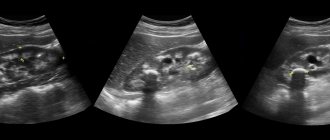
First of all, urolithiasis is diagnosed using ultrasound. If it shows the presence of stones in the kidneys or ureter, then the second diagnostic step is computed tomography (CT). It is this method that is considered the “gold standard” for diagnosing ICD all over the world, since it provides an order of magnitude more information on the number, size, density and location of stones.
For preventive purposes, for early detection of kidney stones, it is recommended to do an ultrasound of the kidneys at least once a year.

