Clinical picture
Deuteranopia is one of three variants of dichromasia - a feature of the color perception of the environment, with only two types of cones available. Other types of dichromasia are protanopia and tritanopia. People with deuteranopia generally do not distinguish some colors of the spectrum, just like people with protanopia, but unlike the latter, they do not have darkening of images. With protanopia, certain dark shades, such as violet, burgundy, purple, and blue, are practically indistinguishable from each other.
The pathology is considered a disease related to color blindness. It usually occurs in men (1% of the population) and is otherwise called color blindness. This term began to be used in honor of the English naturalist J. Dalton, who described this anomaly, which he himself suffered from. When examining the DNA of Dalton's eye cells, preserved in the laboratory, specialists 1.5 centuries after the scientist's death were able to confirm the correctness of the diagnosis that they had made independently.
What is the difficulty of diagnosing color blindness in children?
The baby begins to distinguish colors at the age of 3-4 years, although parents explain their names from the age of one and a half. The child remembers the name of a particular color. Adults have no way of knowing how he actually distinguishes color.
To check his color sense, parents are advised to observe the baby as he draws the world around him. Based on the results of such a drawing, you can assess whether the baby perceives objects correctly.
The main sign of color blindness is when a child replaces red with green or brown. This test is not reliable: he could choose the color based on his imagination. However, if your baby regularly changes from one color to another, it is advisable to consult an ophthalmologist. Only a doctor can diagnose color vision impairment based on special studies.
On topic: Colorblindness in children
Video: Color blindness test
Color vision anomalies
Ophthalmologists consider anomalies of color perception to be all problems and disorders in a person’s determination of not only colors, but also shades of color. They are transmitted according to an autosomal recessive mode of inheritance, i.e. based on linkage to the X chromosome.
Patients with color vision impairment, like people with normal vision, are trichromats. Trichromacy means that three colors must be used to determine the hue of the visible spectrum. True, people who have some deviations in color perception understand the color gamut somewhat differently than trichromats with good color perception.
If we explain the essence of the anomaly using the example of a color comparison test, then they use red and green in different proportions. At the same time, testing using an anomaloscope device shows the following: with protanomaly a person sees more red, with deuteranomaly - green. With tritanomaly, the color perception of yellow and blue is often pathologically altered.
Color vision impairment
Diabetes
Atherosclerosis
1702 28 October
IMPORTANT!
The information in this section cannot be used for self-diagnosis and self-treatment.
In case of pain or other exacerbation of the disease, diagnostic tests should be prescribed only by the attending physician. To make a diagnosis and properly prescribe treatment, you should contact your doctor. Color vision impairment: causes of occurrence, what diseases it occurs with, diagnosis and treatment methods.
Description
Impaired perception of colors and their shades in medicine is called color blindness, or chromatopsia. The ability of the human eye to distinguish many colors and shades opens up enormous possibilities for orientation in space, knowledge of the surrounding world and fine art.
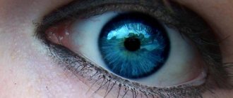
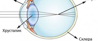
The conductor that helps a person perceive surrounding objects with the help of the organ of vision is light. Light is electromagnetic radiation, visible to the human eye, whose wavelengths range from 380 to 700 nm. Each object can reflect and absorb certain wavelengths of light. Those waves that are reflected pass through the transparent structures of the eye and excite special cells - cones, which are responsible for the perception of color.
The human eye can distinguish almost all color shades resulting from mixing the three basic ones - red, blue and green.
Depending on the percentage of excitation of a particular type of cone, a sensation of color arises in the brain. So, for example, when the brain perceives the color orange, then at that moment an impulse is transmitted from 99% of red cones, 42% of green and 0% of blue, and in the case when the brain perceives green, 31% of red, 67% of green and 36 % blue cones. If all cones are excited equally, then we see white color.
Color vision disorders occur due to a disruption in the excitation of cones and the transmission of impulses to the brain. If this condition is congenital, then a violation of color vision may remain unnoticed for a long time, because a person is guided by brightness and color saturation. Life experience and communication with people provide information about color: grass is green, the sky is blue, strawberries are red. Errors are detected at low brightness and object sizes.
A condition where a person has a reduced or completely absent ability to distinguish all or some colors is called color blindness.
.
Types of color vision impairment
There are congenital (most common) and acquired color blindness. Depending on the clinical manifestations, i.e. Depending on the characteristics of color perception, the following types of color blindness are distinguished:
- Achromatopsia is a complete lack of color vision, when a person perceives everything in black and white. Very rare.
- Deuteranopia is a complete loss of perception of the green part of the spectrum. Most common.
- Protanopia is a complete impairment of recognition of the red part of the spectrum. Deuteranopia and protanopia give similar color perception because... mixing red and blue, blue and green gives rise to a similar interpretation in the brain - swamp green.
- Tritanopia is a complete impairment of the perception of the blue spectrum. Rarely seen.
In addition to color blindness, there is a weakness in color sensitivity, when a person needs much more time and strong color saturation to recognize the color of an object.
Decreased perception of red is called protanomaly, green is called deuteranomaly, and blue is called tritanomaly.
Possible causes of color vision impairment
The most common cause of color vision impairment is damage to the genes involved in color perception on the X chromosome. As you know, men have a chromosome set of 46XY, and women have 46XX. If a boy received a damaged X chromosome from his mother, then there is nothing to compensate for the defective genes (there is no second X chromosome), and a violation of color perception occurs. In women, congenital color blindness is quite rare - in the case when a girl receives a damaged X chromosome from both her father and mother. The color blindness gene can be passed on through generations, manifesting itself in grandchildren and great-grandchildren. To make sure there is no genetic predisposition to color vision impairment, you can take a DNA test. This method is applicable in cases where conventional visual tests cannot be used, for example, for newborns and children in the first years of life.
Acquired types of color vision disorders are relatively rare and occur with various damage to the visual analyzer at one of the stages of receiving, transmitting or processing information.
Among their reasons are the following:
- damage to the retina by ultraviolet light if safety precautions for using UV lamps are not followed;
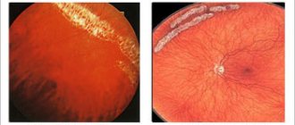
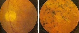
- Diabetes mellitus is one of the common causes of acquired color vision impairment as a result of diabetic macular degeneration. With a constant increase in blood glucose levels, damage occurs to all blood vessels, and especially to the small vessels of the fundus. Due to lack of oxygen, the retina ceases to perform its functions. Along with the symptoms of color vision impairment, distortions of straight lines and blurred vision occur;
- inflammatory diseases of the retina and optic nerve;
- cerebral atherosclerosis;
- taking certain medications (for example, cardiac glycosides can lead to seeing objects in yellow-green tones);
- cataract and glaucoma;
- retinal burns;
- chemical poisoning;
- lack of vitamin A, which results in a change in sensitivity to light and the perception of yellow-blue hues;
- mechanical damage to the retina, optic nerve, as well as traumatic brain injury;
- typhoid fever;
- neurological disorders: multiple sclerosis, previous stroke or cerebral infarction, brain tumors.
Which doctors should I contact if I have color vision problems?
If color vision is impaired, you should consult an ophthalmologist. The doctor will conduct an examination and the necessary tests to evaluate vision and color vision. If acquired color blindness is suspected, he will prescribe a set of laboratory and instrumental research methods and refer you to the necessary specialists.
Diagnosis of color vision impairment
There are several methods for diagnosing color vision disorder:
- tables E.B. Rabkin, or “polychromatic” tables, which consist of circles of different colors and brightness, which a person with color vision impairment will not be able to navigate. The circles are arranged so that some of them form a number or figure that needs to be recognized;
- the Ishihara test involves cards with spots of different colors that form numbers and shapes;
Ishihara test
- anomaloscopy;
- biochemical blood test to determine glucose levels, control cholesterol levels, very low, low, and high density lipoproteins, blood electrolytes - potassium, sodium, calcium, creatinine levels, liver enzymes - ALT and AST, total and direct bilirubin levels);
Dichromats
All known types of dichromatopsia are also transmitted genetically through connection with the X chromosome. The essence of the pathology is that to describe all shades, a person uses only two primary colors. Just like deuteranopes and protanopes, these people have an abnormally altered function of perceiving green and red colors.
For example, with protanopia, it is not possible to distinguish between black and red; people with protanopia are also often confused in describing red in comparison with brown or gray, or less often with green. They see some portion of the color spectrum as achromatic. Tritanopia develops much less frequently. People with tritanopia are unable to distinguish between blue and yellow colors. At the same time, the end of the spectrum of blue-violet shades appears to them as gray-black.
Symptoms and classification
The state of the body's color-perceiving system, in which all colors and shades are fully perceived, is called normal trichromasia
(from Greek chroma - color). In this case, all three elements of the cone system (“red”, “green” and “blue”) work in full mode.
In anomalous trichromats
Color vision impairment is expressed in the inability to distinguish any shades of a particular color. The severity of the changes directly depends on the severity of the pathology. People with mild color anomalies often do not even know about their peculiarity and learn about it only after undergoing medical examinations, which, based on the results of examinations, can introduce significant restrictions on their career guidance and future work activities.
Anomalous trichromasia is divided into protanomaly (impaired red color perception), deuteranomaly (impaired green color perception), and tritanomaly (impaired blue color perception) (Kris-Nagel-Rabkin classification).
Protanomaly and deuteranomaly can have different degrees of severity: A, B and C (in descending order).
With dichromasia
Humans are missing one type of cone and perceive only two primary colors. An anomaly due to which the color red is not perceived is called protanopia, green is deuteranopia, blue is tritanopia.
However, despite its apparent simplicity, it is difficult to understand how people with altered color vision actually see.
, extremely difficult. The presence of one non-functioning receiver (for example, red) does not mean that a person sees all colors except that one. This range is individual in each case, although it has a certain similarity with that of other people with color vision defects. In some cases, a combined decrease in the functioning of cones of various types may be observed, which introduces “confusion” into the manifestation of the perceived spectrum. Cases of monocular protanomalies can be found in the literature.
Table 1
: Color perception in individuals with normal trichromasia, protanopia and deuteranopia.
| Colors perceived by normal trichromats (normal vision) | Colors perceived by persons with color vision impairments | |
| Protanopia | Deuteranopia | |
| Red | Grey | Yellow |
| Green | Yellow | Grey |
| Light red | Dark green | Shades of yellow |
| Light green | Shades of yellow | Dark red |
| Blue | Pink | Violet |
The table below reflects the main differences in the perception of colors between normal trichromats and individuals with dichromasia. Protanomals and deuteranomalies have similar impairments in the perception of certain colors, depending on the severity of the condition. The table shows that the definition of protanopia as red blindness, and deuteranopia as green color blindness, is not entirely correct. Research by scientists has established that protanopes and deuteranopes do not distinguish between red and green colors. Instead, they see shades of grayish-yellow of varying lightness.
The most severe degree of color vision impairment is monochromasia
– complete color blindness. Rod monochromasia (achromatopsia) is distinguished when there are completely no cones on the retina, and when the functioning of two of the three types of cones is completely impaired - cone monochromasia.
In the case of rod monochromasia
When there are no cones on the retina, all colors are perceived as shades of gray.
Such patients also usually have low vision, photophobia and nystagmus. With cone monochromasia,
different colors are perceived as one hue, but vision is usually relatively good.
To designate color vision defects in the Russian Federation, two classifications are simultaneously used, which confuses some ophthalmologists.
Complete color blindness
Anomalies of color perception are quite rare, so absolute non-perception of the color spectrum is diagnosed only in 0.01% of the population. Such people are called monochromats. The colors they can distinguish are black and white. Accordingly, all visible objects appear gray to them, with varying intensities.
Under photopic illumination, such people experience impaired adaptation to color changes. Due to the blinding of the eyes by bright light (natural or artificial), they also have difficulty distinguishing the shapes of objects, which ultimately leads to serious photophobia. Because of this, monochromats have to wear sunglasses during daylight hours in any weather, although during ophthalmological examinations, experts do not find any abnormalities in their retinas. True, there is an opinion that with monochromia, the visual pigment is replaced by rhodopsin.
Diagnostics
Many patients may not complain about impaired color perception for a long time.
In such cases, the pathology is diagnosed during a color vision test during medical examinations. Deviations in color perception can be diagnosed using Rabkin tables, which are pictures made up of circles of different colors. Each such picture contains an encrypted geometric figure or number. There are tables that show both a figure and a number at the same time. A person with normal vision is able to discern what is encrypted on each of the tables. A person with color blindness will either be unable to distinguish or name the wrong shape.
There are a total of 48 pictures for determining color anomalies, which are divided into two groups. The first group includes the main tables, thanks to which the main types of color vision disorders are diagnosed. The second group is the control group. It is necessary to exclude simulation of the patient.
During the examination, the patient should have his back to the light source. The tables in front of him should be positioned vertically at a distance of 0.5-1 meter from the eyes. If the tables are lying on the table, the method for determining color perception is broken and a false result is possible.
When examining color vision, the patient must give an answer within 5-10 seconds - what he sees on the table. All answers are entered into a special form, after which the result is compared with a special table and a final diagnosis is made.
The diagnosis of color vision anomaly can only be made by an ophthalmologist.
It is impossible to completely cure impaired color vision. Treatment of color anomalies is aimed at reducing the severity of symptoms and correcting visual acuity. Treatment is also carried out with the aim of stopping the pathological process. In the absence of adequate therapy, a person can completely lose vision.
To normalize visual acuity, contact lenses or glasses are used. They are recommended to be worn by all patients with impaired color perception. In order to correct the perception of colors, there are tinted glasses and lenses with pigments. Patients are advised to go out in the sun only with sunglasses.
In the case of acquired color vision anomaly, when the cause of this condition is established, the issue of surgical correction can be decided. The operation can be performed on the affected area of the eye or on the part of the nervous system that is responsible for sensitivity to colors. If cataracts are present, surgery to remove them is indicated.
Complex therapy for impaired color vision includes vitamins. All patients are advised to take vitamin complexes with a high content of vitamins A and E.
If the disease occurs as a complication of diabetes mellitus, the patient is recommended to receive appropriate treatment from an endocrinologist.
Anomalies of the rod apparatus
The occurrence of defects in the rod apparatus leads to a decrease in the function of adaptation to twilight lighting. In ophthalmology, this phenomenon is called nyctalopia or hemeralopia. And among the people, such a pathology is called “night blindness.” It develops against the background of insufficient intake of vitamin A into the body. Moreover, it is vitamin A that is the basis for the production of retinol, which is essential for normal vision.
Statistics of color vision disorders
All color vision anomalies are genetically determined and are transmitted as a trait for which the X chromosome is responsible. Thus, men are especially susceptible to the development of these pathologies.
According to statistics, the prevalence of protanomaly in men is approximately 0.9% in the population, deuteranopia is about 1-1.5%, and deuteranomaly is almost 3.5-4.5%, protanopia is 1%. In comparison to women, deuteranomalies make up 0.3% of the population and protanopias make up about 0.5%. Tritanomaly and tritanopia, as color vision anomalies, are extremely rare.
Anomalies of color perception in society
People with color vision impairments (except monochromats) have the same visual acuity as all other people. In this regard, they are not handicapped and do not need social protection. However, there are a number of restrictions for them when choosing a profession and type of employment.
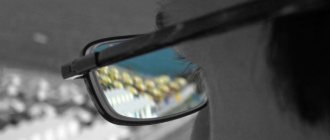
Thus, if color vision is impaired, people are not allowed to work in transport; they cannot be railway workers, pilots, sailors, doctors, chemists, etc.
In Romania and Turkey, persons with diagnosed color vision anomalies are prohibited from being issued a driver's license. According to Russian law, colorblind people with dichromasia (protanopia, deuteranopia) have the right to obtain a driver's license only category A or category B, but do not have the right to work for hire. And in many countries of the European Union there are no such restrictions. Drivers' licenses are freely issued to colorblind people, and traffic lights that are convenient for them have even been developed.
By contacting the Moscow Eye Clinic, each patient can be sure that some of the best Russian specialists will be responsible for the results of treatment. The high reputation of the clinic and thousands of grateful patients will certainly add to your confidence in the right choice. The most modern equipment for the diagnosis and treatment of eye diseases and an individual approach to the problems of each patient are a guarantee of high treatment results at the Moscow Eye Clinic. We provide diagnostics and treatment for children over 4 years of age and adults.
Description of Rabkin tables
In total, Rabkin's table contains 48 pictures. Among them there are 27 main ones, and the rest are clarifying ones. The pictures show numbers and geometric shapes (circle and triangle). They consist of multi-colored small circles, which are selected to reveal deviations in color perception.
Tables 1 and 2 are designed to help the test taker get comfortable with the tasks and understand the principle of the test.
Map 1
The numbers 9 and 6 are recognized by all subjects.
Map 2
The shapes of a circle and a triangle are recognized by everyone.
Map 3
With normal color perception it is recognized as 9, and by people with deviations as 5.
Map 4
Normally - a triangle.
Map 5
In the absence of pathology, these are 1 and 3 (usually read 13), If present, they are recognized as 6.
Map 6
In normal conditions, a triangle and a circle are recognized, but in pathological conditions, the figures cannot be identified at all.
Map 7
When normal, these are 9 and 6 (read 96), but in the presence of pathology, only 6 is recognized.
Map 8
This is 5. It is unrecognizable or poorly distinguishable if there is pathology.
Map 9
This is normally recognized as 9, and in pathology as 6 or 8.
Map 10
Under normal conditions, subjects recognize the numbers 1 and 3 and 6 (pronounce 136), and when impaired, they recognize 66 or 68 or 69.
Map 11
If color vision is normal, then they see a circle and a triangle. Only a triangle is recognized by protanopes, and deuteranopes see a circle or recognize a circle and a triangle.
Map 12
With normal color perception, subjects, as well as deuteranopes, recognize 12, and 13, see protanopes.
Map 13
With normal color perception, the presence of a circle and a triangle are recognized, protanopes recognize the presence of a circle, deuteranopes - a triangle.
Map 14
Under normal conditions, subjects recognize 3 and 0 in the upper half of the table (pronounced as 30), protanopes recognize 1 and 0 at the top, and the number 6 in the bottom half.
Map 15
With normal color perception, subjects recognize a circle (on the left) and a triangle on the right; protanopes recognize 2 triangles (top), a square (bottom), and deuteranopes recognize a triangle (top), square (bottom).
Map 16
Under normal conditions, 9 and 6 are recognized, Only 9 are recognized by protanopes, Deuteranopes recognize only 6.
Map 17
In normal conditions, the presence of a triangle and a circle is recognized, Protanopes - a triangle, Deuteranopes see a circle.
Map 18
Normally, single-color rows (1, 3, 5, 6) are recognized horizontally, and multi-colored rows are recognized vertically.
Map 19
Normally, the numbers 9 and 5 are recognized (pronounced as 95), while Protanopes and Deuteranopes recognize only 5.
Map 20
Under normal conditions, the presence of a circle and a triangle is recognized. Protanopes, as well as deuteranopes, do not detect anything.
Map 21
Normally, single-color rows are recognized vertically, and multi-colored rows horizontally.
Map 22
Normally, the numbers 6 and 6 are recognized (pronounced as 66). Only one 6 is detected by protanopes, as well as deuteranopes.
Map 23
The numbers 3 and 6 are recognized by everyone, except for persons with acquired color vision disorders.
Map 24
Numbers 1 and 4 are recognized by everyone except persons with acquired color vision impairment
Map 25
Persons with acquired color vision impairment do not detect anything; the number 9 is recognized by everyone else.
Map 26
Number 4 is recognized by everyone except those who have an acquired pathology.
Map 27
Under normal conditions, the numbers 1 and 3 are recognized (pronounced as 13). Nothing is detected by protanopes, as well as deuteranopes.
When the presence of color blindness is established, the diagnosis is clarified and its type is determined using additional tables. Congenital color blindness does not cause complications, but acquired color blindness is always associated with an underlying disease or injury and the presence of complications will be associated with them.