Algorithm for determining water balance
Prepare:
- establish a trusting relationship with the patient, make sure that the patient has not taken diuretics during the last 3 days;
— explain the mechanism of the procedure and obtain consent to carry it out;
— medical scales;
— graduated glass containers for collecting urine, vomit and feces;
— water balance sheet;
- handle;
— container with disinfectant solution;
— container for waste;
— disposable wipes and gloves:
Action:
Explain to the patient that at 6.00 it is necessary to release the night portion of urine into the toilet; after this, the patient must collect urine after each urination into a graduated container, record the time and amount of urine, and must also indicate the time of intake and the amount of liquid taken and record the data on a record sheet until 6.00 the next morning; at 6.00 the next day the patient must hand over the registration sheet to the nurse; remove gloves and place them in a container, wash (hygienic level) and dry your hands; record the received data in the medical documentation:
Attention:
— Diuresis is the process of formation and excretion of urine.
— Daily diuresis is the total amount of urine excreted by the patient during the day; in adults it ranges from 800 ml to 2000 milliliters.
— Daily water balance is the ratio between the amount of fluid introduced into the body and the amount of fluid excreted from the body during the day.
— In addition to the liquid drunk, the liquid contained in fruits, soups, vegetables, etc., as well as the volume of parenterally administered solutions, must be taken into account.
— Not only urine, but also the patient’s vomit and bowel movements should be taken into account.
— The expected fluid excreted in urine is calculated using the formula (the amount of fluid drunk is multiplied by 0.8).
— Water balance is considered positive if more fluid is released than introduced and vice versa.
— A positive water balance indicates the effectiveness of treatment.
studopedia.ru
Determination of daily diuresis
| Stages | Rationale |
| I. Preparation for manipulation. | |
| 1 . Prepare everything you need. | |
| 2. Label the measurement of daily urine output: Daily urine output. Last name, first name, patronymic of the patient. Date and time of the start of urine collection. | Ensuring that the procedure is carried out effectively. |
| 3. Attach a label to the graduated container, place it in the toilet room and show the patient or nurse (if seriously ill) where it is. | Ensuring that the procedure is carried out effectively. |
| 4. Establish a trusting relationship with the patient, assess his ability to independently carry out the procedure. | Ensuring informed participation in collaboration. |
| 5. Explain the purpose and progress of the study and obtain the patient's consent to the procedure. | Ensuring the patient's right to information. |
| II. Performing manipulation. | |
| 6. In the evening, explain to the patient (or nurse) the rules for collecting daily urine output. | |
| 7. The patient empties his bladder at 6 a.m., and this urine is not counted. | Reliability of the result. |
| 8. During the day until 6 a.m. the next day, the patient (or nurse) drains the urine into a graduated container, records its amount and pours it into the toilet. | |
| III. End of manipulation. | |
| 9. At the end of the day, the nurse determines the amount of urine excreted and writes down the result of daily diuresis in the corresponding column of the temperature sheet. | Ensuring continuity in nursing care. Information for the attending physician. |
Manipulation No. 38
“Study of arterial pulse (PS).”
Target:
assessment of the functional state of the cardiovascular system, an idea of the general condition of the patient, determination of the basic properties of the pulse.
Indications:
diseases of the cardiovascular system, examination, assessment of the general condition of the patient.
Contraindications:
No.
Equipment:
stopwatch (or clock with a second hand), red pen, temperature sheet, observation sheet, nursing history.
Algorithm:
| Stages. | Rationale. |
| I. Preparation for manipulation. | |
| 1. Prepare everything you need. | |
| 2. Introduce yourself to the patient in a friendly and respectful manner. | Establishing contact with the patient. |
| 3. Clarify how to contact him. | |
| 4. Familiarize the patient with the purpose and progress of the procedure. | Psychological preparation of the patient. Right to information. |
| 5. Obtain the patient's consent to the procedure. | Respect for patient rights. |
| 6. Prepare the necessary equipment | Efficiency of the procedure. |
| 7. Wash and dry your hands. | Ensuring infection safety. |
| II. Performing the procedure: 8. Give the patient a comfortable position (sitting, lying down) and ask him to relax his arm, while the hand and forearm should not be suspended. | Ensuring the reliability of the result. |
| 9. Press the radial arteries on both hands of the patient with II, III, IV fingers (I finger should be on the back of the hand) and feel the pulsation of the arteries. Note: If the pulse is symmetrical, further examination should be carried out on one arm, otherwise on both arms alternately. Remember! You cannot examine the pulse with your thumb, because it has a pronounced pulsation, and you can count your own pulse instead of the patient's pulse! | To determine the symmetry of the pulse (coincidence of pulse beats). Reliability of the result. |
| 10. Take a watch or stopwatch and examine the pulsation of the artery (pulse characteristics). | The accuracy of the study is ensured. |
| 11. Determine the pulse rhythm for 30 seconds. | The accuracy of the study is ensured. |
| 12. Determine the heart rate for 1 minute. | |
| 13. Press the artery harder against the radius and determine the pulse tension. | |
| 14. Determine pulse filling (the degree of filling of the artery with blood). | The accuracy of the study is ensured. |
| 15. Determine the pulse value (by voltage and pulse filling). | |
| III. End of the procedure: 16. Inform the patient the result of the study (pulse rate). | Patient's right to information. |
| 17. Help the patient take a comfortable position or offer to stand. | |
| 18. Wash your hands. | Infection safety. |
| 19. Record the results of the study in a temperature sheet, or an observation sheet, or a nursing medical history, or a nursing process chart. Note: The pulse rate on the temperature sheet is graphically recorded as a red pulse curve. | For documentation of pulse examination results. |
Manipulation No. 39
"Measurement of blood pressure (BP)."
Target:
assess the state of the cardiovascular system, the general condition of the patient, determine blood pressure indicators and evaluate the results of the study.
Indications:
diseases of the cardiovascular system, kidneys, examination of the patient.
Contraindications:
No.
Equipment:
tonometer, phonendoscope, pen with a red rod, temperature sheet (outpatient card, nursing medical history), roller, napkins, 70% ethyl alcohol or disinfectant solution approved for use in this healthcare facility.
Algorithm:
| Stages | Rationale |
| I. Preparation for the procedure: 1. Prepare everything necessary for the manipulation (equipment). | Achieving an effective procedure. |
| 2. Kindly introduce yourself to the patient and clarify how to contact him. | Establishing contact with the patient. |
| 3. Explain to the patient the purpose and progress of the procedure. Get his consent. | Patient's right to information. |
| 4. Wash your hands and dry them. | Ensuring infection safety. |
| II. Performing the procedure: 5. Sit or lie down the patient (depending on his condition), placing the device at chest level. | Reliability of testimony. |
| 6. Place the cuff on the patient’s bare shoulder 2-3 cm above the elbow (clothing should not compress the shoulder above the cuff), secure the cuff so that only one finger passes between it and the shoulder. Note: Do not measure BP on the arm on the mastectomy side, on the weak arm after a stroke, on the paralyzed arm, or on the arm with an IV needle. It is advisable for the patient to sit quietly with the cuff applied for 5 minutes. | Ensuring the reliability of the result. |
| 7. Invite the patient to place his hand correctly; in an extended position of the elbow joint with the palm up (if the patient is sitting, ask him to place a clenched fist of his free hand or a roller under his elbow). | Ensuring the best extension of the limb. |
| 8. Connect the pressure gauge to the cuff and check the position of the pressure gauge needle relative to the zero scale mark. | Make sure the device is working properly. |
| 9. Wipe the phonendoscope membrane with alcohol. | Ensuring infection safety. |
| 10. Find the place of pulsation of the brachial artery in the area of the ulnar fossa (by palpation) and place the phonendoscope membrane in this place. | The reliability of the result is ensured. |
| 11. Ask the patient about his blood pressure readings. | To compare readings. |
| 12. With your free hand, close the valve on the bulb, turn it to the right, and with the same hand quickly pump air into the cuff until the pressure in it exceeds 20-30 mm Hg. Art. the level at which Korotkoff sounds (or radial artery pulsation) disappear. | Discomfort associated with excessive compression of the artery is eliminated and a reliable result is ensured. |
| 13. Release air from the cuff at a speed of 2-3 mmHg. Art./sec., turning the valve to the left, at the same time using a phonendoscope, listen to the tones on the brachial artery and monitor the readings on the pressure gauge scale. When the first sounds (Korotkoff sounds) appear, remember the number corresponding to the systolic pressure. Continuing to release air, note the value of diastolic pressure, which corresponds to the complete disappearance of sounds or their weakening. Remember the number corresponding to diastolic pressure. Note: Monitor the patient's condition while measuring blood pressure. | Obtaining a more reliable result, |
| 14. Tell the patient the measurement result. | Patient's right to information. |
| 15. Repeat the procedure after 2-3 minutes. Note: blood pressure is usually measured 2-3 times, releasing the air from the cuff completely each time. | Reliability of the result. |
| III. Completion of the procedure: 16. Remove the applied cuff. Wipe the phonendoscope membrane with 70% alcohol. Wash the hands. | Ensuring infection safety. |
| 17. Record the measurement data (if necessary, rounding them to “O” or “5”) in the nursing medical history and temperature sheet, making a preliminary correction of the results taking into account the shoulder circumference. Note: see table No. 2. In the nursing history, blood pressure is recorded as a fraction (systolic pressure in the numerator, diastolic pressure in the denominator). In the temperature sheet, blood pressure measurement data is recorded in the form of a column, the upper limit of which means systolic pressure and the lower limit means diastolic pressure. | Documentation of blood pressure measurement results. |
Manipulation No. 40
“Counting the number of respiratory movements (RR).”
Target:
determine the main characteristics of breathing.
Indications:
diseases of the respiratory system and cardiovascular system.
Contraindications:
No.
Equipment:
watch (stopwatch), temperature sheet or nursing observation sheet, pen and paper.
Algorithm:
| Stages | Rationale |
| I. Preparation for the procedure: 1. Introduce yourself kindly and respectfully to the patient. Find out how to contact him. | Establishing contact with the patient. |
| 2. Warn the patient that a pulse examination will be performed. | The ability to control breathing is excluded. |
| 3. Obtain the patient’s consent to perform the procedure. | The patient's rights to information are ensured. |
| 4. Wash and dry your hands. | Ensuring infection safety |
| 5. Ask or help the patient to lie (sit up) comfortably in bed so that you can see the upper part of his chest and abdomen (epigastric region). | To clarify (determine) the type and rhythm of breathing. |
| 6. Determine the type and rhythm of breathing. | The accuracy (reliability) of NPV calculation is ensured. |
| II. Performing the procedure: 7. Take the patient’s hand as for examining the pulse, observe the excursion of the chest or the movements of the epigastric region of the patient’s abdomen. Count your breathing movements in 1 minute. Note: if it is not possible to observe the excursion of the chest, then place your hands (the patient’s and yours) on the chest (in women) or on the epigastric region (in men), simulating the examination of the pulse (while continuing to hold the hand on the wrist) | Determination of NPV |
| 8. Record the result on paper and transfer the data to the nursing observation sheet or temperature sheet. | Ensuring control over the state of the respiratory organs and cardiovascular system. |
| III. Completion of the procedure: 9. Wash and dry your hands. | Ensuring infection safety. |
Manipulation No. 41
“Filling out the temperature sheet.”
Target:
rules for filling out medical documentation.
Indications:
registration of patient examination results.
Contraindications:
No.
Equipment:
temperature sheet, pens (or pencils) with red and blue paste.
Algorithm:
| Stages | Rationale |
| I. Preparation for manipulation. 1. Prepare a standard temperature sheet. 2. Prepare a blue or black pencil (or paste), a red pencil (or paste). II. Performing manipulation. 3. Mark the morning temperature with a dot in column “U”, the evening temperature – in column “B”. 4. Mark the upper limit (systolic) and lower limit (diastolic) blood pressure with a red pencil (or paste). 5. In column “U” mark the results of counting the pulse in the morning with a dot, and in column “B” the results of counting the pulse in the evening. 6. In the “Breathing” column, write down the count of the number of respiratory movements in 1 minute. 7. In the “Weight” column, make a note about the patient’s body weight. 8. In the column “Drinking fluids”, note the amount of fluid that entered the patient’s body per day. 9. In the “Daily amount of urine” column, note the amount of urine excreted by the patient per day. 10. In the “Chair” column, mark the data on defecation with a + sign. 11. In the “Bathtub” column, mark with a + sign the data on the sanitization of the patient. III. End of manipulation. 4. Connect the points of morning and evening temperatures. 5. Connect the dots of the pulse count results. 6. Mark blood pressure in the form of a column with a red pencil. | Rules for filling out medical documentation. Effectively read patient examination results. Reliability of the result. Reliability of the result. Reliability of the result. Information for the attending physician. Ensuring continuity in nursing care. Information for the attending physician. Ensuring continuity in nursing care. Information for the attending physician. Ensuring continuity in nursing care. Information for the attending physician. Ensuring continuity in nursing care. Obtaining a temperature curve. Graphic display of heart rate results. Efficiency of filling out medical documentation. |
Manipulation No. 42
“Drawing up a portion plan and portion requirements.”
The required diet and the duration of its use are determined by the doctor and depend on the disease, the patient’s condition and the tolerability of the prescribed diet. The doctor writes down the diet number in the medical history and prescription sheet. The ward nurse, checking the data on the appointment sheet, collects information daily on the number of different dietary tables (diets), types of fasting and individual diets. She submits this information to the senior nurse, who, based on it, draws up and approves the portion plan (see sample). The portion maker is also approved by the manager. department, after which it is served to the food department.
Information about patients discharged from the department is not included in the portion plan. If patients are admitted in the evening or at night, the nurse on duty prepares the portion plan.
Portion sample
_______________________
(name of institution) f No. 1
PORTION MAN
2. Individual supplementary nutrition
| Name of chambers (departments) | Surnames of patients | Food |
Disabled people of the Great Patriotic War (WWII) - full name. and ID number
Head of Department______________
Senior med. sister__________________
A nutritionist and nutritionist are responsible for drawing up the menu, monitoring the quality of products and storing them.
Manipulation No. 43
"Feeding a patient on bed rest."
Target:
providing dietary nutrition.
Indications:
inability to eat independently.
Contraindications:
No
Equipment:
spoon, bedside table or portable (bedside) table, napkin (towel), cup with cooled boiled water, basin, jug with warm water, soap, kidney-shaped tray, towel.
Algorithm:
| Stages | Rationale |
| 1. Preparation for manipulation. 1. Prepare everything you need. 2. Ventilate the room. 3. Warn the patient about the upcoming meal and obtain his consent to feeding. 4. Free up space on your nightstand or install a portable table. 5. Wipe the table or bedside table with a rag. 6. Help the patient take a position comfortable for eating, a high Fowler’s position, if there are no contraindications. 7. Help the patient wash his hands and dry them with a towel. 8. Cover your neck and chest with a napkin. 9. Bring the food necessary for eating (hot dishes - 60°C, cold ones - must be cold - no higher than 15°C). | Efficiency of the manipulation Providing conditions for eating. Patient's right to information. Ensuring the manipulation. Compliance with dignity hygienic regime. Providing food intake. Prevention of aspiration. Compliance with sanitary and hygienic regime. Prevention of nosocomial infections. Prevention of contamination of underwear. Maintaining food temperature. |
| 10. Familiarize the patient with the contents of the dishes and find out in what order he prefers to take them. 11. Make sure that the food is not very hot by dropping a few drops onto the back of your hand. II. Performing manipulation. 12. Invite the patient to drink a few sips of liquid (preferably through a straw). 13. Feed slowly (without distraction): - name each dish; - fill the spoon 2/3 with food; - bring the spoon to the patient’s mouth and invite him to swallow its contents; - wait until the patient chews and swallows food; - serve a new portion of food in a spoon; - if necessary, offer the patient a drink after a few spoons of food; - wipe your lips with a napkin if necessary. Note: if the patient can take part in feeding, you can give him a piece of bread, a cracker, a cookie, a bun, etc. III. Completion of the manipulation. 14. After eating, ask the patient to rinse his mouth with water. 15. Help the patient take a comfortable position. 16. Remove leftover food and dishes. 17. Wipe down the bedside table or table. | Patient's right to information. Maintaining the patient’s usual eating pattern. Prevention of burns to the oral mucosa (ensuring patient safety). Reduces dry mouth, improves chewing of solid foods. Ensuring adequate nutrition. Patient participation in feeding. Maintaining oral hygiene. Ensuring a comfortable stay in bed. Ensuring infection safety Compliance with sanitary and hygienic conditions. |
| 18. Wash your hands. | Ensuring infection safety |
Sippy cup feeding Purpose:
providing individual dietary nutrition.
Indications:
inability to eat independently.
Contraindications:
unconscious state; lack of swallowing reflex.
Equipment:
sippy cup, napkin, liquid food, towel, boiled water.
Algorithm:
| Stages | Rationale |
| I. Preparation for manipulation 1. Prepare everything you need. 2. Ventilate the room. 3. Warn the patient about the upcoming meal and obtain his consent. 4. Clear some space on the nightstand and wipe it with a rag. 5. Help the patient take a comfortable position for eating, a high Fowler's position (if there are no contraindications). Wash the hands. | Efficiency of manipulation. Providing conditions for eating. Patient's right to information. Compliance with dignity gig. mode. Providing food intake. Prevention of aspiration. |
| 6. Cover your neck and chest with a napkin. 7. Make sure that the food is not hot by dropping a few drops onto the back of your hand. II. Performing manipulation. 8. Fill the sippy cup with liquid food. 9. Feed the patient from the sippy cup in small portions, making sure that the previous portion of food has been swallowed. III. End of manipulation. 10. After eating, invite the patient to rinse his mouth with water and blot his lips with a napkin. 11. Help the patient take a comfortable position. 12. Remove leftover food and dishes and wipe down the nightstand. 13. Wash and dry your hands. 14. Make a note about the feeding in the medical documentation. | Prevention of contamination of underwear. Prevention of burns to the oral mucosa (ensuring patient safety). For feeding the patient. For better absorption of food. Maintaining oral hygiene. Ensuring a comfortable stay in bed. Compliance with dignity gig. mode. Ensuring infection safety. Continuity in the work of medical staff. |
Manipulation No. 44
"Feeding the patient through a nasogastric tube."
Target:
providing individual dietary nutrition.
Indications:
- violation of the act of swallowing;
— unconscious states;
- lack of sucking and swallowing reflex in premature newborns;
- fracture of the jaw bones, diseases of the oral cavity;
- refusal of food in case of mental illness.
Equipment:
sterile nasogastric tube, funnel or Janet syringe, clamp, towel, napkins, gloves, mask, nutritional mixture (t -38-40°C), boiled water (50-100 ml), container with disinfectant. solution, phonendoscope, syringe.
Algorithm
| Stages | Rationale |
| I. Preparation for manipulation 1. Prepare everything you need | Efficiency of manipulation |
| 2. Explain to the patient (if possible) the course of the upcoming manipulation and obtain his consent. | The patient’s right to information and his possible participation in the procedure. |
| 3. Clarify the patient’s understanding of the procedure and his behavior during it. | Ensuring patient participation during manipulation (if possible). |
| 4. Wash and dry your hands. Wear gloves. | Ensuring infection safety. |
| 5. Place the patient in a low Fowler position. Measure the distance from the tip of the nose to the earlobe and down the anterior abdominal wall so that the end of the probe is 2-5 cm below the xiphoid process of the sternum. | Ensuring that the tube enters the stomach. |
| 6. Cover the patient's chest with a towel or napkin. | Prevention of contamination of underwear and bed linen. |
| 7. Lubricate the probe with glycerin. | To facilitate the passage of the probe. |
| 8. Take the probe in your hand like a “writing pen” and carefully insert it through the lower nasal passage to a depth of 15-18 cm. | Ensuring that the probe enters the nasopharynx. |
| 9. Using the finger of your left hand, determine the position of the probe in the nasopharynx and press it to the back wall of the pharynx. | Preventing the probe from entering the trachea. |
| 10. Slightly tilt the patient’s head forward, ask him to drink water in small sips, swallowing the probe advanced by m/s. If the patient develops a cough, cyanosis, or air comes out of the probe during exhalation, immediately pull the probe back and repeat the procedure. | Ensuring the advancement of the probe through the digestive tract. The probe entered the trachea. |
| 11. Insert the probe to the desired mark and apply a clamp to the distal end of the probe. Check that the probe is inserted correctly: - draw air into the syringe; - attach the syringe to the probe; - place the head of the phonendoscope on the stomach area; - remove the clamp; - introduce air through a probe into the stomach under the control of a phonendoscope - you will hear sounds indicating the entry of air into the stomach. This means the probe is in the stomach. If there are no sounds, the probe must be moved. | Control of the tube entering the stomach. |
| II. Performing manipulation. 12. Draw food into the Janet syringe and connect it to the probe (or connect the funnel to the probe). | To introduce food into the stomach. |
| 13. Pour food into the stomach in small portions (30 ml), gradually, with intervals between portions of 1-3 minutes. | For better absorption of food. |
| 14. If a funnel is used, then it is necessary: - lower the funnel to the level of the stomach; — fill it with a nutrient mixture; - slowly raise it above the level of the stomach; - as soon as the nutrient mixture reaches the mouth of the funnel, lower the funnel to the level of the stomach and fill it again with the nutrient mixture. | Ensuring that food reaches the stomach. |
| 15. At the end of feeding, pour in a small amount of boiled water to rinse the probe. | Rinsing the probe from food debris. Prevention of bacterial growth. |
| III. End of manipulation. 16. Disconnect the Janet syringe or funnel and place them in a container with a disinfectant solution. | Prevention of nosocomial infections. |
| 17. Close the probe with a plug (in case of further use as prescribed by a doctor) and secure it. | Preventing the leakage of stomach contents and infection into the tube. |
| 18. Help the patient take a comfortable position. | Ensuring a comfortable stay in bed. |
| 19. Remove gloves and place them in disinfectant solution. | Prevention of nosocomial infections. |
| 20. Wash and dry your hands. | Eliminating the chemical effects of talc on the skin. |
| 21. Make a note about the procedure in the medical documentation. | Ensuring continuity in the work of medical staff. |
Manipulation No. 45
"Feeding a patient through a gastrostomy tube."
Gastrostomy - (gaster - from Greek stomach, stoma - from Greek opening) is an artificial opening in the stomach, created surgically for the purpose of artificial feeding of the patient in case of obstruction of the esophagus or for its functional shutdown.
To feed the patient through a gastrostomy tube, a sterile tube is inserted into the opening of the stomach, which must be fixed. A clamp is placed on the free end of the probe.
Target:
providing individual dietary nutrition.
Indications:
- narrowing or obstruction of the esophagus;
- pyloric stenosis; injuries of the larynx and esophagus;
- severe burns;
- inoperable tumors.
Equipment:
funnel (Zhanet syringe), container with pureed or liquid food (t 50°C), boiled water, sterile wipes, Lassar paste, adhesive plaster, clamp, container with disinfectant solution, gloves (2 pairs).
Algorithm:
| Stages | Rationale |
| I. Preparation for manipulation. 1. Prepare everything you need. 2. Ventilate the room. 3. Warn the patient about the upcoming meal and obtain his consent. 4. Clear some space on the nightstand and wipe it with a rag. 5. Help the patient find a comfortable position for feeding. 6. Wash and dry your hands. Wear gloves. P. Performing manipulation. 7. Remove the clamp (or plug) from the free end of the probe. 8. Attach a funnel or Janet syringe to the probe. 9. Pour the prepared food into the funnel (or inject it with a syringe) in small portions (no more than 30 ml). III. End of manipulation. 10. Rinse the probe with boiled water (30 ml). 11. Place a clamp on the free end of the probe. Disconnect the funnel or syringe and place it in the disinfectant solution. 12. Help the patient find a comfortable position. 13. Remove leftover food and dishes. Wipe down the nightstand. 14. Remove gloves and immerse them in the disinfectant solution. | Efficiency of manipulation. Providing conditions for eating. Patient's right to information. Compliance with sanitary and hygienic conditions. Providing food intake. Infection safety. To introduce nutrition. To introduce nutrition. For better absorption of food. Rinsing the probe from food debris. Prevention of bacterial growth. Prevention of nosocomial infections. Comfortable stay in bed. Compliance with dignity gig. mode. Prevention of nosocomial infections. |
| 15. Wash and dry your hands. 16. Make a note about the manipulation in the medical documentation. Note: administer food 5-6 times a day in a volume of 150-200 ml. As you get used to this volume of food, as prescribed by the doctor, the volume of food is increased to 500-600 ml, and the frequency of feeding is reduced. | Eliminating the chemical effects of talc on the skin. Ensuring continuity in the work of medical staff. |
Question 7. Measuring daily urine output and assessing water balance
Water balance is the ratio between the amount of fluid introduced into the body and the amount of fluid excreted from the body during the day. Normally, 1.5–2 liters of water should be consumed per day, and 75–80% of what you drink should be excreted.
Purpose: determination of hidden edema, monitoring the effectiveness of diuretics.
Equipment: medical scales, graduated glass container for collecting urine, water balance sheet.
Algorithm of actions:
1. Explain to the patient the procedure for the procedure the day before. Make sure that the patient has not taken diuretics for three days.
2. Ask the patient in the morning at 6:00 to empty the bladder into the toilet.
3. Weigh the patient and record the result on the water balance sheet.
4. Explain to the patient:
§ collect urine at each urination until 6 o’clock the next day inclusive in a graduated container, and record the amount in the column “Excreted liquid”;
§ count the amount of liquid consumed during the day, including first and third courses, raw vegetables and fruits (at the rate of 100 g of raw fruit corresponds to 100 ml of liquid), intravenous drips, jet infusions, and record the amount in the “Fluid administered” column.
5. Weigh the patient the next morning (at the same time), and record the result on the water balance sheet.
6. Determine the amount of urine excreted per day and liquid drunk per day.
7. Enter all data into the water balance sheet.
8. Calculate the water balance using the formula: multiply the amount of fluid administered by 0.8 (80%), which is equal to the amount of urine that should be excreted normally.
9. Consider the water balance negative if less fluid is released than calculated (normal).
10. Consider the water balance positive if more fluid is released than calculated.
Assessment: a positive water balance indicates the effectiveness of treatment and the resolution of edema. Negative water balance indicates an increase in edema or ineffective dosage of diuretics.
Question 8. Conclusions
The modern level of development of nursing requires the nurse to be able to independently assess the patient’s condition, conduct proper monitoring of him, and make informed decisions associated with a certain responsibility.
Constant monitoring of the patient will allow the nurse to promptly notice changes in the body and provide competent, systematic care for him.
MINISTRY OF HEALTH OF KHABAROVSK REGION
Regional state budgetary educational institution
studopedia.ru
Taking urine for a sample according to Amburge
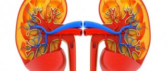
Target. Determination of the number of shaped elements and cylinders. Indications. Inflammatory kidney diseases. Equipment. A clean, dry, clear glass jar; referral to a clinical laboratory; a clean, dry potty (or bedpan for patients on bed rest). Technique for collecting urine for a sample according to Amburge : 1. After selecting prescriptions from the medical history, prepare the dishes and directions. 2. The patient is prepared as follows: “Tomorrow you need to collect urine for research according to Amburge. To do this, at 6.00 am, urinate in the toilet and hold urination for 3 hours until 9.00. At 9.00, after thoroughly toileting your genitals, urinate in the potty and pour all the urine into a jar with a direction. The pot and jar are on the shelf in the toilet.” 3. All urine is sent to the laboratory immediately after urination in a warm state. 4. The result of the study is pasted into the medical history. Notes. If the patient is on bed rest, the nurse performs the washing. Normally, urine when examined using the Amburger test contains: leukocytes - up to 2.5 * 10″3; erythrocytes - up to 1x10″3; cylinders - up to 15.
Determination of water balance
Target:
identification of hidden edema, determination of the amount of urine excreted per day, assessment of the adequacy of therapy, primarily diuretic (diuretic).
Equipment:
medical scales, clean dry 2-3 liter jar, two graduated vessels, water balance sheet, temperature sheet.
Methods:
systematic weighing of the patient, measuring daily diuresis and comparing it with the amount of fluid drunk, repeated measurements of the circumference of the legs, abdomen, palpation.
Measuring daily diuresis and amount of fluid consumed
Algorithm of actions
1. The day before, warn the patient about the upcoming procedure and the rules for collecting urine, give him detailed information about the order of entries in the water balance sheet.
2. At 6 a.m., wake up the patient so that he urinates on his own in the toilet, or releases his urine with a catheter; this portion of urine is not taken into account.
3. The patient must collect all subsequent portions of urine in a jar until 6 a.m. the next day inclusive.
4. During the day, the patient or nurse keeps a record of the fluid introduced into the body in milliliters, including what was drunk, including first courses, parenterally administered and fruits and vegetables eaten in a 1:1 ratio.
5. Using a graduated vessel, calculate the amount of urine excreted per day.
6. Enter the measurement data in a special column on the temperature sheet.
Help with heart pain (angina attack)
Target:
relieve pain immediately.
Algorithm of actions
1. Provide access to fresh air, complete physical and mental rest.
2. Call a doctor immediately.
3. Sit the patient down.
4. Give 1 tablet of nitroglycerin under the tongue.
5. If after 3 minutes the pain has not stopped, repeat sublingual nitroglycerin, but no more than 3 times with an interval of 10 minutes.
6. Place a mustard plaster on the site of the pain projection.
7. Constantly monitor the patient’s condition: respiratory rate, pulse, blood pressure level, skin color.
8. Follow doctor's orders.
studopedia.ru
Is it necessary to supplement a newborn's diet?
- Whether the baby is fed exclusively breast milk or formula, or is on mixed feeding - in any case, he needs an exclusively individual approach. The question about supplementation should be asked to the pediatrician who is observing the child.
Typically, a breastfed baby gets enough fluids through breast milk. If he needs additional fluid, he will simply ask for additional breast milk and quench his thirst with “foremilk,” that is, thinner milk.
The situation with artificial feeding is somewhat more complicated. If there is a lack of fluid, the child should not be given additional formula - this way you can overfeed the child. Therefore, in some situations, a pediatrician may recommend supplementing a bottle-fed baby.
We should not forget that there are certain individual conditions, for example, a hot and dry climate, the special qualities of the mother’s breast milk, as well as various developmental characteristics and health conditions (chronic and acute pathology accompanied by fluid loss). In such cases, the doctor may recommend supplementing the breastfed child with food.
How to understand that a child does not have enough water?
— Water balance disorders in children can occur in various situations. Overheating, heat stroke, fever (increased body temperature) due to various infections, diarrhea, vomiting are the most common causes of dehydration in children. A child's fluid needs are higher than those of an adult, so dehydration can occur much faster.
Signs of dehydration may include lethargy, drowsiness of the child, dry lips and mucous membranes. A deterioration in the child’s condition can develop very quickly. Therefore, if you see that your child has one of these symptoms, you need to see a doctor as soon as possible.