Causes of kidney infarction
- Ischemic renal infarction is ischemic necrosis of the renal parenchyma
- Focal or general
- Acute, subacute or chronic.
Causes of ischemic renal infarction:
- The main cause of ischemic and uric acid infarction of the kidney is acute occlusion of the renal artery or its branches due to:
- Thrombosis: atherosclerosis, periarteritis nodosa, conditions predisposing to the development of thrombosis.
- Embolism: atrial fibrillation, endocarditis, myocardial infarction, direct angiography.
- Trauma: closed injury of the abdominal organs.
Anatomical prevalence of ischemic renal infarction:
- Subsegmental or segmental infarction (occlusion of a subsegmental or segmental artery); one or more infarct sites.
- General infarction (occlusion of the main renal artery).
- Unilateral general infarction: suspected thrombosis/trauma.
- Bilateral multiple segmental (subsegmental) infarction: suspected embolism.
Introduction
Non-traumatic renal rupture, in other words, spontaneous renal rupture, unlike traumatic ruptures, is much less common and in many cases presents diagnostic difficulties [1-3].
In the world literature over the past century and a half since spontaneous renal rupture was identified as an independent pathology, over 1000 observations of spontaneous renal rupture have been published. However, a relatively small number of studies are devoted to studying the mechanisms of occurrence, diagnosis and treatment of spontaneous renal rupture. The main reason for this situation is the rarity of this pathology. Spontaneous renal rupture can be a complication of renal neoplasms, hydronephrosis, renal cysts, observed in periarteritis nodosa, acute pyelonephritis, urolithiasis, aneurysm of the renal arteries, Wegener's granulomatosis, renal infarction, hemorrhagic fever with renal syndrome, etc. Spontaneous kidney rupture may occur in a number of predisposing conditions, such as pregnancy and childbirth. In rare cases, spontaneous renal rupture may occur in an otherwise intact kidney. In turn, spontaneous renal rupture is part of a much larger family of pathological conditions, united in domestic and foreign literature under the general name “spontaneous perirenal bleeding.” Spontaneous perirenal bleeding can also be caused by blood diseases (leukemia, hemophilia, etc.), taking anticoagulants, adrenal rupture, adrenal tumors, aortic aneurysms, etc. [4]. Reviews summarizing cases described in the literature from 1933 to 2002 [5-8] included 448 observation units. The main etiological causes of spontaneous perirenal bleeding are tumors, which, according to various authors, occur with a frequency of 57 to 63% (of which 24-33% are benign and 30-33% are malignant). With a slightly lower frequency of occurrence, the causes of spontaneous renal rupture are vascular diseases (17-26%, of which periarteritis nodosa - 12-13%). The third main cause is infections (4-10%). Currently, there is no uniform diagnostic and treatment strategy for determining spontaneous renal rupture, the severity and prognosis of this condition, despite the fact that spontaneous renal rupture is an urgent condition and requires immediate action. There are frequent cases of incorrect diagnosis, post-mortem diagnosis of spontaneous renal rupture, and unjustified delay of surgical intervention. At the same time, the increasing sensitivity of diagnostic methods such as ultrasound scanning, multislice computed tomography and magnetic resonance imaging significantly increases the surgeon’s ability to timely and accurately determine the cause of spontaneous renal rupture. Based on these data, in most cases it is possible to identify the cause that caused the kidney rupture, assess the feasibility of surgical intervention, and also plan its volume. We present our own observation when, on the basis of a comprehensive examination, we were able to establish a diagnosis of spontaneous renal rupture and choose a rational treatment tactic.
Which method of diagnosing renal infarction to choose: MRI, CT, ultrasound
Selection Methods
- Ultrasound, CT, MRI.
General signs
- The extent of renal infarction can be assessed with contrast-enhanced CT/MRI or color Doppler.
Why is an abdominal ultrasound performed for kidney infarction?
- Color Doppler ultrasound reveals focal or complete absence of blood flow in the renal parenchyma
- It can also be determined by ultrasound using contrast enhancement.
In what cases is a CT scan of the abdominal cavity performed for renal infarction?
Prevalence of renal infarction site:
Subsegmental:
- a clearly demarcated, wedge-shaped area of reduced contrast enhancement;
- the base of the wedge faces the kidney capsule.
Segmental:
- clearly defined
- Caused by occlusion of a segmental artery.
Total:
- complete absence of contrast enhancement of the renal parenchyma
- No contrast agent excretion
- Sometimes, in the presence of collateral circulation, there is an enhancement of contrast like “spokes of a wheel”
- The “cortical ring” symptom indicates the presence of subcapsular blood flow
Kidney infarction. Color Doppler mapping shows a disturbance in the blood supply in the middle third of the kidney.
Ultrasound with contrast. Lack of contrast in the area of infarction.
Distinctive signs of acute and chronic renal infarction
Spicy:
- kidneys of normal size with an even contour
- decrease in contrast intensity in a clearly limited area or throughout the kidney
- absence or decreased excretion of contrast agent
- "cortical ring" symptom.
Chronic:
- unclear outline of the kidney
- Reduction in kidney size due to thinning of the parenchyma
- There is no “cortical ring” sign.
What will intravenous angiography of the kidney show during its infarction?
- Selective renal angiography
- Identification of the site of vessel occlusion.
Why is an abdominal MRI performed for renal infarction?
- Old infarction is isointense on T1-weighted image and hypointense on T2
- Thinning of the parenchyma
- Results obtained with contrast-enhanced T1-weighted MRI are similar to those obtained with contrast-enhanced CT.
Free consultation with a urologist
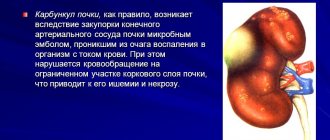
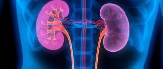
What is a kidney infarction?
Renal infarction is coagulative focal necrosis of one or both kidneys resulting from occlusion of the blood vessels of the kidney. The size and location of the infarction depends on the location of the vessel and its caliber. As a rule, the cortical layer of the kidney is affected, but the medulla can also be involved in the pathological process. Residual renal function depends on the extent of parenchymal damage.
Causes of kidney infarction
The causes of kidney infarction can be divided into two main groups:
- Thromboembolism of the renal artery (90%). Risk factors: atrial fibrillation, myocardial infarction, mitral stenosis, heart failure, cardiac surgery, aortic aneurysm, endocarditis, rheumatic disease, renal artery aneurysm or atherosclerosis. Thus, the main source of emboli is the heart.
- Hemorrhagic renal infarction due to renal vein thrombosis.
Cases of kidney infarction in sickle cell anemia, scleroderma, arterioneprosclerosis, Morphan's syndrome, etc. are described.
Prevalence of renal infarction
The prevalence of renal infarction is low. For example, the largest sample of patients with renal infarction in scientific studies was 50 patients. Since the main symptom of renal infarction is pain in the back or abdominal area, which simulates other pathological conditions, such as nephrolithiasis or pyelonephritis, the diagnosis is often delayed.
What happens during a kidney infarction?
Initial changes in renal infarction:
- Focal necrosis is triangular in shape, the base of which is located in the cortex (Fig. 1).
Fig.1. Kidney infarction – focal necrosis of a triangular shape.
Late changes in renal infarction:
- The kidney is reduced in size;
- Focal atrophy of the kidney parenchyma;
- Cortical atrophy and fibrosis.
Symptoms of kidney infarction
Symptoms of kidney infarction are nonspecific, so diagnosis is usually delayed. The severity of symptoms depends on the extent of the pathological process. The most characteristic symptoms of kidney infarction:
- Acute, sudden, persistent pain in the lower back or abdominal area occurs as a result of stretching of the kidney capsule.
- Nausea or vomiting is always accompanied by pain.
- Arterial hypertension is a common complication that develops several days after a heart attack and is associated with depletion of renal blood flow, leading to activation of the renin-angiotensin system.
- Heart rhythm disturbances.
- Hematuria (blood in the urine), oliguria or anuria (decreased or absent urine output).
- Anorexia.
- Temperature.
Patients with mild focal renal necrosis may not have symptoms of renal infarction.
Diagnosis of kidney infarction
Diagnosis of renal infarction is based on laboratory and imaging studies.
Urinalysis reveals the following changes:
- Proteinuria (high protein content in urine);
- Micro- or macrohematuria;
- Oligo- or anuria.
A biochemical blood test determines an increase in the level of intracellular enzymes (enter the blood due to cell destruction during necrosis of the renal parenchyma): aspartate aminotransferase, alkaline phosphatase, lactate dehydrogenase, creatine kinase. When renal function is significantly impaired, creatinine and urea levels increase.
A general blood test may reveal leukocytosis and an increase in erythrocyte sedimentation rate.
Computed tomography (CT) with contrast injection is one of the most informative diagnostic methods necessary to confirm the diagnosis of renal infarction. CT scan reveals the following signs of renal infarction (Fig. 2):
- A wedge-shaped hypoechoic focus with its base directed towards the kidney capsule;
- Zones of the renal parenchyma where there is no blood flow, corresponding to the course of the occluded renal artery, etc.
Fig.2. CT. The figure clearly visualizes the infarction of the left kidney.
Ultrasound examination of the kidneys allows you to visualize an area of increased echogenicity in the kidney. Doppler ultrasound is also a highly informative research method (Fig. 3).
Fig.3. Dopplerography. Lack of blood flow in the left pole of the kidney.
Angiography reveals :
- Normal intrarenal structure of the venous bed;
- Discontinuity of arterial vessels or defects in vascular filling (Fig. 4).
Fig.4. Angiography visualizes complete proximal occlusion of the left renal artery.
Digital (digital) subtraction angiography (DSA) – renal angiography with computer processing – is the gold standard in the diagnosis of vascular occlusion.
Diagnosis of the causes of kidney infarction
Diagnosis of the causes of renal infarction aims to find the source of the embolus or thrombus and establish the causes of hypercoagulation. Therefore, patients may be prescribed the following studies:
- Echocardiography - allows you to identify blood clots in the heart cavity and a patent foramen ovale.
- Holter monitoring is necessary to detect arrhythmias.
- Examination of the veins of the lower extremities to exclude deep vein thrombosis.
- Study of the hemostasis system (coagulation system).
- Analysis for tumor markers, if there is a suspicion of malignancy, etc.
Treatment of kidney infarction
Treatment of renal infarction can be divided into conservative and surgical.
Conservative treatment of renal infarction
Conservative treatment of renal infarction includes:
- adequate pain relief;
- blood pressure control;
- anticoagulant therapy.
Surgical treatment of kidney infarction
Surgical treatment of renal infarction, depending on the indications, can be carried out in the following ways:
- Nephrectomy is performed for venous infarction. Currently, preference is given laparoscopic operations. Laparoscopic nephrectomy operations are successfully performed.
- Intraarterial thrombolytic therapy.
- Thrombus or embolectomy. Currently, percutaneous thrombus or embolectomy operations are successfully performed using an aspiration catheter and the AngioJet system.
The article is for informational purposes only. For any health problems, do not self-diagnose and consult a doctor!
Author:
V.A. Shaderkina is a urologist, oncologist, scientific editor of Uroweb.ru. Chairman of the Association of Medical Journalists.
‹ Urine cytology Up Laparoscopic kidney resection ›