Before visiting the ultrasound diagnostic room, we suggest that you carefully familiarize yourself with how to properly and effectively prepare for an ultrasound. The main goal of recommendations for preparing for ultrasound is to increase the reliability and accuracy of the study results. Thanks to this you save your time and money.
Try to follow the recommendations below exactly. Performing them allows the ultrasound doctor performing the procedure to clearly and without interference visualize all organs and vessels located in the space being examined. With proper preparation, tissue visualization becomes clearer and the quality of the method is significantly higher. In this case, the patient, in turn, receives confidence that he has received a reliable and complete diagnosis.
Is preparation necessary?
No preparation required:
| Preparation required:
|
Preparation for ultrasound of the gallbladder with determination of function
It is carried out for adults and children as prescribed by a doctor, with the referring doctor indicating the type and quantity of a trial breakfast (eggs, chocolate, banana, etc.).
A week before the ultrasound it is recommended:
- completely abstain from alcoholic beverages;
- exclude gas-forming foods from the menu, since air bubbles accumulating in the intestines interfere with visualization. These foods include raw whole milk, raw unprocessed vegetables and fruits, legumes (such as soy, peas and beans), yeast breads and brown bread.
Three days before the planned procedure, you need to start taking the following medications according to the recommendations of your doctor:
- enzymes – any form of pancreatin. It is best taken in the form of microcapsules in a dosage of at least 10,000 units per meal. The capsule is drunk immediately before or during meals with a glass of water. If you suffer from any diseases of the digestive system, then you should check the dosage regimen with your doctor - you may need a drug with a higher content of enzymes;
- carminatives. The dosage of these drugs is standard, so you can check it with your pharmacist. Take medications every time after meals
- If you suffer from chronic constipation, then start taking lactulose - a tablespoon before bed.
On the eve of an ultrasound to determine the function:
- dinner should be light and nutritious. Let it be cereal porridge with minimal sugar content. Meals should be no later than 20.00
- Before going to bed, be sure to go to the toilet. If necessary, you can use a glycerin suppository. With this type of study, there is no need to cleanse the intestines with an enema.
In the morning, before visiting the doctor:
- Boil the eggs and separate the yolks, which will serve as a choleretic load. You can also use full-fat sour cream or cream for this. Sometimes Sorbitol solution is used for the same purpose.
- don’t even drink water, because the gallbladder will contract prematurely and the test results will be unreliable
- if you are planning the procedure for the afternoon, then breakfast should be at 7.00 and consist of light food.
Indications for ultrasound
The main indications for an ultrasound of the abdominal cavity: flatulence (increased gas production), bitterness in the mouth, nausea, vomiting, pain in the right hypochondrium and abdominal area (epigastric region), jaundice (yellowing of the skin, sclera of the eyes).
It should be done in case of persistent indigestion, chronic constipation or diarrhea, loss of appetite, sudden change in body weight (increase or decrease). Ultrasound of the abdominal organs is performed if there is a suspicion of oncological diseases (cancer of the liver, pancreas), metastases, benign neoplasms (tumors, cysts, adenomas), abscess, pancreatitis, hepatitis, hepatosis, cirrhosis of the liver, cholelithiasis, cholecystitis, ascites, biliary dyskinesia paths (including bending, constriction of the gallbladder).
An abdominal ultrasound is usually performed in a child who complains of pain in the abdomen, lower back (lower back) or girdle pain.
Ultrasound of the retroperitoneal space is prescribed for girdle pain, the presence of blood in the urine, suspected urolithiasis, tumors, pyelonephritis, glomerulonephritis, nephropathy, nephrosclerosis, diseases of the adrenal glands, pathology of blood vessels, lymph nodes.
Ultrasound of the retroperitoneal space allows you to see not only neoplasms, traumatic injuries or inflammatory processes, but also the condition of the blood vessels (the size of the lumen, the presence of atherosclerosis, blood clots, the location of the vessels), as well as an increase in regional lymph nodes.
Preparation
Preparation for ultrasound of the uterus and appendages (gynecological ultrasound, ultrasound of the pelvic organs)
When planning a visit to an ultrasound doctor, please note that ultrasound of the uterus and appendages is performed only on certain days of the cycle. A planned gynecological ultrasound is usually performed at the beginning of the cycle - a maximum of 8-10 days. We recommend that you undergo the study immediately after the end of menstruation, on days 5-7.
Your doctor may also order testing at a different phase of your cycle. This is due to the presence of suspicions of the development of a particular disease. Thus, to diagnose endometriosis, ultrasound of the uterus and appendages is performed in the second half of the cycle, and if bleeding or other complex conditions occur, on any day of the cycle.
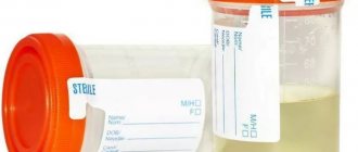
The gynecological ultrasound procedure requires certain preparation from the patient. It consists of eliminating from the diet a few days before diagnosis foods that contribute to increased gas formation in the intestines. These foods include raw whole milk, raw unprocessed vegetables and fruits, legumes (such as soy, peas and beans), yeast breads and brown bread. Immediately before the procedure, you must empty your bladder.
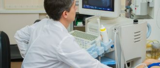
How is ultrasound diagnostics of the kidneys and adrenal glands performed?
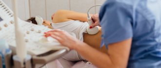
Ultrasound of the kidneys and adrenal glands is a painless procedure that does not require the use of painkillers. The patient lies down on a medical couch and frees the anatomical area of the abdomen from clothing. A special medical gel is applied to it. Its use ensures close contact with the ultrasound scanner sensor and improves the passage of ultrasonic waves.
Having applied the sensor to the treated area, the diagnostician begins to move it and rotate it at different angles, achieving maximum clarity of the image that is displayed on the screen. Penetrating into the soft tissues of the body, ultrasonic waves are reflected from them, which allows the formation of visual images. Based on them, the specialist makes a conclusion about the condition of the organs and existing pathologies.
Preparation for ultrasound of the abdominal organs, ultrasound of the abdominal vessels
The accuracy of the results obtained for diagnosing the abdominal organs depends on the amount of air in the intestinal loops. To reduce its amount and increase the accuracy of the study, preliminary preparation with diet is necessary.
Porridge, beef or poultry, low-fat cheese, soft-boiled eggs, and lean fish are allowed for consumption. Meals are fractional. The volume of liquid taken per day is 1.5 liters.
The study is carried out after eliminating from the diet foods that increase gas formation in the intestines and contribute to increased secretions of the gallbladder:
- Raw fruits and vegetables;
- Raw milk and dairy products;
- Legumes in any form;
- Fatty fish and meats;
- Sweet products;
- Alcohol;
- Carbonated drinks.
Direct ultrasound of the abdominal organs is performed on an empty stomach, six hours after the last meal (preferably in the morning).
What organs are examined by ultrasound of the retroperitoneal space?
The retroperitoneal cavity vertically occupies the area from the diaphragmatic muscle to the conventional border of the small pelvis, therefore ultrasound of the retroperitoneal space represents a one-time examination of tissues and organs located directly in this area.
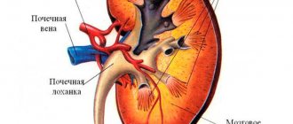
The following organs are located entirely within the retroperitoneal cavity:
- kidneys;
- ureters;
- adrenal glands;
- lymphatic ducts;
- aorta;
- inferior vena cava.
Also, this type of diagnosis is effective in relation to the condition of organs partially located in the retroperitoneal space, including:
- ascending and descending colons;
- duodenum;
- pancreas.
In addition to excluding pathological processes in individual organs, ultrasound of the retroperitoneal space evaluates the condition of fatty tissue, which performs a supporting function, allowing the organs to occupy their location.
The peritoneum should not be confused with the retroperitoneal space. Although, due to the adjacent location, simultaneous ultrasound diagnosis of the abdominal cavity and retroperitoneal space is often performed.
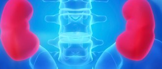
Preparation for ultrasound of the kidneys and adrenal glands
2-3 days before the diagnosis:
- Follow a special diet, especially if you are prone to flatulence. The diet involves excluding from the diet foods that increase gas formation: fresh vegetables and fruits, dairy products, black bread, carbonated drinks, legumes, cabbage, alcoholic beverages;
- Take laxatives if you have problems with bowel movements. They help cleanse the intestines of toxins and gases;
- Maintain normal fluid intake. A sufficient amount is about 1.5-2 liters of liquid. Juices, strong coffee and tea should be excluded.
On the day of the test, also follow the above recommendations. The breakfast menu may include:
- Buckwheat, oatmeal, pearl barley porridge;
- Low-fat hard cheese;
- Boiled lean fish (hake or pollock);
- One boiled egg.
An hour before the kidney ultrasound, you need to drink 2-4 glasses of water. This will ensure that your bladder is filled with enough fluid at the time of the test. If it is difficult to contain your bladder emptying before the examination, you can empty it, but then drink 1-2 glasses of water again.
How to prepare for an ultrasound of the retroperitoneum?
Ultrasound results can be distorted due to improper preparation of the patient for examination of the retroperitoneal space. The preparation is simple and does not differ from that for an ultrasound of the abdominal cavity. Diet. You should start a few days in advance with a diet that excludes ingredients that cause flatulence. For 2 days, it is worth removing from the diet all yeast-based products - bread, kvass, beer, as well as lactic acid products, legumes, sauerkraut, raw vegetables and fruits, carbonated drinks, alcohol. Gas-lowering drugs . During the diet, it is recommended to take activated carbon at the rate of 1 tablet per 10 kg of weight. Coal can be taken at once or divided into several doses. If the patient has increased flatulence, the day before the scan you should start taking medications such as Espumisan, Mezim-Forte. Colon cleansing. If there is a tendency to constipation, then the day before the procedure you will need to take a mild laxative to cleanse the intestines of excess toxins. Starvation. Ultrasound of the retroperitoneal space is performed on an empty stomach, so it is more convenient to do it in the morning. If, however, the procedure is scheduled for the afternoon, then a light breakfast in the form of crackers and tea is allowed. However, you need to remember that the fasting time before diagnosis should be at least 6 hours. Taking water . 30 minutes before the examination, you will need to drink 0.5-0.7 liters of water so that the sensor can better see the bladder, if an ultrasound of the retroperitoneal space and bladder is prescribed.
Previous NextPreparation for ultrasound, TRUS of the prostate gland (prostate)
Preparation for ultrasound of the prostate gland consists of two stages:
- Colon cleansing several hours before the test. To do this, it is recommended to give a cleansing enema in a volume of 1.5 liters of cool water. To cleanse the intestines, you can also use microenemas or special suppositories for insertion into the rectum. This is necessary to cleanse the intestines of feces and gases. Colon cleansing significantly improves the performance of the study;
- Bladder filling. An hour and a half before the examination, drink 1-1.5 liters of liquid (preferably pure water without gas) and do not empty your bladder until the examination.
In some cases, preparation for TRUS of the prostate also includes a special diet, which involves avoiding foods that enhance the process of gas formation: fresh vegetables and fruits, dairy products, brown bread, carbonated drinks, legumes, cabbage, and alcoholic beverages.
Preparation for ultrasound with Doppler (USDG, Dopplerography)
On the eve of the study, you must refuse to eat. A few hours before the examination, eliminate foods and medications containing nicotine from your diet. Nicotine provokes vasospasm, and the examination results may be incorrect.
If you are taking medications, not necessarily of the cardiovascular category, be sure to inform the ultrasound doctor about this before starting the study. Some chemicals included in the preparations can also negatively affect the results obtained.
Preparation for folliculometry
During the entire period of folliculometry, foods that promote and intensify the process of gas formation in the intestines are excluded from the patient’s diet. For example, it is strictly not recommended to eat legumes, fresh vegetables, fruits, carbonated drinks, alcohol, and juices.
Further preparation depends on the method of examination. The ultrasound doctor will inform you about this before starting the diagnosis. So, if the examination is carried out transabdominally, then preparation consists of taking 1-1.5 liters of liquid 1-1.5 hours before the examination. This type of diagnosis assumes a full bladder. If folliculometry is performed transvaginally, then, on the contrary, it is necessary to empty the bladder before the examination.