The purpose of examining the pelvic organs in men is to determine the cause of the disease, including reproductive function and erectile dysfunction. The ultrasound method is completely safe, there are no harmful effects or delayed consequences. In addition, this method provides high reliability, thereby making it possible not only to treat based on the results, but also to monitor dynamics and carry out prevention.
Using ultrasound, as part of a urological examination, the condition of the following organs is studied:
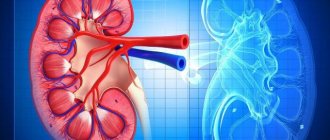
- kidneys, adrenal glands;
- ureters and bladder;
- prostate in men;
- testicles, scrotum.
There are a number of indications for which this examination is prescribed, including:
- Impaired bladder emptying.
- Pain during bowel movements.
- Urging at night without any result.
- Pain in the suprapubic region.
- Blood and/or pus is found in the urine.
- There was injury to internal or external organs.
- Diagnosed with infertility.
- Erectile dysfunction.
- The presence of formations in the pelvic organs.
- Lack of erection or weakening of it.
Also, such an examination cannot be done without such an examination during operations - this is required to accurately determine the features of the structure and location. Ultrasound can also be prescribed after surgery to monitor the condition of the operated organ. In addition, this measure will help prevent complications.
Contraindications for ultrasound:
- Wounds in the area of research.
- Burns.
- The presence of an impressive layer of fat.
All these contraindications apply to the abdominal technique. In this case, a rectal examination is performed. It is carried out through the anus.
How does it feel?
If a transabdominal ultrasound is performed, the patient will likely feel pressure in the bladder and a strong urge to urinate because the bladder is full. The gel applied to the abdomen may feel cold. The patient will feel a slight pressure from the transducer as it passes over the abdomen. If the patient has an injury or pelvic pain, pressure from the transducer may be painful.
Moderate discomfort is usually felt during a transrectal examination. The patient feels pressure from the transducer as it is placed in the rectum.
If a biopsy is performed during an ultrasound, there may be pain when taking the sample.
Indications and contraindications for the procedure
A doctor usually gives you a referral for an ultrasound scan, but if you wish, you can make an appointment yourself. Reasons for examining the adrenal glands may include the following physical manifestations:
- muscle weakness;
- a sharp increase in the amount of hair on the body;
- infertility and unstable menstrual cycle;
- sexual dysfunction;
- sudden change in body weight.
Get your kidneys and bladder checked urgently if you have the following symptoms:
- presence of sediment, pus, blood in the urine;
- decrease or increase in the amount of urine;
- presence of injuries or bruises in the lumbar region;
- pain and pulling sensations in the lumbar region;
- increased body temperature and development of swelling;
- cutting sensation when urinating.
There are no contraindications to the procedure; anyone can undergo examination. However, in order to get an accurate result, you should properly prepare for the examination.
Indications for testing
An ultrasound diagnostic method in men is performed to identify and confirm various diseases of the reproductive and urinary systems. The data obtained using ultrasound helps to accurately determine the diagnosis and make a differential diagnosis of various diseases in complex cases. In addition, ultrasound diagnostics are performed to identify signs of some latent and chronic sexually transmitted infections.
Often indications for this diagnostic method in men may include:
- blood (red blood cells) in the urine;
- spermaturia – the presence of sperm in the urine;
- suspicion or presence of male infertility;
- pain of varying intensity in the pelvic area;
- erectile dysfunction or, conversely, pathologically prolonged arousal (priapism);
- dysuric phenomena, which can lead to pain in the bladder during emptying, high frequency of urination, difficulty or, conversely, urinary incontinence;
- to detect tumors;
- preventive diagnostics, disease screening;
- performing a biopsy under ultrasound guidance.
Preventive research significantly helps reduce mortality from cancer of the genitourinary system in men. They make it possible to detect diseases in the early stages, which increases the efficiency and speed of treatment and recovery.
We can conclude that ultrasound is highly informative for diagnosing reproductive and urogenital problems. Ultrasound is one of the most reliable and accurate methods for diagnosing and identifying various pathologies in the pelvic area.
A pelvic ultrasound may be done for:
- identifying the cause of dysuric problems;
- assessment of the size of the bladder depending on its filling;
- checking whether the bladder empties completely during urination;
- directions for needle placement during biopsy or when draining fluid from a cyst or abscess;
- detection of rectal cancer and its response to treatment.
Method capabilities
Ultrasound of the genitourinary system can detect a wide variety of pathologies.
- Kidneys. Ultrasound can confirm the diagnosis of acute and chronic inflammation, detect stones, abnormalities of kidney development, and various neoplasms. Ultrasound can be used to monitor the effectiveness of treatment.
- Examination of the ureters provides information about the presence of stones and narrowing of the lumen. Occasionally, examination reveals a neoplasm of the ureteral wall. To clearly see this organ, the doctor performs an intracavitary ultrasound. In this case, a miniature sensor is inserted through the bladder into the ureter.
- The doctor may see an accumulation of blood and pus in the bladder. With inflammation, changes in the wall of this organ are visible. Stones, polyps and bladder tumors can be identified.
- In men, the examination includes examination of the prostate, scrotum and penis.
- Ultrasound of the prostate reveals its adenoma. This is a benign growth of gland tissue. It is possible to detect malignant neoplasms and inflammation of the prostate. To examine this organ, a special probe is often inserted into the rectum. This examination is called TRUS (transrectal ultrasound) of the prostate.
- When examining the scrotum, inflammation of the testicles, dilation of the testicular veins (varicocele) or torsion of the spermatic cord are diagnosed. If one or both testicles are absent in the scrotum, it is possible to “search” for it using ultrasound in the abdominal cavity or inguinal canal. The method is also informative for scrotal injuries.
- Ultrasound of the penis is prescribed for injuries to the organ or problems with erection.
The ultrasound machine at the Novomed clinic allows you to perform Doppler ultrasound, which helps to assess the movement of urine through the ureters. Its slowdown or reverse flow indicates a narrowing of the ureter or a stone in it. Sometimes the therapist prescribes Doppler ultrasound of the renal vessels. This can provide valuable information about the causes of severe hypertension.
Preparing for the study
Preparing the patient for this diagnostic examination depends on the ultrasound diagnostic method. The doctor should be informed if the patient has had an x-ray with contrast material (eg barium) within the last 2 days. Barium that still remains in the intestines may subsequently interfere with ultrasound examinations.
The patient may need to remove or lower clothing below the waist before the test.
Preparation for the transabdominal method:
- avoiding foods that contain high amounts of fiber and legumes 2-3 days before the test to minimize gas production;
- it is advisable to refrain from eating for 5-6 hours before the start of the study;
- drink half a liter of still water 2 hours before the start of the study;
- 2 hours before the ultrasound you should refrain from urinating;
- if you have a strong desire, you can urinate a little, but not completely, and after that you need to drink an additional glass of water;
- If the patient cannot drink the required amount of water, it is possible to fill the bladder with water through a thin flexible tube (catheter) inserted into the bladder.
To prepare for the transrectal method, all the above preparation steps are used, but added to them:
- the use of drugs that cleanse the intestines, for this the patient drinks 3-4 packs on the eve of the test;
- if the study is scheduled for the afternoon, then drink two packets in the morning on the day of the study;
- a cleansing enema is an alternative method and is performed in the morning before the examination, and the volume of liquid that is injected into the rectum must be at least 200 ml;
- If a person is having a prostate biopsy, they may be prescribed antibiotics the day before the test.
How it's going
The patient undresses to the waist and lies on his back.
Sometimes the doctor may ask you to turn on your side or stomach. To diagnose kidney prolapse, the patient must be in an upright position. A special gel is applied to the skin. The doctor then moves the sensor over the body, pressing it lightly. In this case, an image of the organs is obtained on the screen. The examination lasts about 30 minutes. The patient does not experience any discomfort. The results can be recorded on any digital media.
Registration at the Novomed clinic is carried out by telephone. Remember that an accurate diagnosis is the basis for effective treatment. Call us at the first alarming symptoms!
Types of Ultrasound
When performing an ultrasound examination of the pelvis in men, there are two main directions: transabdominal ultrasound and transrectal ultrasound. Each method has found its justification and has its own strengths in diagnosing diseases.
Transabdominal method
During a transabdominal ultrasound, a transducer is placed on the anterior abdominal wall. By moving and turning it at different angles, the doctor can examine the structure of the patient’s internal organs in as much detail as possible, assess their condition, blood supply (with additional Doppler examination) and the presence of any changes.
This method is best suited for carrying out preventive studies and does not cause discomfort in humans. In contrast to the transrectal method, the accuracy of the transabdominal examination technique is somewhat lower. In this regard, this method is suitable for determining the presence of a voluminous pathological formation in the pelvis and bladder. It is worth noting that during the transabdominal diagnostic method, the bladder must be in a full state. A full bladder is an organ that conducts ultrasound waves well and makes the examination more effective. If an ultrasound is performed in an emergency, the bladder may be filled with water through a thin, flexible tube (catheter) inserted into the bladder.
Transrectal method
Best materials of the month
- Coronaviruses: SARS-CoV-2 (COVID-19)
- Antibiotics for the prevention and treatment of COVID-19: how effective are they?
- The most common "office" diseases
- Does vodka kill coronavirus?
- How to stay alive on our roads?
Transrectal ultrasound is performed using a miniature ultrasound transducer to minimize patient discomfort. The sensor is inserted directly into the rectum, which must first be cleaned and prepared.
In most cases, the transrectal ultrasound method is used for a detailed examination of the prostate gland and determining the severity of pathology in its tissues. The transducer sends and receives sound waves through the wall of the rectum into the prostate gland, which is located just in front of the rectum. This improves the accuracy of the results. This method allows you to identify even the smallest cysts in the prostate gland - up to 0.2 cm. In addition, this method is the best choice in situations such as urinary incontinence or a small bladder volume, which usually makes transabdominal ultrasound diagnosis very difficult.
Preparation and technique for performing abdominal ultrasound
The examination is carried out in two ways: transabdominal, that is, through the anterior wall of the abdominal cavity, or transrectally, that is, by inserting a sensor through the rectum.
Abdominal diagnosis is carried out using a special sensor, which is moved along the anterior wall of the abdomen. The sensor emits waves that are displayed from the organ being examined; the data is transmitted to the monitor after special processing by a computer.
An important indicator for diagnosis is residual urine; normally its amount is about 50 ml. To determine this indicator, the study is carried out twice:
- the first time with a full bladder;
- the second time immediately after bowel movement.
Preparation for such an examination is necessary and important. You should follow a special diet for a few days. Her requirement is to exclude fatty foods, legumes, cabbage, and whole milk. You should not eat foods that cause gas formation. If there are problems with this, you should take additional medications that promote the release of gases. A mandatory requirement is to fill the bladder. To do this, you need to drink at least 1 liter of water 1 hour before the appointed time. It should not contain gases or sweeteners - just plain clean water.
During the study, the patient lies down on the couch and frees his stomach from clothes. A gel is applied to the skin and diagnostics are carried out using a sensor. Its duration is no more than 20 minutes. At the end of the procedure, you will receive a protocol with which you should go to the attending physician.
Transrectal urological ultrasound: what you need to know about the procedure
The method of examining an ultrasound probe through the rectum is called rectal. Allows you to obtain more detailed information regarding the condition of the pelvic organs in men. It is also prescribed if the traditional method cannot be applied.
Preparation for this study is somewhat less than for the abdominal one, but it is also necessary. The most important thing is to cleanse the intestines. To do this, you need to give 3 enemas - two of them in the evening, one in the morning before the study. The water for the enema is cool, in a volume of 1-1.5 liters.
Since the sensor is inserted into the anus, unpleasant sensations are possible, but the specialist will try to minimize them. To do this, the patient needs to lie on his left side and bend his legs, pull his knees towards the chest. The first step is palpation, only after that the sensor is inserted. To ensure the hygiene of the process, a medical condom is put on it, which is lubricated with a special gel. In order to avoid discomfort, it is important for the patient to relax as much as possible.
Conclusion. Urological ultrasound when examining the organs of the male genitourinary system allows you to find out key data about the condition of the bladder, prostate, and testes. Based on the data obtained, we can talk about the pathology and the degree of its development.
Price for services
| Ultrasound examination (ultrasound) of the bladder | 1 research | 550.00 |
| Ultrasound examination (ultrasound) of the bladder with determination of residual urine | 1 research | 550.00 |
| Ultrasound examination (ultrasound) of the prostate gland transrectally (transrectally and transabdominally) with determination of residual urine | 1 research | 1 100.00 |
| Ultrasound examination (ultrasound) of the urinary tract (kidneys + bladder + prostate gland (transabdominal and transrectal) with determination of residual urine | 1 research | 1 320.00 |
| Transrectal ultrasound examination (US) of the prostate gland (TRUS) | 1 research | 930.00 |
| Ultrasound examination (ultrasound) of the scrotal organs | 1 research | 550.00 |
Where can I get a pelvic ultrasound?
Currently, ultrasound diagnostics is available in almost every medical institution, and when choosing, patients are often guided only by the criterion of how much a pelvic ultrasound costs. But this may lead to additional costs.
Most clinics have low-cost devices installed that can indeed detect serious pathological changes, but only developing processes fall out of sight. Additionally, not all equipment has a Doppler ultrasound sensor, which allows you to assess the condition of the blood vessels.
In any case, if changes are found using a simple device, the doctor may additionally refer you for a clarifying ultrasound of the pelvic organs. The price may depend not only on the class of the device, but also on the professional level of the ultrasound specialist.
Therefore, if you are given this price and you are guided by how much a pelvic ultrasound costs, then compare the class of equipment and the training of specialists.