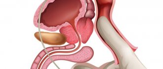
The prostate is often called “a man’s second heart.” But not all men know what important functions their “second heart” performs. The testicles produce sperm, this is necessary for procreation. What about the prostate gland? Its secretion makes sperm more mobile and viable. Prostate dysfunction can result in infertility. In addition, during the ejection of semen, this organ, like a valve, closes the urethra. An enlarged prostate gland, for example, with prostatitis or benign hyperplasia, constantly puts too much pressure on the urethra - hence difficulty urinating and frequent trips to the toilet at night.
Our expert in this field:
Isaev Artur Ramazanovich
Oncologist, urologist, surgeon
Call the doctor
In order to understand the nature of the pathological processes occurring in the prostate gland, an analysis of its juice is sometimes necessary. Taking prostate secretions is not a very pleasant procedure, but in some cases it is necessary.
Detailed description of the study
The study of prostate secretions is used to determine pathologies of the prostate gland.
The secretion of the prostate gland is a biological fluid that is part of semen. Necessary for the normal functioning of sperm outside the male body (preserving their motor activity and fertilizing ability).
The study of prostate secretions includes determining:
- consistency;
- colors;
- transparency;
- cellular composition of biological fluid.
The study of the cellular composition of prostate secretion is aimed at detecting fungi, leukocytes, erythrocytes, epithelial cells, trichomonas and other microorganisms. Microscopy is important for the identification of lecithin grains, amyloid bodies, macrophages, cholesterol crystals and Trousseau-Lallemand bodies.
The biomaterial used for research is the secretion of the prostate gland, which is placed in a dry and sterile container with a volume of 3–5 ml. Since, due to the anatomical location of the prostate, the procedure is quite complex in terms of execution, urine is allowed to enter the biomaterial.
Study of prostate secretion (cellular composition, microflora)
Study of prostate secretion (cellular composition of microflora)
- bacterioscopic examination, which is performed to identify leukocytes, erythrocytes, epithelial cells, gonococcus and other bacteria, as well as trichomonas and fungal mycelium in the prostate secretion;
which is carried out to diagnose various clinical forms of prostatitis. Bacterioscopic examination
Bacterioscopic examination is the study of bacteria under a microscope. Bacterioscopy is widely used when an infectious process is suspected and the presence of purulent-inflammatory manifestations of the disease, as well as during clinical examinations in gynecological and obstetric practice.
Normal human microflora
Normal human microflora is a set of microbiocenoses that occupy numerous ecological niches on the skin and mucous membranes at the points of contact of the human body with the environment.
Leukocytes in prostate secretion
Normally, the number of leukocytes in the test sample should not exceed 10 per field of view. A higher number indicates the presence of an inflammatory process. Depending on whether microorganisms can be identified, prostatitis is divided into bacterial and non-bacterial. Normally, prostate secretion is a sterile environment. However, if it enters a non-sterile urethra, the sample may become contaminated, as a result of which microorganisms can be detected during microscopy, but their number does not exceed one in the field of view. A large number of bacteria indicates bacterial prostatitis. Most often, it is possible to identify gram-negative bacilli (E. coli, Enterobacter, Proteus, etc.).
In patients under 35 years of age, the most common causes of prostatitis are Neisseria gonorrhoeae and Chlamydia trachomatis. The detection of a large number of leukocytes and gram-negative diplocococci may indicate the presence of gonorrhea, but bacteriological culture is performed to make a final diagnosis. On the other hand, microscopic findings may provide the basis for empirical therapy before the final culture result is available. Identification of Chlamydia trachomatis by microscopic or bacteriological examination is difficult, especially in chronic infections. The detection of mycelium or pseudomycelium of the fungus, as well as trichomonas in the prostate secretion, is always a pathological sign that requires close attention from a doctor.
The absence of microorganisms in the prostate secretion in a patient with pain syndrome indicates the presence of a special clinical form of prostatitis - chronic non-bacterial prostatitis, or chronic pelvic pain syndrome. Also, as in the case of bacterial prostatitis, the final diagnosis can be established only after obtaining the results of bacteriological culture.
Red blood cells in prostate secretion
Another significant clinical and laboratory indicator assessed using microscopy is the presence of red blood cells and their number. Normally, single red blood cells in the field of view may be present in the secretion of the prostate gland. Their elevated levels are typical for prostatitis and prostate adenocarcinoma. It should be noted that any inflammatory and oncological diseases of the genitourinary system can be accompanied by the appearance of red blood cells in the urethra and prostate secretions. Unlike kidney diseases, prostate pathology is accompanied by the appearance of unchanged red blood cells, which, however, is also characteristic of diseases of the bladder and urethra. However, microscopic examination of prostate secretions is not used as a screening for prostate cancer.
The study of prostate secretion (cellular composition of microflora) allows us to assess the state of the prostate microflora. Based on the data obtained on the number of leukocytes, erythrocytes, epithelial cells, as well as the presence or absence of microorganisms, prostatitis can be classified as one of the 4 main clinical forms of this disease: acute and chronic bacterial prostatitis, chronic pelvic pain syndrome and asymptomatic chronic prostatitis.
Indications
For symptoms of prostatitis: incoming dysuria, pain or discomfort in the perineum and groin areas, impotence, painful ejaculation, as well as unmotivated weakness and discharge from the urethra.
Preparation
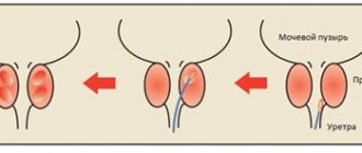
The material is collected by a doctor. They obtain the secretion through prostate massage. The secretion released from the urethra is collected in a sterile tube. Sometimes the secretion of the prostate gland does not end up in the urethra, but in the bladder. Then the centrifuge of the washing fluid released from the bladder after massage of the prostate gland is subject to examination.
Interpretation of results
Reference values for various types of microorganisms depend on their location (point of collection of biological material).
Reasons for detecting a large number of leukocytes and bacteria:
- acute and chronic bacterial prostatitis;
- other infectious and inflammatory diseases of the genitourinary system: urethritis, cystitis;
- benign prostatic hyperplasia;
- prostate adenocarcinoma.
Reasons for detecting a large number of red blood cells:
- acute bacterial prostatitis;
- mechanical trauma or surgical manipulation (cystoscopy);
- prostate adenocarcinoma.
Negative result (absence of leukocytes, erythrocytes and microorganisms):
- norm; control of treatment against the background of the disease;
- incorrectly taking a secret for research.
References
- Vorobiev A.V. Prospects for prevention, diagnosis and staging of prostate cancer / A.V. Vorobyov, P.I. Krzhivitsky // Practical oncology. – 2008. – No. 9 (2). – pp. 71–82.
- Kurbatov D.G., Kuznetsky Yu.Ya., Algorithm for diagnosing chronic prostatitis/chronic pelvic pain syndrome. file:///C:/Users/1/Downloads/8530-13448-1-PB%20(2).pdf
- Anderson RU, Weller C. Prostatic secretion leukocyte studies in nonbacterial prostatitis (prostatosis). J Urol 1979; 121: 292–294.
- McNaughton Collins M., Stafford R., O, Leary M., Barry M. How common is prostatitis? A national survey of physician visits. J Urol 1998; 149: 1224—1228.
- Naber KG EUA Guidelines on urinary and male genital tract infections.2002. P. 49-55.
Preparation for analysis of prostate secretions
Prostate secretion for analysis when prepared according to the recommendations of a urologist will be most informative for laboratory examination.
However, there are universal rules that will be useful to all patients.
- 5-7 days before your visit to the laboratory, you should avoid drinking alcohol, visiting a bathhouse or sauna, or taking a bath.
- It is also undesirable to take medications during this period, therefore, when taking a test against the background of any therapy, the attending physician should be informed about the fact of taking medications.
- When conducting an analysis of prostate secretions, preparation includes abstaining from sexual intercourse and masturbation 2-3 days before the test.
- Directly on the day of taking prostate secretions for analysis, preparation consists of performing a douching (enema) of the rectum and careful hygiene of the groin and gluteal area. In addition, you should urinate before extracting prostate juice.
Normal indicators
The laboratory report is deciphered by the attending physician. In making a diagnosis, he relies on the morphophysiological qualities of the prostate secretion, using the medical standard as a basis.
It provides the following indicators:
- The amount of material should vary from 0.5 to 2 ml. If the amount is less, which is caused by difficult secretion of juice, then this signals inflammation.
- The color should be similar to the tone of milk diluted with water. But a white color with a yellow tint, or even complete transparency, hints at prostatitis.
- Normal density is 1.022 units. There should not be any deviations from the indicated figure, and if they do occur, then they speak of a developing pathology.
- The environment's reaction remains neutral. In rare cases, a minimal deviation towards the alkaline indicator is allowed. But oxidation is the first sign of the inflammatory process. Indirect evidence will tell you that the fertilizing qualities of sperm are gradually disappearing.
- When examined microscopically, lecithin grains should completely cover the entire field of view. The standard is over 10 million units of lipoid bodies per milliliter of juice. If the number is far from the typical level, then this signals inflammation. Sometimes the grains are not visible at all, which indicates an extreme stage of the disease. Such indirect evidence often indicates impaired sperm fertility.
A separate assessment of leukocytes is carried out, for which specialists use special devices. An ideal, or close to it, count should show about three hundred cells per 1 μl of biological material. But if the analysis is studied using a standard microscope, which has a volume magnification factor of up to 280, then the indicator should be equal to 10 leukocytes. So many elements must come into view at one time. But if you twist the magnification factor to almost the maximum in clinical laboratories of 400 units, then the number should drop to five cells in one field of view.
If the laboratory technician cannot detect a single leukocyte at all, this does not indicate any abnormalities in the patient’s health. But an increase in the approved standards by 1 μl is the first alarm bell.
The absolute absence of red blood cells looks quite normal, but if a few units still appear in the field of vision, then this is also not a reason for concern. But exceeding one level is sometimes a strong argument in favor of additional testing for cancer markers. The emergence of a malignant tumor, which quickly metastasizes into the gland tissue, without surgical treatment threatens to damage neighboring internal organs.
Sometimes the abundance of red blood cells indicates another, but no less dangerous disease - a pronounced change in the prostate, which is caused by severe forms of prostatitis.
The cells of the desquamated epithelium of the excretory ducts deserve close attention. If there are no more than a couple of them in the field of view, then everything is in order. But an increase in the direction of ten indicates that an inflammatory mechanism of the descamatous type has started in the body. This means the activation of pathological peeling of the epithelial lining.
In a healthy man, giant cells cannot be detected in tests. But if the patient is sick, then they may manifest themselves, which indicates chronic inflammation. The same will happen if a person has congestion.
The situation with Böttcher crystals is a little different. This is the name for inclusions that are formed during the solidification and subsequent drying of a mixed biological product of various secretions of the male reproductive system. There is no clear framework for the normal state, so the crystals do not have significant diagnostic value.
This means that although it is possible to determine the average number of inclusions, the information received will be of little use, since inflammation occurs both with and without them.
Close attention should be paid to the possible presence of pathogenic microorganisms such as:
- trichomonas;
- gonococci.
In general, they should not be in the results, but since they have shown themselves, this is practically direct evidence of prostatitis.
Also, the mycelium of the fungus should not make itself felt if everything is fine with male sexual health. As soon as it is sown, this automatically means a fungal infection of the gland tissue, or a mixed infection, which usually takes longer to cure.
The results must include a note about opportunistic microflora. The standard includes a single number of cocci, as well as varieties of certain rods that are part of the human body. But exceeding the maximum permissible parameters indicates that a process of nonspecific inflammation has started in the body.
If necessary, the doctor may request an additional examination of the secretion, which means checking for the presence of a fern symptom. To do this, the laboratory assistant adds a microscopic dose of physiological sodium chloride solution to the patient’s juice collected on a glass slide.
The measure aims to confirm some possible deviations. But to do this, you will first need to dry the preparation and then examine it using a light microscope. If the composition of the secretion has any atypical physical or chemical deviations, then sodium chloride molecules precipitate. At the same time, they form a pattern that to some resembles the imprints of a fern leaf.
How is prostate secretion analyzed?
An analysis of prostate secretions is carried out by a medical professional (usually a urologist).
How is prostate secretion analyzed? After treating the gluteal area with an antiseptic, the doctor inserts a finger 3-4 cm into the anus, where the prostate gland is located under the intestinal wall. The lobes of the gland are massaged, and the secretion obtained from the urethral opening is collected in a test tube.
How long does it take to analyze prostate secretions? The procedure itself takes only a couple of minutes, so the patient usually does not feel much discomfort.