Treatment of brain cancer
The choice of treatment for a brain tumor depends on the type, size and location of the tumor, as well as the patient's general health.
After a diagnostic examination, consultation with a neurosurgeon and radiosurgeon, radiation therapist and chemotherapy, your attending physician will develop a treatment regimen with maximum effectiveness and minimal risk.
Therapy of brain tumors is complicated by the following circumstances:
- In the same lesion, there are usually many clones of abnormal cells that grow at different rates, respond differently to therapy, etc.
- Many drugs cannot reach malignant cells due to the physiological characteristics of the brain.
- If in other parts of the body cancer is removed along with part of the healthy tissue for preventive purposes, then during surgery to remove a brain tumor this cannot be done, since every millimeter must be protected here. Moreover, it is also almost impossible to completely cut out the tumor itself, because it usually does not have clear boundaries.
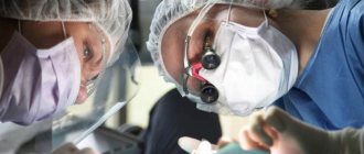
Surgery to remove a brain tumor
During surgery, the neurosurgeon tries to remove as much abnormal tissue as possible. Such operations are justified because even after partial removal of a brain tumor, the signs and symptoms of the disease may decrease. Astrocytomas and other neoplasms located in accessible places, away from vital centers, nerve plexuses and blood vessels, are surgically removed. Recently, alternative non-surgical methods to classical surgery have been actively developing.
Stereotactic radiosurgery is a radical non-surgical method
Therapy with this method occurs using computerized Gamma Knife, Cyber Knife and TrueBeam installations. In some cases, stereotactic radiosurgery is the only possible treatment for brain tumors. This gentle method is often used instead of surgery to remove a brain tumor and when there are no direct contraindications for classical surgery.
Treatment of a brain tumor with a cyberknife
The peculiarity and main advantage of stereotactic radiosurgery is the high precision of irradiation. This allows you to increase its intensity several times, while simultaneously reducing the radiation dose to healthy tissue. Such directed beams easily reach places where the surgeon’s hands cannot reach. And they destroy cancer cells, causing virtually no harm to the patient. Stereotactic surgery is effective in treating cancer metastases from other organs, various types of gliomas, etc. This method successfully removes acoustic neuromas, pituitary adenomas, chordomas, and hemangiomas.
Treatment with Gamma Knife
Gamma Knife has the highest focusing accuracy, but there is a limitation on new growths of no more than 3-4 cm, and they must be of the correct shape. Cyberknife, thanks to a unique irradiation technique, allows you to destroy malignant neoplasms of irregular shape and fairly large size (up to 6 cm). At the same time, the effect of the CyberKnife itself does not cause any discomfort in the patient.
Tomotherapy
This is a gentle and highly effective method of treating inoperable tumors with a directed proton flow. This method can be used, for example, in the destruction of tumors of the optic nerve, or neoplasms in young children and for other indications. Tomotherapy can be done in Russia in only a few centers. Tomotherapy is also carried out at the Voronezh Interregional Oncology Center.
Radiation therapy
In certain cases, the patient is prescribed external beam radiation therapy (RT), and extremely rarely, brachytherapy (contact irradiation). External beam radiation, for example, is indicated for large tumors, as well as when it is necessary to irradiate the entire brain (for example, for secondary cancer). Radiation therapy is often used after brain surgery to stabilize the results.
Chemotherapy
Chemotherapy drugs can be prescribed in tablets or injected into a vein. Sometimes, during surgery, implant capsules with carmustine are placed in the tumor bed.
Chemotherapy may be effective in the complex treatment of brain tumors such as:
- anaplastic astrocytoma,
- astrocytoma grade 4,
- oligogastrocytomas and oligodendriogliomas with a high degree of malignancy.
Targeted drug therapy
This method is characterized by a targeted effect directly on cancer cells, and it is increasingly used in combination with other methods of treating brain tumors.
Treatment of brain lipoma
If the neoplasm does not cause discomfort to a person and is not a malignant tumor, then doctors only try to monitor the progression of the disease. The patient is not limited in anything. Periodically, the patient must appear at the hospital for tests. A course of special injections is also prescribed. Diprospan is prescribed for injections. Using a thin needle, the medicine is injected into the tumor.
When an increase in formation occurs, this means that at one point the fatty lump inside the skull will begin to grow and go from neutral to a dangerous cancerous formation.
The best period for getting rid of lipomas is the early stage, when the tumors are small and do not create much inconvenience.
How to treat a lipoma if it spreads to both hemispheres? Chemotherapy and radiation will inevitably be required. Surgery for a wen is very difficult, so this method is rarely used. Doctors prescribe each type of surgical intervention, depending on the complexity of the case.
Craniotomy
The essence of the method is that a small piece of the skull is cut out from the skull. And through this hole the tumor is removed. This is a very traumatic procedure. However, its effectiveness has been proven by the practically absence of relapses, since the source of pain is localized and completely removed.
From the editor: Symptoms and dangers of VSD in adolescents
Endoscopic phenomenon
A hole large enough to insert the endoscope is drilled into the skull. Using cameras, doctors get to the tumor and remove it with special instruments. Interhemispheric lipoma of the brain is highly treatable. Lipoma is difficult to remove if it is intertwined with blood vessels and nerves. Any careless action can cause brain dysfunction, which will lead to disability for the patient.
Puncture-aspiration method of removal
With this method, a hole is also made in the skull and a needle is inserted. Having reached the lipoma, the adipose tissue is pumped out. But the likelihood of a new fat capsule appearing is high.
The direction for surgical intervention and removal of the lipoma along with the capsule is only an increasing benign tumor in size and pressure, which gradually increases and produces irreversible changes.
Every time, faced with a lipoma and, especially, the need to remove it, a specialist is obliged to choose the least invasive method of intervention in the human brain.
Removal occurs on an outpatient basis. After the operation, you will remain in the hospital for another 10-14 days. It all depends on the complexity of the procedure.
Scientists have found that this type of tumor is not susceptible to high X-ray radiation and to drugs used in chemotherapy.
If treatment is carried out in the early stages, complete relief from the disease is possible. If formations have begun their intensive growth, this leads to negative consequences.
A lipoma in the head never leads to death, being only a benign formation. However, it is in the brain that it is possible to develop into the opposite quality. This may occur due to lack of early diagnosis. The patient appears in the hospital when only severe consequences remain to be treated. In such situations, surgical intervention has saved many lives.
The method of pain relief depends on how intense the pain is. If the pain is not severe, then a simple and cheap painkiller will do. To relieve more serious symptoms, take No-shpa.
Rehabilitation
In the postoperative period after surgery for a brain tumor, the care of family and friends is very important. Often, a person needs to learn again things familiar from childhood, such as walking
It is important to help him take care of himself, get to the toilet on his own, relieve himself, get dressed/undressed, go to bed, etc.
Rehabilitation after removal of a brain tumor
How long recovery after surgery will take will depend on several factors:
- Psychological and physical condition of the patient;
- age category;
- location of the tumor body;
- degree of tissue damage.
On average, recovery time is 3-4 months. The rehabilitation process includes a set of activities such as physiotherapy, massages, exercise therapy, and sessions with a psychotherapist. During rehabilitation, the patient must give up heavy physical labor, contact with pesticides, and avoid stressful situations. Patients need to go through several stages of recovery.
If the recovery process is successful, after 2 months, rehabilitation after removal of the OGM continues at home.
Purpose of rehabilitation therapy
Rehabilitation after tumor removal is aimed at restoring the functionality of the brain centers, with the goal of helping the patient return to a normal lifestyle. If the consequences of tumor removal do not allow the complete restoration of brain tissue structures, the primary task of recovery is to adapt the person to the emerging limitations.
The rehabilitation program is drawn up separately for each case. To begin with, set short-term goals. First of all, the patient is taught to sit up on the bed without assistance. After completing the first task, a new one is assigned to the person. The methodology for setting short-term goals allows us to adequately assess the dynamics of recovery. You should not refuse psychological support from a qualified specialist. Psychological therapy is often needed not only by the patient, but also by his family.
Physiotherapy
Physiotherapeutic effects are aimed at restoring the functionality of the musculoskeletal system. Under the supervision of a physiotherapist, the patient learns to move independently or in a wheelchair.
Massage
Massage therapy is aimed at improving blood circulation in tissues, which promotes the rapid restoration of damaged cells. Massage can increase sensitivity and tone muscles, improve tissue structure.
Massage courses are repeated several times. At the beginning of the recovery period, all procedures are carried out under the supervision of doctors; subsequently, massage is done at home.
Exercise therapy
Physical activity is indicated before and after trepanation. In the first case, physical therapy is aimed at increasing muscle tone, training the cardiovascular system, as well as the respiratory system. In the postoperative period, exercise therapy is prescribed to restore the functionality of the body as a whole. The level of stress largely depends on where the tumor is located.
At the beginning of therapy, the exercises are performed lying down. Basically, this is breathing exercises. If there are no contraindications, the list of motor exercises is gradually expanded. Over time, the exercises begin to be performed while sitting, and then standing. Exercises should not cause pain or fatigue.
Forecast and consequences
In the majority of people who have lost the ability to move due to AMS, motor functions are restored after surgical manipulation. According to statistics, recovery is successful in 60% of patients. Vision is restored in 86% of patients. Consequences of operations such as mental disorders are extremely rare. Most often, pathological conditions appear in the first 3 years after surgery.
Disturbances of the central nervous system occur extremely rarely, only in 6% of patients. In such cases, people lose the ability to speak and take care of themselves. The worst consequence is relapse. Its appearance cannot be predicted. The likelihood of relapse depends entirely on the stage of the cancer and its type.
From the editor: Symptoms and features of a microstroke
The survival rate depends on the age category. In 50-90% of cases, people aged 22 to 25 years can live 5 years after surgical procedures. Survival rate among patients 45-55 years old decreases by 1/3. A period of 5 years is considered minimal; most people, in the absence of relapses, can live after surgery for more than 20 years. https://www.youtube.com/watch?v=E8yV6vQXcto My way to avoid relapse is described here.
Postoperative period and recovery
After the intervention, the patient is taken to the intensive care unit or recovery room, where doctors carefully monitor the function of vital organs. On the second day, if the postoperative period is successful, the patient is transferred to the neurosurgery department and spends there for up to two weeks.
It is very important to control the discharge through the drainage, as well as the hole during resection trepanation. Bulging of the bandage, swelling of the facial tissues, bruising around the eyes may indicate an increase in cerebral edema and the appearance of a postoperative hematoma.
The consequences of craniotomy can be various neurological disorders when the meninges, vascular system and brain tissue are damaged: disorders of the motor and sensory sphere, intelligence, convulsive syndrome. A very dangerous complication of the early postoperative period is the leakage of cerebrospinal fluid from the wound, which is fraught with the addition of infection with the development of meningoencephalitis.
The long-term result of trephination is deformation of the skull after resection of a section of bone, the formation of a keloid scar when regeneration processes are disrupted. These processes require surgical correction. To protect brain tissue and for cosmetic purposes, the hole after resection trepanation is closed with synthetic plates.
Some patients after craniotomy complain of frequent headaches, dizziness, decreased memory and performance, feeling tired and psycho-emotional discomfort. There may be pain in the area of the postoperative scar. Many symptoms following the operation are associated not with the intervention itself, but with the pathology of the brain, which was the root cause of trephination (hematoma, bruise, etc.).
For intense pain, analgesics are indicated; in case of seizures, anticonvulsants are indicated; the doctor may also prescribe sedatives for severe anxiety or agitation. Conservative treatment after surgery is determined by the nature of the pathology that brought the patient to the operating table.
If various parts of the brain are damaged, the patient may have to learn to walk, speak, restore memory and other impaired functions. Complete psycho-emotional rest is indicated; it is better to avoid physical activity. An important role at the rehabilitation stage is played by the patient’s relatives, who, already at home, can help cope with some inconveniences in everyday life (taking a shower or cooking, for example).
In case of severe brain damage with paralysis and paresis, disorders of speech, thinking, memory, etc., the patient needs additional care and cannot not only go to work, but also take care of himself independently. Of course, such cases require the establishment of disability. After craniotomy, the disability group is determined by a special medical commission of different specialists and depends on the severity of the patient’s condition and the degree of disability.
Removal of a brain tumor is a fairly serious operation, after which the patient may be unable to work for a long time.
Therefore, it is extremely important to think through all the nuances of restoring the functions of the patient’s body.
Rehabilitation after brain surgery depends on the severity of the disease, the age of the patient, and the individual characteristics of the body. The following specialists take part in the rehabilitation process:
- exercise therapy instructor;
- neurosurgeon;
- oncologist;
- speech therapist;
- neurologist;
- psychologist;
- ophthalmologist;
- physiotherapist.
Rehabilitation after removal of a brain tumor begins almost immediately after surgery. Even the slightest delay can affect the restoration of brain activity, and the damage will become irreversible.
Drug therapy is also used for recovery. As a rule, the list of medications consists of drugs that prevent relapses.
The use of supporting agents will help speed up the rehabilitation process after radiation therapy. Since this treatment method can negatively affect the functions of the hematopoietic organs, it is useful to take substances that increase hemoglobin levels and have an antianemic effect - gelatin preparations, folic acid, vitamin B12.
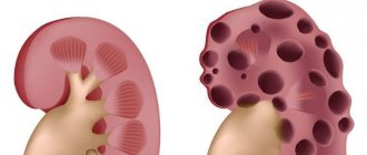
Manifestations of benign brain tumors
The initial manifestations of benign brain tumors may be erased and not raise suspicions about oncology until it reaches such a size that compression of certain parts of the brain occurs. One or more symptoms may appear in the case of a benign brain tumor. These symptoms are not specific and can also occur with other diseases.
- Impaired vision, hearing, smell.
- Balance and coordination problems
- Mental impairment, such as problems with attention, concentration, memory or speech
- Sudden seizures
- Skeletal muscle twitching
- Frequent nausea and/or vomiting for no particular reason
- Partial or complete facial paralysis
- Frequent headaches
- Numbness of the limbs
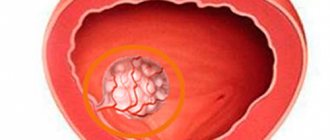
Causes of tumor formation
Any tumor arises and grows due to the spontaneous growth of abnormal cells resulting from mutation. If the tumor is benign, their growth and division gradually slow down, and they may remain in a “sleep state” for a long period. Malignant cells are characterized by constant activity, a high rate of division and gradual germination into tissues located nearby.
It is impossible to say for sure what triggers the growth of a tumor, but doctors note a number of factors that contribute to their division:
- genetic predisposition. A brain tumor can be diagnosed in a newborn, in rare cases - in an embryo during the prenatal period,
- exposure to electromagnetic waves (including mobile communications),
- infrared and ionizing radiation,
- GMOs in consumed products,
- radioactive exposure is the most common cause of tumors according to medical observations,
- prolonged contact with toxic chemicals: contact with mercury, lead, arsenic, etc.,
- human papillomatosis viruses.
https://youtube.com/watch?v=Le6T-EEGTr4
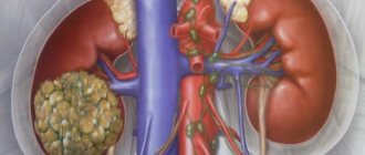
Classification of the disease
In addition to dividing into benign and malignant, there is a classification into primary and secondary tumors - in the second case, they arise as a result of the spread of malignant processes from other organs.
For treatment, it is important to know in which cells and tissues the neoplasm occurs. Common primary tumors include the following:
- developed directly from brain tissue;
- meningiomas - tumors of the membranes;
- Neuromas are formations in the nerves of the skull;
- pituitary adenoma.
Based on their location, neoplasms can be located in the upper or lower sections, hemispheres, midline structures, or base of the brain.
Types of surgeries to remove a brain tumor
- endoscopic trepanation;
- craniotomy (open craniotomy);
- stereotactic radiosurgery.
Depending on the type of operation, the neurosurgeon makes a decision on the course of the intervention, the type of anesthesia, calculates possible risks and warns the patient about them.
From the editor: Anatomical features, causes, symptoms of liquorodynamic disorders
Craniotomy
The clear consciousness of the patient during surgery allows the neurosurgeon to control the psychological and physical state of the patient while working on the brain. For example, control of the sensitivity of the limbs, preservation of vision, speech, hearing.
The operation begins with dissection of the soft tissues of the head. Next, it is necessary to remove the bone fragment at the site of the tumor. Excision of the tumor is carried out with a laser, which allows you to simultaneously stop the blood supply to the tumor and stop the bleeding.
After the tumor is completely removed, the bone fragment is placed in place and fixed with metal structures (screws, plates). If cancer cells grow into the thickness of the skull bones, all affected areas are removed, and the hole is closed with an artificial implant (titanium, porous polyethylene).
Brain surgery can last several hours. For the purpose of constant monitoring, the patient is recommended to spend about 2 weeks in the intensive care unit for timely assistance in case of deterioration of the general condition.
Endoscopic trepanation
The location of the tumor in a hard-to-reach place does not allow its removal using an open method. The operation is performed by inserting an endoscope through an incision in the upper palate or nasal passage. A tumor in the brain, for example, in the area of the pituitary gland, is removed with a special attachment.
The excision process is controlled by a sensor on the endoscope. Readings from the sensor are displayed on the monitor, which minimizes damage to surrounding brain tissue.
After the endoscope is removed from the skull, minor hemorrhages may be detected. If the bleeding does not stop, the surgeon decides on the need for craniotomy. A favorable outcome of the operation is characterized by the patient emerging from anesthesia without any complications.
Stereotactic radiosurgery
This method of treating brain tumors allows you to remove the tumor without craniotomy and without anesthesia. The neurosurgeon acts on the tumor with a directed beam, which consists of gamma radiation (gamma knife), a beam of photons (cyberknife) or a stream of protons.
The tumor removal procedure lasts about an hour until all malignant cells are destroyed. The duration of exposure depends on the size of the tumor. The advantage of this method of treatment is that it is not tied to the location of the tumor.
The tumor removal procedure involves exposing cancer cells to ionizing radiation. A course of treatment using a cyberknife is carried out over several days (3-5), depending on the size of the tumor. Each procedure lasts between one and a half hours.
In this case, the patient does not feel any discomfort. Hospitalization for the duration of treatment is not required, since the patient can visit the clinic at the appointed time, undergo a course of radiation exposure and go home. The Gamma Knife procedure is performed under local anesthesia, but hospitalization is also not required.
Complete removal of skull bone fragments
Such surgical intervention is practiced only after consultation with other specialists - otolaryngologist, plastic surgeon, maxillofacial surgeon.
Treatment of brain meningioma
The Burdenko Neurosurgery Center offers a range of services for the diagnosis and treatment of brain meningioma of any location and complexity. Removal of meningioma in the center is carried out using microsurgical instruments and an operating microscope under general anesthesia. If necessary, the clinic’s doctors perform cranioplasty, which allows you to hide skull defects after surgery. Neurosurgeons at the Burdenko Clinic use the latest technologies and programs for the treatment of brain cancer that meet international standards. Prices for treatment are developed individually; they are established by the current legislation of the Russian Federation. At the clinic you can apply for a quota for free treatment, but to do this you need to collect a whole package of documents and get a decision from the medical board.
When is surgery scheduled?
Brain surgery is prescribed in several cases:
- The tumor body grows rapidly;
- education is in an easily accessible place and does not put pressure on the brain centers that regulate vital processes;
- the age category and physical condition of the patient allow manipulation;
- when squeezing the brain.
Surgery to remove a brain tumor is considered the preferred method of treatment and, in the absence of contraindications, is prescribed first. In the initial stages, it is extremely rare to observe damage to nearby areas by metastases. Surgery to remove a brain tumor is not performed if the patient refuses treatment. Contraindications are multiple metastases and localization of the OGM in close proximity to the centers that regulate the vital functions of the body.
Diagnosis of a benign brain tumor is an indication for trepanation. Even though the cyst does not metastasize and grows slowly, it can compress the capillaries, resulting in impaired blood circulation. When the functioning of the circulatory system is disrupted, neurons begin to die, which is why mental disorders are often observed with OGM.
Is treatment necessary?
If the ongoing process is benign, it still poses a threat, since as the tumor grows it begins to put pressure on vital neighboring structures.
Because of this, intracranial pressure increases and various neurological problems appear - from headaches and dizziness to epileptic seizures, impaired coordination of movements, sensitivity of the limbs, memory and speech. All this can lead to disability and death. If we are talking about malignant neoplasms of the brain, the patient’s condition rapidly deteriorates. The prognosis in this case is extremely unfavorable. Therefore, when neoplasms are detected, regardless of their nature, prompt medical intervention is required.
How much does it cost to remove a brain tumor?
In Russia, every resident with cancer who has a compulsory medical insurance policy is given the right to have a cancerous tumor removed free of charge and receive subsequent medical support. Free surgery is almost the only way to save patients with low and medium incomes, since its average cost is tens of thousands of dollars. The disadvantage in this case is the lost time, since in most cases the promised quota can be expected for years.
In private domestic and foreign clinics, the tumor can be removed immediately after diagnosis, but the operation and rehabilitation will have to be paid for out of pocket. In Russian clinics, the cost of tumor removal, depending on the type of operation, varies within the following range:
- craniotomy method – from $2300 to $7700;
- stereotactic method – from $700.
Is something bothering you? Illness or life situation?
Describe your problem to us, or share your life experience in treating the disease, or ask for advice! Tell us about yourself right here on the site. Your problem will not go unnoticed, and your experience will help someone!
Removal of a brain tumor using the endoscopic method is practiced in foreign clinics. Depending on the country and the characteristics of the tumor, the cost of such an operation will vary from $1,500 to $20,000.