Thoracic cavity
The thoracic cavity is the space in the body that is located inside the chest. The chest and abdominal cavities separate the internal organs that they contain from the skeleton and muscles of the body, allowing these organs to move smoothly inside relative to the walls of the body. Organs located in the chest cavity: heart, blood vessels and nerves, trachea, bronchi and lungs; The esophagus passes from the chest cavity to the abdominal cavity through an opening in the diaphragm. The abdominal cavity contains the stomach and intestines, liver, kidneys, spleen, pancreas, numerous blood vessels and nerves.
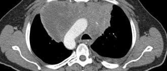
The photo shows where and what organs of the chest cavity are located. The heart, trachea, esophagus, thymus, large vessels and nerves are located in the space between the lungs - in the so-called mediastinum. The dome-shaped diaphragm, attached to the lower ribs, the back of the sternum and the lumbar vertebrae, forms a barrier between the human thoracic and abdominal organs.
Abdominal composition
The abdominal cavity is occupied by organs responsible for the digestive tract process.
There is also the pancreas, as well as the liver and kidneys. Next to them is the spleen, stomach, just below the kidneys and genitals, namely:
- The stomach is part of the body of the digestive system. It continues with the esophagus and is separated by a valve. In appearance it resembles a bag. The walls of the stomach produce mucus, and juice breaks down food.
- The gastrointestinal tract includes the intestines. It is large in size and length. The intestines begin after the outlet of the stomach. Body parts include intestines of different types. Thin - or in other words - duodenum, which gradually turns into a thick one, and then into a straight one. The intestines perform an important function - digest crushed foods and rid the body of food debris.
- The largest gland in size is the liver . She is involved in digestion. One of the main tasks of the organ is to carry out metabolism. The liver is located under the diaphragm and has two lobes. Thanks to a passing vein, the liver is connected to the duodenum.
- The spleen is a part of the body located above the diaphragm. It is responsible for important functions, among them hematopoiesis and protection of the body. Depending on the flow and accumulation of blood, the size of the spleen changes.
- Kidneys are organs that come in two pairs. They occupy the lumbar zone and are responsible for the regulation of homeostasis and the release of urine. The shape of the buds resembles beans. The organs are located in a fibrous capsule. Parts of the body consist of parenchyma; they include systems responsible for the accumulation and excretion of urine. The outside of the kidney is covered with a dense sheath. The outer layer of the cortical component makes up the parenchyma, and the inner part includes the medulla. The small calyces of the kidneys serve to collect urine, then the general collection of urine occurs by draining the fluid. The end of the renal pelvis is near the ureter.
- Above them are the adrenal glands . They belong to paired endocrine glands. Parts of the body regulate metabolism and also prepare the human body for stressful conditions. The adrenal glands have a medulla where adrenaline is stored. Thanks to the action of this hormone, heart contractions become faster and larger. Blood pressure begins to rise, the pupils become dilated, and glycogen quickly turns into glucose.
Heart
The most active muscle in the human body is the heart or myocardium. The heart moves blood steadily, with a certain rhythm, without stopping - approximately 7200 liters daily. Different parts of the myocardium synchronously contract and relax with a frequency of approximately 70 times per minute. During intense physical work, the load on the myocardium can triple. Heart contractions are triggered automatically by a natural pacemaker located in the sinoatrial node.
The myocardium works automatically and is not subject to consciousness. It is formed by many short fibers - cardiomyocytes, interconnected into a single system. Its work is coordinated by a system of conductive muscle fibers consisting of two nodes, one of which contains the center of rhythmic self-excitation - the pacemaker. It sets the rhythm of contractions, which can change under the influence of nerve and hormonal signals from other parts of the body. For example, under heavy workload the heart beats faster, sending more blood to the muscles per unit of time. Thanks to its efficiency, about 250 million liters of blood are passed through the body over 70 years of life.
The brain is an important organ
The brain is one of the main organs in the human body. The work of this part of the body is aimed at mental activity. The brain consists of the cerebral hemispheres, the cerebellum, and two pons. All thought processes are carried out with the help of the cerebral hemisphere, so a person consciously controls movements:
- The cerebellum occupies the back of the brain and is responsible for balance. Under the influence of the cerebellum, the reflexes of the muscle structure are controlled.
- The pons is located at the base of the skull and is also located below the cerebellum. Its action consists of a simple function: through it the reception and transmission of nerve impulses occurs.
Just below the listed organs there is another bridge, which is attached to the spinal cord. It receives and transmits signals located in other departments.
Trachea
This is the first of the organs of the human thoracic cavity. This organ is designed to pass air into the lungs and is located in front of the esophagus. The trachea originates at the height of the sixth cervical vertebra from the cartilage of the larynx and branches into bronchi at the height of the first thoracic vertebra.
The trachea is a tube 10-12 cm long and 2 cm wide, consisting of two dozen horseshoe-shaped cartilages. These cartilaginous rings are held in place anteriorly and laterally by ligaments. The space of each horseshoe ring is filled with connective tissue and smooth muscle fibers. The esophagus is located immediately behind the trachea. Inside, the surface of this organ is covered with a mucous membrane. The trachea, dividing, forms the following organs of the human chest cavity: the right and left main bronchi, descending into the roots of the lungs.
Structure of the bronchi
The main bronchi diverge to create 5 lobar bronchi. 10 segmental bronchi continue from them, each time decreasing in diameter. The smallest branches of the bronchial tree are bronchioles with a diameter of less than 1 mm. Unlike the trachea and bronchi, bronchioles do not contain cartilage tissue. They consist of many smooth muscle fibers, and their lumen remains open due to the tension of elastic fibers.
The main bronchi are located perpendicularly and rush towards the gates of the corresponding lungs. At the same time, the left bronchus is almost twice as long as the right one, has 3-4 more cartilaginous rings than the right bronchus, and seems to be a continuation of the trachea. The mucous membrane of these organs of the thoracic cavity is similar in structure to the mucous membrane of the trachea.
The bronchi are responsible for the passage of air from the trachea to the alveoli and back, as well as purifying the air from foreign impurities and removing them from the body. Large particles leave the bronchi during a cough. And small particles of dust or bacteria that have penetrated the respiratory organs of the chest cavity are removed by the movements of the cilia of epithelial cells, propelling bronchial secretions towards the trachea.
Lungs
In the chest cavity there are organs that everyone calls the lungs. This is the main paired respiratory organ, which occupies most of the chest space. The right and left lungs are divided according to location. In their shape, they resemble cut cones, with the apex directed towards the neck, and the concave base towards the diaphragm.
The top of the lung is 3-4 cm above the first rib. The outer surface is adjacent to the ribs. The bronchi, pulmonary artery, pulmonary veins, bronchial vessels and nerves lead to the lungs. The entry point of these organs is called the portal of the lung. The right lung is wider, but shorter than the left. The left lung has a niche in the lower front part for the heart. The lung contains a significant amount of connective tissue. It has very high elasticity and helps the contractile forces of the lungs, which are necessary with each inhalation and exhalation.
Location of the pelvic organs
Consider what is located below the abdominal cavity:
- The bladder is a special bag that collects urine. The urethra helps in this. Part of the body, together with the reproductive system, is located in the pelvis. In the same place in men there are seminal vesicles with the prostate gland. The organ consists of elastic muscle tissue that has the ability to stretch and contract. The shape of the bladder changes after it fills with urine. These organs are separated by special boundaries that extend to the umbilical point.
- The uterus is the female part of the body that borders the bladder and is located in the middle of the pelvis. The length of the elastic organ reaches 7 cm. During pregnancy, the size of the uterus increases. After the bladder fills, the uterus moves. The lower part of the uterus is round in shape.
- The ovaries are the paired part of the body in the female body; the process of childbirth depends on it. The ovaries are involved in the production of sex and steroid hormones.
- Seminal vesicles - in structure they resemble twins and are located in the rear side part. The organs have a predominant excretory function; it nourishes and promotes sperm. Bubbles are involved in ejaculation.
- The prostate gland is found only in the male body in the lower region of the front part of the body. In appearance it looks like a chestnut and has furrow divisions. The prostate is involved in the secretion of the base found in sperm.
Diaphragm
The respiratory muscles rhythmically increase and decrease the volume of the lungs, changing the size of the chest cavity. The main work is done by the diaphragm. As it contracts, it flattens and descends, increasing the size of the chest cavity. The pressure in it drops, the lungs expand and draw in air. This is also facilitated by the raising of the ribs by the external intercostal muscles. Deep and rapid breathing involves accessory muscles, including the pectoral and abdominal muscles.
The mucous membrane of these organs of the chest cavity consists of epithelium, which, in turn, consists of many goblet cells. The epithelium of the branches of the bronchial tree contains many endocrine cells that control the blood supply to the lungs and maintain bronchial muscles in tone.
To summarize all of the above, it is necessary to note that the organs of the human chest cavity are the basis of his life. It is impossible to live without a heart or lungs, and disruption of their functioning leads to serious illnesses. But the human body is a perfect mechanism, you just need to listen to its signals and not harm, but help Mother Nature in its treatment and restoration.
View of body parts from the back
The arrangement of human organs from the back is slightly different and only some parts of the body are visible. Closer to the central zone is the spleen. The small and sigmoid colon are clearly visible from the side. The location of the cecum and along with it the ascending colon are clearly visible. The liver is visible, as well as the pancreas.
The adrenal glands and kidneys are located in the posterior region of the human body. The location of the lungs is visible from the back of the back.