Laboratory work "STRUCTURE OF VEGETATIVE AND GENERATIVE KIDS, THEIR LOCATION ON THE STEM"
"_____" ______________ 20_____
Laboratory work No. 7
Practical
STRUCTURE OF VEGETATIVE AND GENERATIVE KIDS, THEIR LOCATION ON THE STEM
Goal of the work:
study the structure of the kidneys; prove that the bud is an embryonic shoot.
Materials and equipment:
leafless shoots of rowan (lilac, horse chestnut, maple or other plants), a magnifying glass, a dissecting needle, glass slides, a blade or scalpel.
Progress
- Consider the shoots of the plants offered to you.
Find the apical and lateral buds.
Sketch part of the shoot, indicating the apical
and lateral buds.
- Determine what type of bud arrangement is typical for the plants given to you. Draw diagrams of the location of the kidneys. Below the diagrams give examples of plants with different types of bud arrangement.
Types of bud arrangement on the stem
- On a maple (or other plant) branch, look for smaller elongated (vegetative) and larger rounded (generative, or floral) buds.
- Separate the buds from the shoot and examine them. What adaptations help the kidneys withstand adverse conditions?
_______________________________________________________________________________________________________________________________________________________________________________________________________________________________________
- Cut the vegetative bud lengthwise and examine it with a magnifying glass. Find the rudimentary stem, at the top of which there is a growth cone, rudimentary leaves, and covering scales. Draw a cross-section of a vegetative bud. Label the names of its parts.
Structure of a vegetative bud:
_______________________________________________________________________________________________________________________________________________________________________________________________________________________________________________________________________________________________________________________________________________________________________________________________________________________________________________________________________________________________________________
- Carefully cut a large generative (flower) bud lengthwise and examine its internal structure with a magnifying glass. Find the flower primordia on the rudimentary stem. Draw a diagram of the structure of a generative bud.
Structure of the generative bud:
____________________________________________________________________________________________________________________________________________________________________________________________________________________________________________________________________________________________________________________________________________________________________________________________________________________________________________________________________________________________________________________________________________________________
Compare the structure of vegetative and generative buds. What do their structures have in common and how do they differ?
_______________________________________________________________________________________________________________________________________________________________________________________________________________________________________
- Compare the structure of a bud and a typical shoot (see Fig. 110 on p. 132 of the textbook). Prove that the bud is a rudimentary shoot.
Conclusion:________________________________________________________________
____________________________________________________________________________________________________________________________________________________________________________________________________________________________________________________________________________________________
Give short answers to the questions
- How can you prove that plants have dormant buds?
__________________________________________________________________________________________________________________________________________________________________________________________________________________________________________
- Trees and shrubs are pruned in populated areas. For what purpose are they doing this? Is it possible to do this kind of pruning annually?
______________________________________________________________________________________________________________________________________________________________________________________________________________________________________________________________________________________________________________________________________________________________________________________________________
- During an excursion in the fall, students noticed that from the stumps of maples and poplars cut down in the spring, young shoots up to 1 m high grew in one summer, and the shoots of these plants growing nearby do not exceed 10-15 cm. Explain what stump shoots develop from and why does it grow faster than seedlings?
________________________________________________________________________________________________________________________________________________________________________________________________________________________________________________________________________________________________________________________
Complete the task
Place a currant branch (gooseberry, cherry or any other bush or tree) in the water. Observe the development of shoots from the buds. Note when the buds swell and the green leaves appear and unfold. Watch the growth of the shoot in length. Also note the formation of adventitious roots. Enter the observation data into the table.
Development of shoots __________________ from the buds
| Start of experience (date) | Swelling of the kidneys (days from the start of the experiment) | Leaf appearance (days) | Leaf unfolding (days) | Formation of adventitious roots (days) |
Draw a conclusion about how the shoot and bud develop.
Conclusion:
_________________________________________________________________________
___________________________________________________________________________________________________________________________________________________________________________________________________________________________________________________
Structure and diversity of angiosperms.
57. Fill out the diagram. Organs of angiosperms: Vegetative - root, shoot; Generative - flower, fruit with seeds.
58. After completing the laboratory work “Structure of seeds of dicotyledonous plants” (see p. 93 of the textbook), label the parts of a bean seed in the picture.
59. Complete the laboratory work “Structure of the grain of wheat” (see p. 94 of the textbook). Label the parts of a wheat grain in the picture.
1 - pericarp fused with the seed coat; 2 - endosperm; 3 - cotyledons; 4 - kidney; 5 - stem; 6 - spine; 7 - embryo; Conclusion: The embryo contains many organs. Embryonic root, stalk, bud and cotyledons.
60. Fill out the table “Comparison of seeds of dicotyledonous and monocotyledonous plants.”
61. Compare the parts of a seed and a sprout. Show with arrows on the diagram from which parts of the seed the corresponding parts of the seedling developed.
Conclusion: The embryo is the rudiment of the future plant. Each organ of the embryo is very important; the organs of the future plant develop from it.
62. Look at the pictures. Indicate the type of root systems of the plants shown. 1 - rod; 2 - fibrous.
63. After completing the laboratory work “Tap and fibrous root systems” (see p. 97 of the textbook), fill out the table.
Conclusion: In dicotyledons the root system is taprooted, while in monocotyledons it is fibrous.
64. What agricultural technique is shown in the figure? For what purpose is it used? More tubers, more lateral roots are created during hilling. And there is more harvest. Hilling is very beneficial for plants.
65. Consider the image of a longitudinal section of a young root. Indicate which parts of the root are indicated by numbers. 1- root cap; 2 - division zone; 3 - growth zone; 4 - suction zone; 5 — holding area; 6 - root hairs.
After completing the laboratory work “Root cap and root hairs” (see p. 101 of the textbook), indicate what is indicated by numbers.
1 - shell; 2 - vacuole; 3 - peephole; 4 - root cap.
66.Compare the structure of onion skin cells and root hairs shown in the figure. Connect the identical parts of these cells with arrows.
Conclusion: They have similar features: ocellus, vacuoles, cytoplasm and membrane.
67. Fill out the table “Relationship between the structure of root zones and the functions they perform.”
Conclusion: The root has many zones, and each zone does its own job.
68. Look at the picture. What agricultural practice is shown on it? For what purpose is it used? Picking - pinching off the tip of the root when planting young plants using a pointed peg - peaks.
69. Fill out the table “Modifications of roots.”
70. Complete the definition. A shoot is a stem with buds located on it.
71. Label the parts of the shoot indicated in the pictures.
1 - apical; 2 - axillary; 3 - internode.
72. Having completed the laboratory work “Structure of the kidneys. Location of buds on the stem” (see p. 109 of the textbook), sketch the location of the buds on the stem. 1 - next; 2 - opposite.
Label the parts of the kidneys in the picture. Indicate which of them is vegetative and which is generative.
1 - kidney scales; 2 - leaves; 3 - kidney; 4 - stem. Conclusion: in a generative bud, the bud is larger.
73. What devices help the kidneys withstand unfavorable conditions? Kidney scales.
74. Finish filling out the diagrams. Types of buds by structure: Vegetative and generative. Types of buds by location on the stem: alternate, opposite and whorled. Structure of vegetative buds: Kidney scales, leaves are conceived. , bud and stem. Structure of generative buds: Bud scales, leaves are conceived, bud, stem is conceived.
75. Look at the drawing. Compare the structure of the bud and shoot. Connect with arrows the corresponding parts of the bud and shoot.
Conclusion: Each organ of the bud grows and becomes an organ of the future plant.
76. Look at the drawing. Label what kind of leaves these are based on the way they are attached to the stem and what their parts are.
77. Look at the drawing. Write down separately the numbers that indicate simple leaves and compound leaves. Simple leaves: 1, 4, 6, 8, 7. Compound leaves: 2, 3, 5.
78. Look at the drawing. Determine what type of venation these leaves have.
79. Complete the laboratory work “Simple and compound leaves, their veining and leaf arrangement” (see p. 115 student), fill out the table.
80. Look at the drawing. What does it show? Sign what is indicated by numbers.
Stomata with surrounding skin cells. 1 - Guard cell; 2 - Stomatal fissure; 3 - Chloroplast; 4 - skin cells.
81. After completing the laboratory work “Structure of the skin of a leaf” (see pp. 116-117 of the textbook), make drawings and captions for them. Conclusion: the composition of the leaf skin includes the stomatal fissure, skin cells, chloroplast, and intercellular space.
82. The figure shows a cross section of a sheet. After completing the laboratory work “Cellular structure of a leaf” (see pp. 118-119 of the textbook), make signatures.
83. The picture shows a light and a shadow leaf. What structural features are characteristic of each of them? 1 – light sheet 2 – shadow sheet. The shade leaves are thinner and have a darker green color. Light leaves have a lighter color.
84. Indicate which plants are shown in the picture and what their modified leaves are turned into.
85. Look at the drawing. Indicate the types of stems according to the direction of growth.
86. In the picture, label the layers on the trunk of a cut tree.
87. In the picture, look at the cross section of the branch. Label its main parts.
88. Fill out the table.
89. After completing the laboratory work “Internal structure of a tree branch” (see pp. 128-129 of the textbook), make drawings and captions for them.
90. After completing the laboratory work “Structure of a tuber” (see pp. 131-132 of the textbook), in the figure, connect the tuber section with the cross section of the stem using arrows. Label the corresponding layers in the picture.
Conclusion: in both pictures the structure is the same, although they look different.
91. Complete the laboratory work “Structure of the onion” (see p. 133 of the textbook). In the picture, label its main parts.
1 – scales 2 – modified leaves 3 – buds 4 – bottom 5 – adventitious roots Conclusion: onions have a fairly simple structure, these are: scales, modified leaves, buds, bottom, adventitious roots.
92. Fill out the table “Functions of modified shoots.”
93. In the picture, label the names of the parts of the flower.
94. In the picture, compare cherry and tulip flowers. Label their main parts. What are the similarities in the structure of these flowers? What is the difference?
Conclusion: the first flower has a double perianth, and the second one has a simple one.
95. After completing the laboratory work “Structure of a flower” (see p. 138 of the textbook), sketch the parts of the flower and label their names.
Flower formula Ch5 L5 T∞ P1
96. Compare the cabbage and viola flowers in the picture. What is their difference? Write what these flowers are called.
In the correct one you can draw several planes of symmetry, but in the incorrect one - only one.
97. Fill in the missing words. Flowers that have both stamens and pistils are called bisexual. A flower that has only stamens is called staminate, and a flower that has only pistils is called pistillate. If a plant develops both staminate and pistillate flowers, it is called monoecious. If staminate flowers are located on some plants, and pistillate flowers are located on others, then such plants are called dioecious.
98. Fill out the table “Features of the structure of inflorescences.”
99. Complete the laboratory work “Inflorescences” (see p. 141 of the textbook).
Conclusion: the biological significance of inflorescences is that small, often inconspicuous flowers, collected together
100. Finish filling out the “Classification of Fruits” diagram. Fruits: 1) dry – single-seeded, multi-seeded; 2) succulent – single-seeded, multi-seeded.
101. Complete the laboratory work “Classification of fruits” (see p. 146 of the textbook). Based on the results of your work, fill out the table.
102. Fill out the table.
103. Solve crossword number 4.
Leaf and flower buds, their location on the stem.
The stem is the part of the plant that connects the main nutritional organs - the root and leaves.
The stem with leaves or buds located on it is called a shoot. Each shoot develops from a bud. Remember: leaves can be arranged alternately, oppositely and whorled. The places where the leaves are located are called nodes, and the areas between the two closest nodes are called internodes.
There are elongated shoots with long internodes and shortened shoots with short internodes and close nodes.
The angle between the leaf and the internode lying above it is called the leaf axil.
At the top of the shoot there is usually an apical bud, and in the axils of the leaves there are axillary, or lateral, buds.
Rice. 70. Location of the kidneys: 1 - alternate; 2 - opposite.
While studying seed germination, you observed the development of a shoot from the embryonic bud of the seed
beans. How does a shoot develop from a bud located on the stem? Consider a shoot of a poplar or apple tree. At the top of these shoots there is one apical bud. On the sides of each branch, in the places where the leaves were, there are lateral buds.
The lateral buds are located on the stem in the same way as the leaves. On the branches of poplar and apple trees, the buds are located singly, one after the other. This arrangement is called sequential. Many trees and shrubs have a regular arrangement of buds: cherry, birch, bird cherry, hazel and others.
If we look at the shoots of lilac, elderberry, jasmine, honeysuckle or indoor fuchsia plant, we will see a completely different arrangement of buds. They are located in twos, one against the other - opposite. Let's see what structure the kidneys have.
The outside of the bud is covered with dense leathery scales that protect it from rain, wind and other adverse conditions.
Let's cut off one of the buds from the shoot and examine it with a magnifying glass. Under the scales, a rudimentary stem located in the very center of the bud and small, wrinkled rudimentary leaves are clearly visible. In the axils of these leaves there are rudimentary buds, similar to grains, barely noticeable even with a magnifying glass. Thus, the bud is a rudimentary shoot.
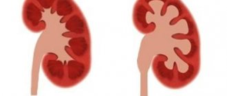
If you cut several buds, you can see that inside some of them there are only rudimentary leaves around the embryonic shoot. Such buds are called vegetative or leaf buds. Rudimentary buds are visible inside other buds. These are flower buds.
Flower buds can be distinguished from vegetative ones by their appearance. They are larger than vegetative ones and have a rounded shape.
Rice. 71. Apical (1) and lateral (2) buds.
Rice. 72. Flower bud (1) and leaf bud (2).
By the location of the buds on the shoots, their shape, size, color, pubescence and some other characteristics characteristic of each type of plant, you can determine the name of the tree or shrub even in winter.
When identifying trees and shrubs in winter, pay attention to the following signs characteristic of buds.
The buds are usually located directly on the stem. The exception is alder. In alder they sit on special legs, and by this feature, as well as by earrings and small cones, alder can be easily distinguished from other trees. Poplar is recognized by its sticky, resinous, pointed buds, which have a peculiar, pleasant odor. The willow bud is covered with only one cap-like scale. Buckthorn has no kidney scales at all. The oblong large buds of rowan are pubescent and therefore clearly distinguishable from the buds of other trees.
Bird cherry and black currant buds have a pleasant fragrant smell. The oppositely located elderberry buds, on the contrary, have an unpleasant odor. By smelling them, you will immediately distinguish elderberry from other shrubs.
If you find an error, please select a piece of text and press Ctrl+Enter.
so UNT / Biology / Laboratory work in biology
Topic: ESCAPE
25.09.2014 11459 0
Laboratory work 10.
The structure of vegetative and generative buds
Goals:
show the structural features of the vegetative and generative buds of a plant, compare the structure of the vegetative and generative buds of plants; continue to develop the skill of performing biological drawings.
Equipment:
live shoots of lilac, elderberry, maple, chestnut, magnifying glass, tweezers, dissecting needle, scalpel.
Progress:
1. Consider the location of the buds on the shoot. Find the lateral and apical buds. What is the location of the kidneys?
2. Tear off one of the side buds of the shoot. Carefully remove the kidney scales from it and count their number.
3. Carefully cut the side bud lengthwise. Examine the rudimentary leaves and rudimentary stem. Count the number of rudimentary leaves. What kind of bud is this - vegetative or generative?
4. Sketch the appearance and internal structure of the kidney. Sign what kind of kidney is this? Label its main parts.
5. Examine the apical bud of the shoot. Count the number of kidney scales.
6. Carefully cut the kidney lengthwise. Find the rudimentary stem, rudimentary leaves, rudiments of flowers. How many rudimentary leaves do you see? What kind of bud is this - vegetative or generative?
7. Sketch the appearance and internal structure of the kidney. Sign what kind of kidney is this? Label its main parts.
8. Compare the buds you saw, draw a conclusion about the similarities and differences between these buds. How are they related to the functions they perform?
Laboratory work 11.
External structure of modified underground shoots - rhizomes and bulbs
Goals:
show the structural features of the rhizome and bulb, compare the structure of these modified shoots, and conclude that these parts of the plants are indeed modified shoots; continue to develop the skill of performing biological drawings.
Equipment:
potato tubers, onion bulbs, magnifying glass, tweezers, dissecting needle, scalpel.
Progress:
1. Consider the external structure of the bulb. Find the dry filmy scales that cover the bulb. What is their main function?
2. Carefully cut the onion lengthwise. Find and examine the modified stem, modified leaves, apical and lateral buds. Consider the roots growing from the stem. What are these roots called? What kind of root system do they form?
3. Draw a longitudinal section of the bulb. Label its main parts.
4. Consider the external structure of a potato tuber. Find apical and axillary buds (“eyes”), find leaf scars (“edbs”). Count the approximate number of eyes on the tuber.
5. Draw the external structure of a potato tuber, label its main parts.
6. Draw a conclusion about the similarities and differences in the structure of a potato tuber and an onion bulb. List the signs that prove that potato tubers and onion bulbs are indeed modified shoots.