Kidney cysts are a common abnormality of kidney development, manifested by the appearance of round formations containing fluid in the kidney tissue. The size of kidney cysts ranges from very small (1-2 cm) to gigantic ones, larger than the kidney itself (10 cm or more). The causes of kidney cysts are an abnormal development of the kidney structures, when the glomeruli do not connect to the tubules, resulting in their expansion and filling with fluid.
This pathology is often detected accidentally and does not require treatment. Treatment of kidney cysts is carried out strictly according to indications, among which are:
- Pain in the lumbar region
- Persistent increase in blood pressure that cannot be treated with tablets.
- Compression of the urinary system of the kidney
- Large cysts that pose a risk of rupture
- Rapidly growing kidney cysts
- Cysts suspicious for kidney cancer
- Suppurating kidney cysts
Indications for cyst removal
If a pathology is detected, the patient should regularly see a doctor and undergo examinations. An ultrasound examination of the organ is mandatory at least once a year. Most often, with a kidney cyst, careful observation is sufficient, but cases when it is necessary to remove a cyst on the kidney cannot be ruled out.
Each patient diagnosed with this pathology must clearly identify the main signs that are an indication for surgical intervention. If you do not start treating a kidney cyst in time, the likelihood of developing dangerous complications increases. First of all, the growing formation compresses the blood vessels supplying the kidneys with blood, as well as the tissues of the organ. As a result, the normal functioning of the kidneys is disrupted and the load on the organ increases. In addition, removal of a kidney cyst is prescribed in the following situations:
- violation of the free passage of urine through the ureters;
- increased blood pressure;
- severe pain in the area of the affected organ;
- rupture or inflammation of the cyst;
- the appearance of blood in the urine;
- symptoms of malignancy of the cyst.
In the latter case, a biopsy is mandatory. If a positive result is obtained, nephrectomy is often prescribed, since removal of part of the kidney often ends in recurrence of the pathology. To treat a kidney cyst correctly, you need to conduct a thorough examination of the patient. In addition to ultrasound examination, magnetic resonance imaging with ascertaining is prescribed.
But the detection of a cyst does not always become the reason for turning to surgeons. In some cases, surgery is not prescribed. These are polycystic disease in the acute phase, asymptomatic kidney cyst, diseases of the cardiovascular and respiratory systems, blood clotting disorders, as well as diabetes mellitus in the decompensation stage.
Our doctors
What kind of disease is this?
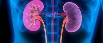
Kidney tissue has a special structure that has its own functional tasks: it filters blood, retains necessary components and removes toxic metabolic products.
A congenital kidney cyst can form in the prenatal period, which is influenced by genetic factors and adverse external influences. The development of an acquired kidney cyst can be caused by certain provoking factors. The size of the cyst varies from one millimeter to several centimeters. The biggest problem is represented by multiple cysts in the kidneys. Such formations are dangerous because, penetrating the kidney tissue, they disrupt the functioning of a large volume of the filtration system. Due to dysfunction of part of the parenchymal tissue, the activity of the kidneys is disrupted.
Methods for removing kidney cysts
In order to choose the most appropriate method for removing the formation, you should evaluate its location, size, presence or absence of signs of inflammation, as well as the general condition of the patient. Modern surgeons most often use the following surgical methods:
- removal of a kidney tumor using the open method;
- percutaneous removal of the formation (percutaneous);
- laparoscopy;
- retrograde intrarenal intervention.
In rare cases, open surgery is prescribed. It consists of creating access to the kidney through an incision. This is the most traumatic method. After such surgery, patients recover for a long time in a hospital setting.
The percutaneous method is used in cases where the cyst is localized on the posterior surface of the kidney. An endoscope is inserted through a minimal skin incision into the area of formation. Thanks to the presence of a camera, it is possible to carefully examine the pathological focus and excise all affected tissue using the least traumatic method.
Laparoscopic kidney surgery requires making three minimal incisions in the abdominal wall through which a camera and instruments are inserted to remove the mass. This method is indicated when the cyst is located on the anterior and lateral parts of the organ. In addition, with a similar method of treating kidney cysts, excision of several formations in polycystic disease can be performed at once.
Retrograde intrarenal intervention consists of providing access through the urethra. Through the urinary canal and bladder, the endoscope is brought to the kidney. Thanks to the laser beam, a neat incision is created in the kidney tissue and subsequent excision of the cyst with its contents. After this, the wound is sutured. Due to less trauma, the rehabilitation period after surgery is minimal.
The duration of surgery depends on the complexity of the operation and the number of cysts. On average, kidney resection lasts 40-120 minutes.
UROLOGY IN OMSK
The main points of puncture treatment of large kidney cysts:
- Visualization of the cyst using an ultrasound machine with a puncture sensor.
- Cyst puncture.
- Cystography, dilatation of the puncture tract and installation of drainage.
- Aspiration of cyst contents.
- Introduction of sclerosing solution
Operation set:
- puncture needle
- Plastic bougies
- String
- Drainage (puncture nephrostomy type Pigtail), metal conductor for drainage
- Syringe for cystography
- Syringe for local anesthesia
- Puncture adapter for ultrasound probe
- Container for solution (treatment of the surgical field)
- Container for saline solution
- Container for novocaine solution 0.5%
- Tools for fixing drainage, scalpel.
Visualization of the cyst using an ultrasound machine with a puncture sensor.
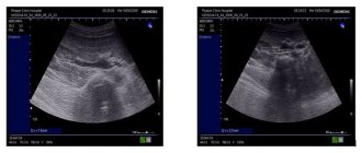
After processing the surgical field, the cyst is visualized using an ultrasound sensor with a puncture adapter, the puncture site and the puncture course are specified, which should not affect the renal parenchyma. Tissue infiltration is carried out with a solution of novocaine 0.5%.
Cyst puncture
Phone in Omsk +79095377482. Urology on Berezovaya.
The puncture does not present technical difficulties when localizing the cyst in the lower and middle segment along the lateral edge of the kidney or along the posterior surface. After removing the inner part (stylet) of the puncture needle, the contents of the cyst are aspirated for cystological examination. A mixture of radiopaque contrast agent and saline solution is injected into the cyst cavity.
Cystography, dilatation of the puncture tract and installation of drainage.
Cystography is carried out in the process of percutaneous puncture treatment of a simple cyst to clarify the location of the cyst and determine its relationship with the pyelocaliceal system. This allows you to avoid sclerosing solution entering the renal cavity system and the development of complications.
Subsequently, a fine drainage (from 5 to 9 according to Ch) is installed into the cyst cavity using the Seldinger method.
A string is inserted into the cyst cavity through a puncture needle under X-ray control, and the needle is removed. A small skin incision is made with a scalpel (about 3 mm).
Then, plastic bougies are carried out sequentially with rotational movements.
The operation ends with the installation of a self-fixing drainage on a metal conductor along a string, which is additionally fixed with two skin sutures. Additionally, an aseptic dressing is applied.
Introduction of sclerosing solution
For sclerotherapy, an ethyl alcohol solution is used. About 25% of the volume of the drug from the initial volume of the cyst is injected into the cyst cavity. Administration of a smaller volume of the drug is reliably ineffective (Perugia and associates). The patient alternates body position, turning on the right side, on the back, on the left side and on the stomach, spending 10 minutes in each position. This ensures that the entire inner surface of the cyst is treated with a sclerosing solution. With a technique without drainage of the cyst cavity, when only puncture and aspiration of the contents are performed, such a result cannot be achieved. After sclerotherapy, the drainage is opened and the patient is observed for 24 hours.
If there is no discharge through the drainage, the latter is removed.
After surgery to remove a kidney cyst
Regardless of the method of intervention, the patient must remain in bed on the first day. From the second day, short walks around the ward are allowed. Patients return to their normal lifestyle after 1.5-2 months.
Diet is a must. For several days after surgery, it is recommended to reduce the consumption of spicy, smoked, fried foods, but the amount of liquid should be increased.
Depending on the method of surgical intervention, the duration of inpatient treatment ranges from three days to three weeks. Antibiotics and anesthetics are mandatory during rehabilitation. To prevent a cyst on the kidney from forming again, it is recommended to undergo an annual examination by a nephrologist after surgery.
Features of the operation in our clinic
Clinic of Urology named after. Frontshteina is equipped with modern equipment that allows diagnosing kidney cysts at an early stage of development. Experienced specialists will help cure the kidney and reduce the likelihood of recurrence of the pathology to zero. In our clinic you can perform a robot-assisted nephrectomy or remove a kidney using the laparoscopic method. It is also possible to perform an operation to remove part of the kidney (kidney resection) using laparoscopy or a modern robot-assisted method. From the moment you contact us, determine the method of treatment for the cyst, and until complete recovery, you will be under the close attention of doctors and medical personnel, who will create the most comfortable conditions for successful rehabilitation.
Causes of tumors in the kidneys
Despite the fact that kidney cyst is a disease that occurs quite often, experts have not yet figured out the reliable causes of this pathology. As a result of traumatic, hereditary or infectious factors, abnormal tissue growth is possible, which leads to the formation of a kidney cyst. Accordingly, this disease can be classified as either acquired or congenital.
Experts believe that a genetic predisposition to cysts in the kidneys occurs due to a lack of connective tissue in the body. If complications arose during the mother's pregnancy (there were injuries, infections), then the likelihood of the child developing a kidney cyst increases significantly.
Acquired kidney tumors can occur both at the site of a hematoma due to trauma, and when the site of infection is not stopped in a timely manner.
The age factor is also important, playing a role in the formation of kidney cysts. Most often, people over 50 years of age suffer from this disease.
So, the provoking factors for the development of the disease are:
- age-related changes;
- hereditary predisposition;
- pyelonephritis;
- urolithiasis disease;
- kidney tuberculosis;
- malignant tumors;
- injuries,
- prostate adenoma in men.
The risk of developing a kidney cyst increases in people suffering from hypertension and urolithiasis, frequent colds, which are complicated by inflammation of the kidney parenchyma.
Make an appointment
