Live broadcasts on Instagram every Sunday at 12:00
Subscribe so you don't miss out! Subscribe
- home
- Urology
- Kidney cyst
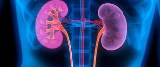
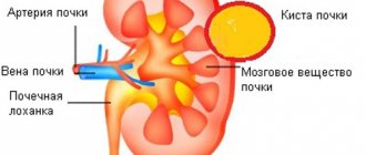
A kidney cyst is a benign, thin-walled, voluminous formation that develops from the kidney parenchyma and usually contains serous fluid. A kidney cyst is a spherical cavity. Most often, patients are diagnosed with single renal cysts, up to 3 cm in diameter, located subcapsularly in the renal parenchyma. Cysts occur with approximately the same frequency in both the right and left kidneys. Large single cysts, renal sinus cysts, and multiple renal cysts are quite rare.
Symptoms, signs and clinical picture of kidney cysts
The most characteristic symptoms of a simple kidney cyst are:
- dull pain in the hypochondrium or lower back, intensifying after physical activity;
- palpable mass formation in the lumbar region;
- persistent rise in blood pressure without dynamics;
- total hematuria.
A simple kidney cyst does not have typical clinical signs, and in 70% of patients it is asymptomatic and does not cause any clinical manifestations for years or even decades. That is why it cannot be reliably diagnosed based on clinical symptoms alone. Often, a simple cyst is accidentally discovered when examining a patient for a disease that has nothing to do with it (other urological or neurological diseases, etc.). With an increase in the size of the cyst and compression of the parenchyma of the kidney or urinary system, characteristic clinical manifestations appear.
The intensity of pain depends on the size of the cyst, its location, and the condition of the ligamentous apparatus of the kidney. A palpable formation in the kidney area can only serve as a presumptive sign of a simple cyst, since a true kidney tumor or the lower segment of a displaced kidney can be mistaken for a cyst.
Diagnosis of kidney cyst
Rice. 1. The number of punctures in the abdominal wall at the site of trocar insertion during excision of a cyst on the right. Position of the patient and the operating team.
The diagnosis of a kidney cyst is made based on the patient’s complaints, clinical picture and instrumental examination data. If a renal cyst is suspected, it is necessary to perform an ultrasound examination of the kidneys, intravenous excretory pyelography and radioisotope renal scintigraphy.
When determining the indications for surgery (especially in the case of a multi-chamber cyst or the presence of septa) and choosing a surgical procedure for a kidney cyst, in addition to ultrasound and excretory pyelography, it is necessary to conduct a computed tomography of the retroperitoneal organs with contrast to exclude oncological disease.
Diagnostics
- Survey and excretory urography
- Ultrasound, Dopplerography
- CT (computed tomography)
- Diagnostic puncture and cystography
X-ray methods: It is not possible to establish an accurate diagnosis of a simple renal cyst only on the basis of survey and excretory urography. With the help of excretory urography, it is possible to identify the “sickle” symptom or the “open mouth” symptom, which are characterized by the spreading of the kidney calyces without their “amputation”.
Ultrasound examination (ultrasound) reveals a simple renal cyst as an echo-negative formation of a round or oval shape, with clear, even, continuous contours and thin walls. Ultrasound allows for dynamic observation and use as a screening test. Dopplerography is a method that allows you to study the blood supply to the kidney. This diagnostic method is especially important for the combination of kidney cysts and arterial hypertension.
Computed tomography (CT) in some cases cannot provide 100% confidence in the accuracy of the diagnosis, especially with parapelvic cysts and tumors in the cyst. This somewhat reduces its diagnostic value. The CT scanning criteria for cystic renal lesions are similar to those used in echography: sharp, thin, distinct, smooth walls and edges; round or oval shape; homogeneous content. Density ranges from -10 to +20 HU, similar to that of water, and no enhancement should occur after intravenous injection of contrast medium.
Percutaneous puncture cystography is currently not the main diagnostic method, but can be used in the process of percutaneous puncture treatment of a simple cyst to clarify the location of the cyst and determine its relationship with the pyelocaliceal system.
Treatment of kidney cyst
What to do if a kidney cyst is detected? There are no therapeutic methods for treating kidney cysts. Until the mid-90s, before the widespread introduction of endoscopic kidney surgery methods into medicine, patients with single or multiple kidney cysts were under dynamic observation and received only symptomatic treatment. In the case of small single cysts, patients underwent surgery for sclerosis of the kidney cyst under ultrasound guidance. When the kidney cyst grew rapidly, patients underwent open kidney surgery.
Since, unlike a tumor, a kidney cyst is a benign disease, a cyst can be monitored in 70% of cases without resorting to active surgery. Patients whose kidney cyst does not exceed 5 cm in diameter, is located on the periphery of the kidney and does not cause disturbances in the kidney’s blood circulation and urine outflow, usually require dynamic monitoring using ultrasound once every 6–12 months.
Surgical treatment of a kidney cyst is not performed in cases where the cyst does not bother the patient, does not cause a disturbance in the outflow of urine from the kidney, as well as in severe concomitant conditions of the patient and blood clotting disorders.
To determine the type of kidney cyst, its localization to the main structures of the organ and indications for surgery, as well as choosing the correct surgical treatment tactics, you need to send me a complete description of the kidney ultrasound, MSCT data of the kidneys with contrast, indicate your age and main complaints to your personal email address. Then I will be able to give a more accurate answer to your situation.
Indications for surgical treatment for kidney cysts:
Organ-preserving surgery - laparoscopy of a kidney cyst. More than 250 operations were performed.
- compression of the kidney cyst of the urinary tract
- compression of the cystic tissue of the kidney parenchyma
- infection of the cyst cavity and abscess formation
- rupture of kidney cyst
- large cyst sizes and/or rapid growth of the kidney cyst
- pain caused by a cyst
- hematuria from the kidney from the side of the cyst
- arterial hypertension
- erythremia
- the presence of blood in the punctate from the cyst or hemorrhage into the cyst
- cyst rupture or danger thereof
- cancer in the cyst (based on radio-radiological, echographic data or examination of puncture fluid from the cyst).
During the surgical treatment of a kidney cyst, interventions of varying scope can be performed:
- percutaneous therapeutic and diagnostic puncture of a kidney cyst with sclerosis;
- enucleation of the cyst (husking);
- cyst resection;
- kidney resection with cyst;
- nephrectomy.
Watch a video of operations performed by Professor K.V. Puchkov. You can visit the website “Video of operations of the best surgeons in the world.”
Renal puncture is currently the first step in the treatment of simple single (solitary) uncomplicated kidney cysts with a diameter of up to 3–5 cm and located subcapsularly in the renal parenchyma. Currently, percutaneous therapeutic and diagnostic puncture of a kidney cyst is carried out only with sclerosing, since puncture of a kidney cyst without the use of a sclerosing agent in most cases leads to recurrence of the cyst and the need for repeated intervention. In some cases, there may even be an increase in the volume of the cyst compared to its original size. Percutaneous therapeutic and diagnostic puncture of a kidney cyst is carried out under ultrasound control, evacuation of the contents and introduction of an adhesive substance into its cavity.
The introduction of sclerosant into the cavity of the cyst significantly reduces the frequency of relapses (up to 2%) within a year after percutaneous therapeutic and diagnostic puncture of the kidney cyst. Within three years, cyst recurrence after sclerotherapy is observed in 7–15% of patients.
It must be taken into account that even with small sizes of single kidney cysts, technically, not all cysts can be punctured, which is determined by the localization of the cyst in the kidney.
Currently, in the USA and Europe, treatment of renal cysts using laparoscopic endovideosurgical surgery is the main method. Laparoscopic surgery for kidney cysts is a modern and low-traumatic method for radical removal of cysts. The laparoscopic technique for treating solitary (single) kidney cysts has great advantages over both puncture methods and open operations.
On the one hand, a radical operation is performed - excision of the cyst with histological examination, and on the other hand, the principle of minimally invasiveness is maintained (3 incisions of 5 mm each remain on the abdominal wall). Laparoscopic access allows you to remove a kidney cyst of any size and location. An enlarged image on the monitor makes it possible to see the subtle anatomical formations of the kidney, which makes the operation delicate and safe, and the use of modern ultrasonic surgical scissors and the device for dosed electrothermal tissue ligation “Liga sure” (USA) allows for bloodless excision of the cyst.
For large cysts (diameter more than 5 cm), with a predominantly intraparenchymal location (most of which are located inside the kidney), it is necessary to excise the “roof” of the cyst (its upper part). In this case, the inside of the cyst is treated with argon-enhanced plasma (Force Triad device, USA) to prevent cyst recurrence.
Open surgical treatment of simple kidney cysts is resorted to only when complications occur , such as suppuration or rupture of the cyst, its malignant degeneration, for cysts that have led to significant or complete loss of kidney parenchyma and hypertension. In addition, from the previously existing large list of indications for open surgery for simple renal cysts, there are also concomitant urological diseases that require open surgery, for example, some clinical forms of urolithiasis, stricture of the ureteropelvic segment. Open surgical treatment of a kidney cyst consists of nephrectomy, kidney resection, enucleation of the cyst or excision of its free wall.
Multiple cysts are sometimes confused with congenital polycystic kidney disease; the treatment of these diseases differs significantly from each other.
Polycystic kidney disease is a congenital kidney disease that is usually inherited. The disease first appears in childhood or young age and progresses gradually with a slow increase. Sudden manifestation occurs extremely rarely, against the background of a cold or weakening of the body. In case of acute onset, polycystic kidney disease is manifested by a sharp rise in blood pressure and impaired excretory function of the kidneys. During examination, kidneys of enormous size are often discovered with degenerated renal tissue (parenchyma), which is compressed by multiple cysts.
In congenital polycystic kidney disease, the feasibility of excision of cysts is questionable. Patients with polycystic kidney disease need surgical treatment only in cases of suppuration of the cysts. In general, therapeutic symptomatic (palliative) treatment is preferable for them to limit the growth of cysts, preserve kidney function and postpone the need for continuous hemodialysis and kidney transplantation for as long as possible. Even small single kidney cysts can cause the development of malignant arterial hypertension, which is not amenable to conservative treatment. Large cysts can cause atrophy of the kidney parenchyma and chronic renal failure.
- Magazine archive /
- 2020 /
Simple and peripelvic cysts of the kidneys
DOI: https://dx.doi.org/10.18565/urology.2020.3.121-127
ON THE. Gerasimenko, V.A. Zhmakin, G.E. Krupinov, A.V. Amosov
1) Institute of Urology and Human Reproductive Health of the Federal State Autonomous Educational Institution of Higher Education “First Moscow State Medical University named after. I. M. Sechenov" Ministry of Health of the Russian Federation (Sechenov University), Moscow, Russia; 2) Institute of Clinical Medicine named after. N.V. Sklifosofsky Federal State Autonomous Educational Institution of Higher Education “First Moscow State Medical University named after. I. M. Sechenov" Ministry of Health of the Russian Federation (Sechenov University), Moscow, Russia; 3) International School “Medicine of the Future” Federal State Autonomous Educational Institution of Higher Education “First Moscow State Medical University named after. I. M. Sechenov" Ministry of Health of the Russian Federation (Sechenov University), Moscow, Russia
The article covers the topic of cystic kidney formations. The localization and structure of solitary, parapelvical and multiple renal sinus cysts are described. The Bosniak classification (1986) is presented, as well as the updated Bosniak classification, which in 2021 received a number of additions. Modern diagnostic methods and factors that allow you to adhere to the tactics of dynamic observation are described. Modern methods of surgical treatment are presented.
Key words: dynamic observation, BOSNIAK classification, cystic kidney formations, modern diagnostic methods, surgical treatment
The full text of the article is available in the Doctor's Library
Literature
1. Javad-zade MD Polycystic kidney disease. M., “Medicine”, 1964. P. 243. Russian (Javad-zade M.D. Polycystic kidney disease. M.: Medicine. 1964. P. 243).
2. Pytel AL The multivolume guide to surgery. V. 9. Gos. izd, "Medicinskaya literatura" 1959 y. Russian (Pytel A.L. Multivolume manual of surgery Vol. 9. State edition: “Medical Literature”, 1959).
3. Ackerman LV Tumors of the retroperitoneum, mesenteric and peritoneum. Washington; 1954, 136 p.
4. Zakhmatov Yu.M., Trofimov NS Minimally invasive treatment methods of simple renal cysts. Rossiyskiy meditsinskiy zhurnal. 2002; 5:40–43. Russian (Zakhmatov Yu.M., Trofimov N.S. Minimally invasive methods for the treatment of simple kidney cysts. Russian Medical Journal. 2002;5:40–43).
5. Krichevsky MA, Smolyannikov AV, Sarkisov DS Pathology of human tumors. A guide for doctors in 2 volumes. V.2. M., "Medicina". 1993. Russian (Krichevsky M.A., Smolyannikov A.V., Sarkisov D.S. Pathological diagnosis of human tumors. A guide for doctors in 2 volumes. T. 2. M.: Medicine. 1993).
6. Markosyan TG Diagnosis and treatment of fluid-filled kidney masses. Diss. kmn M., 2006. 264 p. Russian (Markosyan T.G. Diagnosis and treatment of fluid formations of the kidneys. Diss. candidate of medical sciences. M., 2006. 264 p.).
7. Mitkov VV A practical guide to ultrasound diagnostics. M., "Vidar." 1999. Russian (Mitkov V.V. Practical guide to ultrasound diagnostics. M.: Vidar. 1999).
8. Daniel-Beck KV Retroperitoneal tumors. M., “Medicina”. 1976. 191 p. Russian (Daniel-Beck K.V. Retroperitoneal tumors. M.: Medicine. 1976. 191 p.).
9. Samsonov VA Kidney and renal pelvis tumors. M., "Medicina" 1970.240 p. Russian (Samsonov V.A. Tumors of the kidneys and renal pelvis. M.: Medicine. 1970. P. 240).
10. Steg A. Les affections kystiques du rein de 1 adulte 65th section. AFU Paris. Oct. 1975. JU Nephrol. 1975;81:21.
11. Lopatkin NA, Maso EB Simple renal cyst. M., “Medicina”, 1982. 140 p. Russian (Lopatkin N.A., Mazo E.B. Simple renal cyst. M.: Medicine. 1982. 140 p.).
12. Helenon O., Correas JM, Balleyguier C., Ghouadni M., Сornud F. Ultrasound of renal tumors. Eur Radiol. 2001;11(10):1890–1901.
13. Helenon O., Andre M., Correas JM, Ghouadni M., Khairoune A., Merran S., Balleyguier C. Characterization of renal masses. J Radiol. 2002;83(6 Pt 2):787–804.
14. Halabi DA Diagnosis and treatment of parapelvic renal cysts. Urologiia and Nephrologia. 1983; 2:20–26. Russian (Halabi D.A. Diagnosis and treatment of parapelvic kidney cysts. Urology and Nephrology. 1983;2:20–26).
15. Halabi DA, Ufimtseva AG Changes in kidneys in patients with simple and parapelvic renal cysts. Urologiia and Nephrologia. 1984; 1:13–16. Russian (Halabi D.A., Ufimtseva A.G. Changes in the kidneys with simple and parapelvic cysts of the kidneys. Urology and Nephrology. 1984;1:13–16).
16. Dedola G. Sulle cisti sierose. Arch. Urol. 1961;34(JV6):471–485.
17. Pytel Yu.A., Amosov AV Multiple renal sinus cysts. Sovetskaya medicina. 1986; 6:25–28. Russian (Pytel Yu.A., Amosov A.V. Multiple cysts of the renal sinus. Soviet Medicine. 1986;6:25–28).
18. Khitrova AN Differential diagnosis of renal sinus cysts and hydronephrosis by multiparametric ultrasound study. M. Diss. kmn 1995;150. Russian (Khitrova A.N. Differential diagnosis of renal sinus cysts and hydronephrosis using the method of complex ultrasound examination. M. Diss. Ph.D. 1995;150).
19. Scholl AJ Peripelvic lymphatic cysts of the kidney. JAMA 1948;136:4.
20. Winter P., Brühl P., Vogel J., Schmitz G. Parapelvic cysts: difficulties in differential diagnosis. Zeit. fur Urol, und Nephrol. 1989;82(5):237–245.
21. Mazin VV, Buylov VM, Turzin VV A differential diagnosis of peripelvic cysts and hydronephrosis using X-ray and ultrasound. Vestnik rentgenologii i radiologii. 1993; 3:49–53. Russian (Mazin V.V., Builov V.M., Turzin V.V. Differential X-ray and ultrasound diagnostics of parapelvic cysts, calicopyelectasia and their combinations. Bulletin of radiology and radiology. 1993;3:49–53).
22. Kissane IM Conginetal malformation. In: Pathology of the Kidney. Edited by RH heptistall. Boston. 1966;63117.
23. Lopatkin NA, Maso EB Simple renal cyst. M. Medicina. 1982; 140 p. Russian (Lopatkin N.A., Mazo E.B. Simple kidney cyst. M.: Medicine. 1982; 140 p.).
24. Trapeznikova MF, Urenkov SB, Ba UR Diagnosis and treatment of simple renal cysts. Moscow. 1997; 197. Russian (Trapeznikova M.F., Urenkov S.B., Ba U.R. Diagnosis and treatment of simple kidney cysts. M., 1997; 197).
25. Abdusalamov AF Diagnosis of upper urinary tract disorders using virtual endoscopy and three-dimensional reconstruction. Diss. kmn Moscow. 2004; 177 p. Russian (Abdusalamov A.F. Diagnosis of diseases of the upper urinary tract using virtual endoscopy and three-dimensional reconstruction. Diss. Ph.D. M. 2004; 177 p.).
26. Prokop M., Galanski M. Spiral and multislice Computed Tomography of the Body. Thieme. Stuttgart New York. 2003;1090.
27. Reiser ME, Takashi M, Modic M, Bruning R. Multislice CT Springer, 2001;23:34.
28. Chaly ME Assessment of blood flow in renal masses using color Doppler study. Abstract. Diss. kmn M. 1999; 24 p.m. Russian (Chaly M.E. Assessment of blood circulation in kidney space-occupying lesions using color echo Dopplerography. Abstract of thesis. Ph.D. M. 1999; 24 p.).
29. Zubarev AV, Nasnikova II, Kozlov VP, et.al. Ultrasound angiography: new perspectives in diagnosis of renal mass lesions. Ter Arkh. 2001;73(8):46–50.
30. Alyaev Yu.G., Amosov AV, Krupinov GE Endoluminal ultrasound of the upper urinary tract and urethra. Moscow Publishing House R. Valent. 2002; 136 p. Russian (Alyaev Yu.G., Amosov A.V., Krupinov G.E. Endoluminal echography of the upper urinary tract and urethra. M.: Publishing house R. Valent. 2002; 136 p.).
31. Krupinov GE Endoluminal ultrasound of the upper urinary tract and urethra. Diss. kmn Moscow. 2002. Russian (Krupinov G.E. Endoluminal echography of the upper urinary tract and urethra. Abstract of thesis. Candidate of Medical Sciences, M. 2002).
32. Ba UR A contemporary approach to the diagnosis and treatment of simple renal cysts. Diss. kmn Moscow. 1996; 118 p. Russian (Ba U.R. Modern approach to the diagnosis and treatment of simple kidney cysts” Diss. Ph.D. M., 1996; 118 p.).
33. Gaffurov RG Emission computed tomography for the diagnosis of kidney parenchyma tumors and cysts: Avtoreferat diss…kmn M. 1991; 22 p.m. Russian (Gaffurov R.G. Emission computed tomography in the diagnosis of tumors and cysts in the renal parenchyma: Abstract of dissertation candidate of medical sciences M., 1991; 22 p.).
34. Grigoriev NA Diagnostics of urological diseases using magnetic resonance imaging: diss... dmn M. 2003; 383 p. Russian (Grigoriev N.A. Diagnosis of urological diseases using magnetic resonance imaging: diss. d.m.s. M. 2003; 383 p.).
35. Alyaev Yu.G., Ternovoi SK, Sinitsyn V.E., et al. Magnetic resonance urography: opportunities and prospects. Urologiia. 2001; 4:7–11. Russian (Alyaev Yu.G., Ternovoy S.K., Sinitsyn V.E., et al. Magnetic resonance urography: possibilities and prospects. Urology. 2001;4:7–11).
36. Ignashin NS Invasive ultrasound study for the diagnosis and treatment of urological diseases. Diss. dmn Moscow. 1989; 389 p. Russian (Ignashin N.S. Invasive ultrasound interventions in the diagnosis and treatment of urological diseases. Diss. d.m.s. M. 1989; 389 p.).
37. Ignashin NS, Martov AG Diapeutics in urology. Moscow IPO "Poligran".. 1993; 197 p. Russian (Ignashin N.S., Martov A.G. Diapeutics in urology. M.: IPO Polygran. 1993; 197 p.).
38. Eissa A., Sherbiny A., Martorana E., et.al. Non-conservative management of simple renal cysts in adults: a comprehensive review of literature. Minerva Urol Nephrol. 2018;70(2):179–92.
39. Hélénon O., Crosnier A., Verkarre V., et.al. Simple and complex renal cysts in adults: Classification system for renal cystic masses. Diagnosis Interv Imaging. 2018;99(4):189–218.
40. Liu W., Zhang C., Wang B., et.al. Randomized study of percutaneous ureteroscopic plasma column electrode decortication and laparoscopic decortication in managing simple renal cyst. Transl Androl Urol. 2018;7(2):260–265.
41. Hu J., Dirie NI, Yang J., et.al. Percutaneous ureteroscopy laser unroofing-a minimally invasive approach for renal cyst treatment. Sci Rep. 2017;7(1):14445.
42. Mao X., Xu G., Wu H., Xiao J. Ureteroscopic management of asymptomatic and symptomatic simple parapelvic renal cysts. BMC Urol. 2015;15:48.
43. Shen J., Chen Y., Wang R. Efficacy and Complication of Flexible Ureteroscopic Holmium Laser Incision for Simple Renal Cysts: A Retrospective Study. J Endourol. 2019;33(11):881–886.
44. Yu W., Zhang D., He X. et.al. Flexible ureteroscopic management of symptomatic renal cystic diseases. J Surg Res. 2015;196(1):118–123.
45. Toriu N., Hoshino J., Kobori S., et.al. Transcatheter Arterial Embolization Therapy for Huge Renal Cysts: Two Case Reports. Case Rep Nephrol Dial. 2018;8(1):82–89.
46. Yonguc T., Sen V., Aydogdu O., et.al. The comparison of percutaneous ethanol and polidocanol sclerotherapy in the management of simple renal cysts. Int Urol Nephrol. 2015;47(4):603–607.
47. Nalagatla S., Manson R., McLennan R., et.al. Laparoscopic Decortication of Simple Renal Cysts: A Systematic Review and Meta-Analysis to Determine Efficacy and Safety of this Procedure. Urol Int. 2019;103(2):235–241.
48. Efesoy O., Tek M., Bozlu M., Doruk HE Comparison of single-session aspiration and ethanol sclerotherapy with laparoscopic de-roofing in the management of symptomatic simple renal cysts. Turk J Urol. 2015;41(1):14–19.
49. Ozcan L., Polat EC, Onen E., et.al. Comparison between Retroperitoneal and Transperitoneal Approaches in the Laparoscopic Treatment of Bosniak Type I Renal Cysts: A Retrospective Study. Urol J 2015;12(4):2218–2222.
50. Crisan N., Andras I., Telecan T., et.al. Retroperitoneal laparoendoscopic single-site approach for renal cyst decortication – first experience and a review of literature. Clujul Med. 2018;91(3):346–350.
About the authors / For correspondence
AUTHORIZATION: N.A. Gerasimenko – 6th year student, Institute of Clinical Medicine named after. N.V. Sklifosofsky Federal State Autonomous Educational Institution of Higher Education “First Moscow State Medical University named after. I. M. Sechenov" Ministry of Health of the Russian Federation (Sechenov University), Moscow, Russia; e-mail: [email protected]
Similar articles
- Current methods for early diagnosis of prostate cancer
- Transurethral monopolar enucleation of benign prostatic hyperplasia. Our experience
- Unusual mass in the scrotum
- Fibroepithelial polyps of the pelvis and ureter
- The volume of the contralateral testicle with unilateral non-palpable cryptorchidism is a diagnostic criterion for optimizing surgical tactics