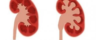
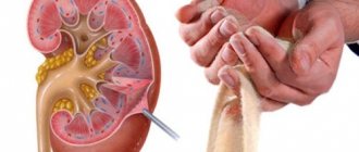
Kidney disease in dogs is considered one of the most dangerous diseases. Danger awaits an individual of any breed, height or age. According to statistics, kidney diseases develop in pets due to poor living conditions for the animal. Therefore, all veterinarians recommend prevention and careful monitoring of your four-legged pet.
Any unusual behavior may indicate the development of the disease. Kidney failure often develops as a complication of infectious or viral diseases. Why is kidney disease dangerous and what are its signs? A number of reasons causing the progression of kidney disease in dogs. Let's talk about all this in more detail.
Basic kidney functions
Kidney diseases are in second place among the ten most dangerous ailments that lead to the death of an animal. The main problem when the basic functions of the kidneys are impaired is the inability to restore already damaged organ tissue. Therefore, treatment as such only helps to support kidney function, especially in advanced cases. The kidneys perform a number of important functions:
- Cleansing the body's blood of toxins produced as a result of processing food, water, air, etc.
- Removing poisons, such as those acquired from food or from inhaling dangerous toxic gases.
- Excess water is also eliminated through the kidneys.
The kidneys are involved in the production of essential hormones, one of which is erythropoietin, which is responsible for the production of red blood cells. The work of the kidneys balances the salt and water content in the animal's body. The kidneys are like a well-functioning mechanism; if one of the systems is damaged, the functioning of the others gradually deteriorates.
Important: The main problem is the timely detection of kidney disease in an animal, because the symptoms of the disease appear already in an advanced state. And sometimes the level of organ damage, after diagnosis, is 60%. There are two courses of kidney disease:
- Spicy.
- Chronic.
The acute form manifests itself in the form of a sharp progression of the disease in one of the functional systems of the kidneys. Chronic kidney disease in a pet can be asymptomatic for several years. Much depends on the animal's immunity. Usually dogs are diagnosed with the chronic form.
Urolithiasis in cats. It is also known as urolithiasis and is much more severe than in dogs. Clinically, this disease initially manifests itself in the form of chronic cystitis. The terminal phase of ICD is difficulty or impossibility of urination. The animal often sits down, there is pain, but urine does not pass away at all or is released in scant drops, often mixed with blood. This is a threatening condition. It must be remembered that delaying urination for more than a day poses a danger to the life of the animal. Urinary retention is caused by stones and sand in the bladder. It would seem that by removing this mechanical obstacle we will solve the problem. But not everything is so simple... You need to know that KSD is the main cause of kidney loss in humans and animals on planet Earth. Kidneys fail due to ICD. The causes of ICD are not fully understood. Genetic and feeding factors have their place, but they are not the main ones. Treatment of ICD is symptomatic. For mild forms - therapy and optimization of feeding. For conditions of moderate severity - catheterization and medications. In severe cases - surgery. Shortening urethrostomy. Through surgery, the urethra becomes short and thick and allows sand and small stones to be expelled from the bladder. It must be understood that urethrostomy is a palliative operation, and only alleviates the course of urolithiasis, but does not eliminate the cause of stone formation. The disease itself, even after surgery, requires continuous monitoring and long-term treatment. You need to know that animals after shortening perineal urethrostomy become more susceptible to ascending genitourinary infections. The short and thick urethra makes it easier for infection to get to the bladder.
The characteristic pose of a cat that cannot urinate and a schematic illustration of why. The stone got stuck in the urethra, thereby causing swelling of the mucous membrane and complete obstruction of the urethra with the impossibility of emptying. As an emergency measure in such cases, catheterization of the bladder is intended to restore the outflow of urine.
Inserting a catheter into the bladder. Catheterization The procedure most often takes place under sedation or superficial anesthesia. Shifting the stone or sand that caused the blockage is quite painful. Stone or sand during catheterization scratches and injures the inflamed mucosa. The procedure should be performed by a qualified doctor with experience in such manipulations. Do not apply excessive force if the catheter does not pass. In such cases, you can use a pass-through catheter with a guidewire of a smaller diameter, and then install a normal-sized catheter over it, or gradually erode the stone plug with the pressure of a sterile antiseptic. Incorrect, rough, forceful catheterization, or installation of a catheter of the wrong diameter can lead to disastrous consequences: injuries and ruptures of the urethra, bleeding, and infection of the urethra. Sometimes the consequences of incorrect catheterization cannot be corrected even by surgery!
The catheter breaks up the sand plug or, if it is a stone, pushes it back into the bladder. This must be remembered! If the blockage is caused by a stone with a diameter of more than 1.5 mm, then we use a catheter to push it back into the bladder and it will not come out through the catheter. That is. After removing the catheter, urinary retention may occur again, since we have not eliminated the cause. The reason is a big stone. We only eased the situation for a while. Therefore, in case of acute urinary retention, the most important and informative study is ultrasound. It must be carried out to assess the morphological state of the bladder and determine the size of stones. If the delay is caused not by sand but by stones with a diameter of more than 1.5 mm, then surgical treatment is considered, namely the operation of shortening perineal urethrostomy.
The procedure for ultrasound examination of the abdominal organs and bladder.
The sand in the bladder looks like a scattering of flying snowflakes. When shaken, they fly up in a circle and gradually settle to the bottom of the bubble.
The stone in the bladder is of impressive size. Its configuration is visible - round, oval or with an uneven shape. He leaves an acoustic shadow behind him.
However, the presence of an acoustic shadow directly depends on the size of the stone and the frequency of the sensor. If the stone is not large in size and a low-frequency sensor is used for research, then the stone is clearly visible, but does not leave behind a dark stripe - an acoustic shadow.
A stone with uneven edges. On ultrasound it looks like a starfish. This form of stones is the most traumatic for the mucous membrane of the bladder. The presence of stones of this shape causes severe pain in the lower abdomen and constant bleeding.
Stones in the kidneys. This is much worse than bladder stones. In such a situation, the development of acute renal failure is possible - the organ actually stops working due to blockage of the tubules and injury to the epithelium of the renal pelvis. Often stones at points become the cause of pyelonephritis.
Stones in the bladder and kidneys are clearly visible on x-rays. Moreover, using the X-ray method, you can determine their mineral density - is it a solid stone or compressed sand.
A number of urological diagnostic procedures are performed using X-rays. For example, excretory urography. A special contrast agent, visible in X-rays, is injected into the animal’s blood, and through a series of photographs, it is observed how it accumulates in the kidneys and enters the bladder. The rate of staining of the kidneys and the intensity of the color are of diagnostic importance. Using the method of excretory urography, you can determine: the shape and size of the structural units of the kidney, its excretory ability, detect stones or tumors, and determine whether there are narrowings or compression of the upper urinary tract.
Another diagnostic technique performed using x-rays is retrograde contrast cystography. This method determines the tightness of the bladder, its shape, the morphological nature of the internal walls and the presence of tumors or cysts. To perform retrograde cystography, the patient's bladder is catheterized and a contrast agent, visible on x-ray, is injected into it. The bladder is filled with contrast in real time along with a series of x-rays.
Kidney removal. Kidney removal in dogs and cats.
Indications for removal of kidneys in animals may be: tumor growth, polycystic disease, hydronephrosis, infarction with severe hemodynamic disturbances, necrosis, trauma incompatible with the viability of the organ, pyelonephritis not responding to drug therapy, technically unrepairable ureteral rupture, incurable damage to the renal pelvis with ICD. Scientifically, removing a kidney is called nephrectomy. If we compare incurable kidney diseases that require nephrectomy, then in cats the list is much wider and they have to undergo this operation more often than in dogs. However, the decision to remove a kidney must be carefully considered. Everything needs to be double checked at least twice. And with any chance to preserve the organ and its functionality, we must fight. The nephrectomy operation is considered as an extreme and forced step. And even during an operation to remove a kidney, all output data that led to the decision to remove it is visually and diagnostically rechecked. We can say that after accessing the organ, the doctor puts the instrument aside three times before performing surgery. More often in veterinary practice, complete removal of the kidney is performed. But in cases where portioned resection is possible, partial removal of the kidney is performed. The functional status of the kidney “for removal” and the other kidney must be checked using the method of excretory urography. Based on the accumulation and rate of removal of contrast, a conclusion is drawn about the performance of the excretory system with the percentage of health of the left and right kidneys. The method of excretory cystochromography is less commonly used. A special dye is injected into the patient’s blood and the amount of it released from each kidney is visually observed. Other mandatory preoperative studies include: ultrasound, biochemical and clinical blood tests, urinalysis, x-rays. CT and ECG can be prescribed as auxiliary methods.
Surgical stage of kidney removal.
In general, as we see, there are plenty of urological diseases in animals, as well as diagnostic methods for detecting them and treatment regimens for getting rid of them. You just need to remember that the vast majority of urological diseases are asymptomatic for the time being, that is, nothing bothers the patient. But when the disease enters its decisive phase, there is often a threat to life. For the prevention and early detection of urogenital diseases, it is recommended to undergo a mini-dispensary examination once a year, including blood, urine and ultrasound tests.
Finally, an interesting fact: in animals with a high tendency to form stones on their teeth, the risk of urolithiasis is significantly lower. And vice versa - animals with urolithiasis, as a rule, have good and clean teeth.
Doctor of Veterinary Medicine M. Shelyakov
Classification of kidney diseases
Common kidney diseases:
Pyelonephritis occurs as an internal inflammation of the connective tissue of the organ and the renal pelvis. It develops due to bacterial infection of the organ, for example:
- coli,
- Pseudomonas aeruginosa,
- staphylococcus, etc.
The disease can also develop as a complication of cystitis or other inflammation of the genital and urinary organs. The presence of a tumor of any internal organ also provokes this disease. A distinctive feature is that both kidneys are affected. Pyelonephritis progresses so quickly that the animal dies within 24 hours if the disease worsens.
Glomerulonephritis is a non-infectious kidney disease. Develops as a complication from previous illnesses:
- acute allergic reaction,
- poorly treated wound on an animal,
- severe inflammation of internal organs,
- severe infectious disease.
When the functioning of the renal tubules, which are responsible for the elimination of toxins and protein metabolism in the animal’s body, is disrupted, nephrosis develops. Kidney failure is the last stage of the disease. The gradual failure of each kidney function leads to uncontrolled degradation of the organ. If the dog has been given this particular diagnosis, then the animal’s further life will be reduced to the constant presence of the pet under a drip and injections.
Attention! A thorough examination must be done to determine the accuracy of kidney disease. Insist on a detailed clarification of the cause of renal failure; the correctness of the therapy selected by the veterinarian to treat the animal depends on this.
Causes of kidney disease development
There are a number of reasons for the development of kidney disease:
- Poor nutrition with a lack of nutrients leads to vitamin deficiency and a decrease in the dog's immune system.
- Presence of hereditary diseases. Purebred pets are the most susceptible to this condition. The disease, acquiring a chronic form indirectly, provokes renal failure.
- Serious infectious or bacterial diseases.
- Weak immune system.
- The presence of tumors in the animal's body.
- Accumulation of toxins.
- Acute poisoning.
- Severe dehydration of the animal's body, leading to poor blood supply to the kidneys.
It is important to prevent the progression of the disease and consult a doctor at the first manifestations of unusual behavior in your pet.
Main symptoms of kidney disease in dogs
Symptoms of kidney disease can take many weeks to appear, gradually worsening your four-legged friend's condition. You should not make a diagnosis based solely on visual signs of a dog’s illness. After all, the symptoms of many health problems are similar. Here is a list of the main signs of the disease:
- A sharp decrease in appetite or complete refusal to eat.
- Increased thirst, so it is important that the dog always has a full bowl of fresh water.
- There is a frequent urge to go to the toilet, and the amount of urine is either small or large.
- The animal may vomit.
- Nervous condition.
- The color of urine changes, depending on the cause of the disease, it can be bloody, colorless or cloudy, with the presence of other impurities.
- The smell of urine becomes stronger.
- The previously clean dog begins to walk around small in various places: at home, in the car.
- The smell of ammonia from the mouth indicates the accumulation of a large amount of toxins in the animal’s body.
- Diarrhea.
- A peculiar gait. Due to constant pain, the animal begins to arch its back unnaturally.
- Swelling of the dog's paws appears. Other parts of the body may also swell: the abdominal region, the upper eyelids of the animal.
- Pain and whining of your pet when urinating.
- If this is a male, then when going to the toilet he sits down instead of lifting his paw up.
- A brown coating can be observed on the dog's tongue.
The presence of several signs should immediately alert the dog owner. The sooner you see a doctor, the greater the chance that your pet will survive.
Diagnosis and treatment
To make an accurate diagnosis, a comprehensive diagnosis should be performed. The accuracy of treatment depends on the nature of the disease. Only a veterinarian can determine what kind of kidney disease has affected your pet.
Treatment for kidney disease in dogs can take a long time. Much depends on the neglect of a particular case, determining the form of the disease. After clarification of all the nuances, drug therapy is prescribed. It is important to be careful and accurately calculate the dosage of the medicine so as not to harm the animal. First, you need to provide access to water to avoid dehydration.
In the chronic form, it is impossible to completely cure a dog; drug therapy only slows down the symptoms, thereby prolonging the animal’s life. It is important to establish the cause of the development; the dog’s therapy will be based on this. Compliance with a special diet, which is prescribed by a doctor based on the results of the examination. The duration of therapy depends on the progress of the disease and the condition of the animal. Each case of the disease is individual, and treating a dog at home without consulting a specialist risks the pet’s imminent death.
Treatment of pyelonephritis
The main task of a veterinarian treating pyelonephritis is to eliminate the cause that provoked the inflammation. Antibiotics of various groups are most often prescribed. If necessary, drugs are selected that are suitable for the animal's sensitivity (for example, when using urine culture).
If pyelonephritis occurs as a result of urolithiasis, then initially the specialist determines the type of stones and chooses a method for eliminating them. Additionally, antispasmodic and diuretic drugs, as well as immunostimulants, are used. Do not try to prescribe medications yourself - follow the instructions of a specialist to avoid complications.
The duration of therapy is at least 14 days. In especially severe cases, treatment of pyelonephritis takes several months.
If dog owners turn to the veterinarian only in the last stages of pyelonephritis, complications in the form of abscesses and hydronephrosis cannot be ruled out. They clog the tubules and stop the flow of urine. As a result, the only possible treatment for pyelonephritis remains surgical intervention - removal of the kidney.
In severe cases of the disease, accompanied by swelling and delayed urine output, a specialist may prescribe up to 48 hours of a fasting diet. Gradually, protein foods can be introduced into the animal’s diet, increasing the serving size each time, but without adding salt.
The best medicine is prevention
To prevent the development of kidney diseases, follow a number of preventive measures. They will protect your four-legged friend and help you live a long life.
- Don't let your dog play with stray dogs. There is a high probability of catching some disease from them.
- Get vaccinated in a timely manner to avoid contracting serious illnesses that lead to complications.
- Don't let your dog eat unhealthy foods and make sure your dog gets all the vitamins and minerals it needs through its food.
- Get a preventive examination from a veterinarian to identify possible illnesses in time.
Monitor your pet's behavior; if the dog begins to behave strangely or begins to consume more water than usual, then it is worth checking it for diseases. Caring for your four-legged friend will help you avoid a number of troubles related to the animal’s health. Even if your pet gets sick, you should immediately contact a veterinarian, this will help you begin treatment sooner and reduce the likelihood of complications developing in your dog.
Currently reading:
- Prognosis and treatment of kidney failure in dogs
- Thyroid dysfunction in dogs (hypothyroidism)
- What blood pressure is considered normal in dogs and how to measure it
- The first symptoms of a stroke and its treatment in a dog
Chronic renal failure (CRF) in dogs
Chronic renal failure is a pathological condition of the body, characterized by irreversible kidney damage, impaired excretion of nitrogen metabolism products from the body and a disorder of many types of homeostasis (that is, the relative constancy of the internal environment of the body).
This disease can be considered as the final stage of progression of a wide variety of kidney diseases: congenital defects, glomerulonephritis, amyloidosis, pyelonephritis, nephrolithiasis, polycystic disease and many others. Most of these diagnoses can only be made by biopsy (taking a piece of an organ for histology), so in most cases the conclusion is about chronic bilateral nephropathy.
As mentioned above, damage to more than 75% of the mass of the renal tissue leads to disruption of the kidneys: the concentration function decreases (which leads to a decrease in the density of urine), the excretion of nitrogen metabolism products is delayed (this is the final stage of protein metabolism in the body), and at a late stage Chronic renal failure in dogs develops uremia - poisoning of the body with decay products. The kidneys also produce the hormone erythropoietin, which is responsible for the synthesis of red blood cells, so when the kidneys fail, hormone synthesis decreases and anemia gradually develops.
As in the case of acute pathology, the diagnosis of chronic renal failure is made on the basis of anamnesis and characteristic examination results: hypoplastic anemia, increased creatinine and blood urea nitrogen, hyperphosphatemia, acidosis, and hyperkalemia are revealed. Decreased urine density (in dogs below 1.025 g/l), moderate proteinuria is also possible (increased protein in urine).
In case of renal failure in dogs, an X-ray may reveal an uneven structure of the kidneys and a decrease in their size; an ultrasound may reveal a heterogeneous structure, sclerosis of the parenchyma, complete loss of layers (impaired corticomedullary differentiation), and a decrease in the size of the organ.
Based on the value of creatinine concentration in the blood serum, 4 stages of chronic renal failure in dogs are distinguished:
- Non-azotemic stage - this includes any impairment of kidney function without a clearly identified cause associated with the presence of nephropathy. Initial changes in the kidneys can be detected by ultrasound, in the urine - an increase in the amount of protein and a decrease in density. According to blood biochemistry, a persistent increase in creatinine content is noted (but within normal limits).
- Mild renal azotemia - serum creatinine values are 125-180 µmol/l. The lower threshold of creatinine values may be a variant of the norm, but at this stage the pets already experience any disturbances in the functioning of the urinary system. Symptoms of kidney failure in dogs may be mild or absent.
- Moderate renal azotemia - serum creatinine values are 181-440 µmol/l. At this stage, as a rule, various clinical signs of the disease are already present.
- Severe renal azotemia – creatinine values above 441 µmol/l. At this stage, severe systemic manifestations of the disease and pronounced signs of intoxication are observed.


