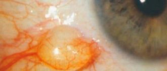
Malignant tumor, development. From embryonic retinal cells. Age, usually 1st 2 years of life. Initially, a grayish flat lesion appears on the retina. Gradually, the retina deepens due to the growth of the tumor node. Its surface takes on a pink tint.
Rapid necrotization occurs due to the rapid growth of the tumor. Then blindness occurs, the pupil dilates and acquires a yellowish glow and is called “amaurotic cat's eye.”
The tumor metastasizes to the liver, lungs, skull bones, orbit, and brain.
Treatment: Surgical, chemotherapy, radiation therapy.
50.Method of taking a smear from the conjunctival cavity.
This manipulation should be carried out in the morning before washing and instilling drops.
Hold the pipette with the thumb and index finger of your right hand.
Draw up the anesthetic solution from the bottle.
Ask the patient to look up.
Place a cotton ball on your lower eyelid with your left hand and pull it down slightly.
Place the medicine dropper with the tip down at an angle of 45° at a distance of 2-5 cm from the eye so that it does not touch the eyelashes and eyelids.
Instill 1-2 drops of anesthetic three times with an interval of 1-2 minutes. into the lower fornix of the conjunctiva.
Sterilize the platinum loop over the flame of an alcohol lamp.
The smear is taken using a loop or a regular probe, which is sterilized in advance over a fire.
After the loop has cooled, it is necessary to pull back the lower eyelid and pass the loop along the lower transitional fold.
After everting the upper eyelid, you need to take a smear from the upper transitional fold. The material taken from the patient is applied in a thin layer to a glass slide, which is first wiped with alcohol. After the preparation has dried, it is fixed over the burner flame. Then the smear site is outlined with a pencil, the patient’s name is indicated in the empty space and sent to the laboratory.
If it is necessary to conduct a bacteriological study, then the material is collected in a similar way. Then the edges of the test tube are burned over the flame of an alcohol lamp and the loop with the material is placed in a test tube with a nutrient medium.
Ticket No. 11
Features of taking a smear from the conjunctiva of the eye
Smears from the urethra, vagina and cervix in women
Urethral swabs in men
Throat and nasopharynx swabs
If diphtheria or sore throat is suspected, material is taken from the throat or nasopharynx. Before taking a smear, the patient should not gargle with disinfectant solutions. A smear is not taken due to the risk of vomiting immediately after eating. To take a smear, press the tongue with a spatula and take the discharge using a sterile cotton swab, which is then lowered into a closed sterile tube. It is advisable that the time between taking the material and sowing does not exceed 5-6 hours to avoid drying out the swab.
To take material from the urethra of men, clean, fat-free glass slides are prepared in advance. Discharge from the urethra is taken in the morning before the first urination. The external opening of the urethra is wiped with cotton wool moistened with a sterile isotonic sodium chloride solution or 1% chloramine solution. If the discharge is scanty, lightly press on the back wall of the urethra, remove the protruding drop and prepare smears for bacterioscopy with a wire loop.
In women, discharge from the urethra, vagina and cervix is taken before urination. In order to obtain discharge from the urethra, insert a finger into the vagina and press on the back wall of the urethra. The smear is taken with a forceps, metal spatula or wire loop. The discharge is smeared into a thin layer on a glass slide. Vaginal discharge is taken with a wire loop, a grooved probe or a blunt spoon. Slides with smears are marked with the letters C (cervix) and V (vagina). Dried smears are held 1-2 times for fixation over a burner flame and sent to the laboratory for microscopy. Material for inoculation is taken in sterile tubes in compliance with all sterility rules. In the accompanying referral, indicate the surname, first name, patronymic of the patient, department or site, purpose of the study, surname of the person sending, and date.
A smear from the conjunctiva of the eye is taken in the morning before washing and instilling drops using a platinum loop or probe, which is sterilized over a red-hot fire. The material is taken with a cooled instrument from the lower transitional fold of the retracted lower eyelid. It is advisable to turn out the upper eyelid and take mucus from the upper transitional fold. The material is applied in a thin layer onto a clean, grease-free glass slide. When the smear dries, it is fixed over the flame, and the place where it is located is outlined with a glass graph. To inoculate, a loop of material is placed over a burner flame into a sterile tube containing agar or broth. After distributing the material in the nutrient medium, the test tube is capped over a flame.
Features of selection and examination of an eye smear
Indications
Viral infections that affect the nasopharynx often cause purulent eye infections. Neutral corynebacteria can also cause inflammation under certain conditions. Fungi of the genus Aspergillus and Candida appear after treatment with antibiotics. A common cause of illness is gonococci. Microflora culture is prescribed when it is necessary to identify the pathogen, as well as to determine which medications are contraindicated during therapy. Samples of discharge are taken if necessary to establish pathogenic bacteria or viruses, confirm or exclude the following diseases:
- blepharitis;
- conjunctivitis;
- keratitis;
- dacryoadenitis;
- dacryocystitis;
- demodicosis;
- iridocyclitis;
- molluscum contagiosum;
- retinitis;
- scleritis;
- endophthalmitis;
- episcleritis;
- barley.
How is a smear taken?
If the patient has conjunctivitis, then the material is collected with the eyelid retracted.
If the patient was treated with medications, then their use is canceled 8-12 hours before the procedure. The flora discharge is collected immediately after waking up, before water procedures, adhering to a strict methodology:
- Microflora is collected using a sterile cotton swab in each eye in turn.
- A smear for conjunctivitis is done with the eyelid retracted so as not to touch the eyelashes.
- Dry blepharitis crusts are removed with tweezers and material is taken from the opened erosions.
- In case of keratitis, the manipulation site is first anesthetized.
- The material is also taken from contact lenses if the patient wears them.
- Swabs, separately for each eye, are placed in containers, labeled and immediately sent for examination. If necessary, the test tubes are stored at a temperature not exceeding +8 °C.
Failure to comply with sanitary rules when using contact lenses often leads to purulent-inflammatory eye diseases.
Types of analyzes
To accurately determine which microbiological pathogen is the cause of the disease, a smear on the culture tank is examined in several ways:
The growth rate of the pathogenic agent can be determined by inoculating material taken from the patient on a nutrient medium.
- Cytological analysis. The material is stained and examined under a microscope.
- Bacterioscopic. It is carried out according to the same principle as the first one.
- Bacteriological. Sowing the material on a nutrient medium to determine the growth rate of the pathogen.
Bacteriological seeding of eye discharge is carried out in case of purulent-inflammatory processes of the eyes - conjunctivitis, blepharitis, keratitis. Keratoconjunctivitis caused by the normal microflora of the conjunctiva, naopharynx, and oral cavity is often preceded by a viral infection of the upper respiratory tract, allergic rhinitis, trauma, surgical interventions, and the use of rinsing solutions.
A weak inflammatory reaction can also be caused by non-pathogenic microorganisms of the genus Corynebacterium.
Long-term local use of antibiotics leads to the release of fungi of the genus
Kandila and Aspergilus .
Inflammation is most often localized on the conjunctiva, mucous membrane of the eyelids, lacrimal sac, and less often affects the cornea. One of the common causes of acute purulent conjunctivitis is gonococci.
Preparing for the study
No special preparation is required for the study. The collection of biological material is carried out strictly before the start of the use of antibacterial and chemotherapeutic drugs or no earlier than 10-14 days after their discontinuation.
Most often ordered with this service
| Code | Name | Term | Price | Order |
| from 5 w.d. | 1500.00 rub. | |||
| from 4 days | 840.00 rub. | |||
| from 6 days | 2640.00 rub. | |||
| The information provided is for reference only and is not a public offer. For up-to-date information, contact the Contractor's medical center or call center. |
Bacteriological seeding of eye discharge is carried out in case of purulent-inflammatory processes of the eyes - conjunctivitis, blepharitis, keratitis. Keratoconjunctivitis caused by the normal microflora of the conjunctiva, naopharynx, and oral cavity is often preceded by a viral infection of the upper respiratory tract, allergic rhinitis, trauma, surgical interventions, and the use of rinsing solutions.
A weak inflammatory reaction can also be caused by non-pathogenic microorganisms of the genus Corynebacterium.
Long-term local use of antibiotics leads to the release of fungi of the genus
Kandila and Aspergilus .
Inflammation is most often localized on the conjunctiva, mucous membrane of the eyelids, lacrimal sac, and less often affects the cornea. One of the common causes of acute purulent conjunctivitis is gonococci.
Taking material
At least 5-6 hours before taking material for bacterial culture, all medications and procedures are canceled.
The material is directly collected after local anesthesia. If the patient uses contact lenses, it is necessary to examine their inner surface.
Using a sterile swab, material is taken from the affected areas at the height of the inflammatory process. Be sure to follow the rules of asepsis. Place in a transport medium or apply to a nutrient medium. Storage conditions:
Decoding
- I degree of growth (very meager growth) - single colonies up to 10 (
- II degree of growth (small amount) - up to 20 colonies (10 3 CFU/tampon)
- III degree of growth (moderate amount) - more than 21, but less than 100 colonies (10 4 CFU/tampon)
- IV degree of growth (large number) - more than 100 colonies (> 10 5 CFU/tampon)
III and IV growth degrees indicate the etiological significance of this microorganism.
Many eye diseases cause not only discomfort and blurred vision, but also noticeable discharge from the eyes. In case of inflammatory processes, to determine their nature and find the most effective method of treatment, patients are prescribed an analysis and bacterioscopic examination of an eye smear.
During preliminary preparation, 5-8 hours before taking a smear, doctors usually cancel all medical procedures and the use of prescribed medications. The material released from the eye is taken immediately after sleep before washing from the place of its greatest accumulation on the conjunctiva of the eye.
According to the method, a smear is taken separately from each eye using a dry sterile swab-probe or a platinum loop, which is heated red-hot in an alcohol lamp for sterilization. The lower eyelid is pulled back and a cooled loop or swab is passed along its lower transitional fold. Having turned out the upper eyelid, a lump of mucus is collected from its transitional fold.
A clean glass slide is wiped with alcohol and a thin layer of material taken from the eye is applied to it. The dried smear is fixed over the burner and, for convenience, its contours are marked using a glass graph.
Inoculation is carried out by lowering a loop with the collected secretion into agar or broth in a sterile test tube over a burner flame, which is closed with a stopper.
Material for smear analysis can be stored at temperatures no higher than 8° C and is recommended to be delivered to the laboratory as soon as possible. A complete analysis of an eye smear includes the following types:
Cytological analysis, in which the infectious or allergic nature of the inflammation is revealed by examining a stained smear under a microscope.
Bacterioscopic analysis, in which the microbial composition of a stained smear from the eye is studied under a magnifying device. This method quickly and accurately identifies streptococci and staphylococci, Escherichia coli and diphtheria coli, chlamydia, gonococci and Aspergillus fungi.
Bacteriological analysis consists of inoculating a smear on nutrient media in order to grow colonies of inflammatory pathogens.
In any case, a certain number of opportunistic microorganisms live on the conjunctiva of the eye, therefore, to determine the nature of the disease, it is necessary not only to isolate all the microbes in the smear, but also to count their exact number.
The bacteriological method is effective when it is necessary to confirm that purulent inflammation is caused by excessive growth of one of the representatives of the native microflora of the eye. Clarification of the level of sensitivity to antibacterial drugs.
Staphylococcus is a gram-positive bacteria. Currently there are 27 varieties of them. About 14 types of staphylococcus are found on human skin and mucous membranes, but only some of them can cause disease. To identify this bacterium in the body, a microbiological research method is used.
Instructions
Some types of staphylococcus are dangerous to the body because they can weaken its immunity. Bacteria act directly against cells of the immune system and facilitate access to other pathological microorganisms. Staphylococcus can cause allergic reactions, its enzymes have a negative effect on other cells, and its poisons poison the body. These bacteria can be especially dangerous for pregnant women; staphylococcal infection leads to damage to internal organs and the fetus; in newborns it manifests itself as pustular wounds and tumors. An analysis for staphylococcus is prescribed in the following cases: if there is a suspicion of an infection caused by staphylococcus (sore throat, pharyngitis), for bacterial carriage, before antibacterial treatment against an infection caused by staphylococcus, for nosocomial infections, during regular preventive examination of medical staff and catering workers, during pregnancy. Staphylococcus can be detected in the body by submitting biomaterial for analysis. The study is carried out using the microbiological method. The following biomaterial is used for analysis: a swab from the nose, oropharynx, breast milk, a single portion of urine, sputum, a swab from the conjunctiva, ear discharge, a urogenital swab, a rectal swab, feces. 8-12 hours before donating sputum, drink a large amount of liquid (water). Do not take diuretics for 2 days before urine collection. Avoid taking laxatives, administering rectal ointments, suppositories, limit taking medications that affect intestinal motility (pilocarpine, belladonna, etc.) and stool color (bismuth, iron, barium sulfate) for 3 days before collection feces Do research before taking antibiotics and other antibacterial drugs. Women should take a urogenital smear or urine test before their period or 2 days after their end. Breast milk is collected from the left and right breasts into separate containers. Men are not recommended to urinate for 3 hours before submitting urine or a urogenital smear. The analysis result indicators are reflected in the number of colony-forming units in 1 ml of material. In healthy people with strong immunity, carriage of staphylococcus can be detected. In this case, the test result can be up to 10 CFU/ml. In people with reduced immunity, this figure can be over 10 CFU/ml, in which case staphylococcus can cause a severe inflammatory process. Print
How to take an eye swab
www.kakprosto.ru
Eye conjunctival smear
An eye disease such as conjunctivitis (inflammation of the outer mucous membrane of the eye) is the most common in the world. It affects several million people every year.
It is not always possible to prescribe adequate treatment for this infectious disease, especially in its chronic form, without special diagnostics, since there can be many reasons for its occurrence:
Bacterial damage;
Chlamydia;
Allergic reaction.
Self-medication often does not give the desired result, since special laboratory tests are required to diagnose conjunctivitis. To identify the causative agent of the infection, the doctor takes a smear from the patient for microflora and scrapings from the conjunctival sac. In accordance with the data obtained, treatment is subsequently prescribed to achieve the best effect.
Laboratory diagnostics. Bacterial culture
A swab from the conjunctiva of the eye is taken in the morning - it is important that the patient does not wash his face before taking the test. The taken material is placed in a test tube with a nutrient medium for 6-7 days, after which the colonies of microorganisms grown on it are examined. It can be:
Bacteria (staphylococci, streptococci, pneumococci, gonococci, E. coli, diphtheria bacillus);
If culture from the eye confirms the presence of pathogenic microflora, additional studies are carried out to determine their sensitivity to various antibacterial drugs and phages. Thus, an eye smear allows you to obtain information not only about the nature of the causative agent of the disease, but also to select the most suitable means for treatment. When allergic conjunctivitis is detected, treatment consists of identifying the allergen, eliminating it from the patient’s body and prescribing antihistamines.
yasnoe-oko.ru
MICROBIOLOGICAL STUDIES OF DISCHARGE FROM THE EYE
| Cat. No. | Name | Type | Price* | Regular | Urgent** | Order |
| b0308 | Culture for microflora and determination of sensitivity to antibiotics of discharge from the eye (right) | semi-quantitative | 850 rub. | 4 w.d. | — | Order |
| b0309 | Culture for microflora and determination of sensitivity to antibiotics of discharge from the eye (left) | semi-quantitative | 850 rub. | 4 w.d. | — | Order |
| b0408 | Culture for Candida spp. and determination of sensitivity to antimycotic drugs of discharge from the eye (right) | semi-quantitative | 745 rub. | 4 w.d. | — | Order |
| b0409 | Culture for Candida spp. and determination of sensitivity to antimycotic drugs of discharge from the eye (left) | semi-quantitative | 745 rub. | 4 w.d. | — | Order |
| b0608 | Culture for Neisseria gonorrhoeae and determination of sensitivity to antibiotics of discharge from the eye (right) | semi-quantitative | 920 rub. | 7 w.d. | — | Order |
| b0609 | Culture for Neisseria gonorrhoeae and determination of sensitivity to antibiotics of discharge from the eye (left) | semi-quantitative | 920 rub. | 7 w.d. | — | Order |
| b1808 | Culture for Staphylococcus aureus and determination of sensitivity to antibiotics in eye discharge (right) | semi-quantitative | 745 rub. | 3 w.d. | — | Order |
| b1809 | Culture for Staphylococcus aureus and determination of sensitivity to antibiotics in eye discharge (left) | semi-quantitative | 745 rub. | 3 w.d. | — | Order |
| b2108 | Culture of microflora and determination of sensitivity to antibiotics of discharge from the eye (right) (extended spectrum) | semi-quantitative | 975 rub. | 4 w.d. | — | Order |
| b2109 | Culture of microflora and determination of sensitivity to antibiotics of discharge from the eye (left) (extended spectrum) | semi-quantitative | 975 rub. | 4 w.d. | — | Order |
| b2208 | Microflora culture without determining the sensitivity to antibiotics of discharge from the eye (right) | semi-quantitative | 720 rub. | 2 w.d. | — | Order |
| b2209 | Microflora culture without determining the sensitivity to antibiotics of discharge from the eye (left) | semi-quantitative | 720 rub. | 2 w.d. | — | Order |
| b2308 | Culture for microflora and determination of sensitivity to bacteriophages of discharge from the eye (right) | semi-quantitative | 825 rub. | 4 w.d. | — | Order |
| b2309 | Culture for microflora and determination of sensitivity to bacteriophages of discharge from the eye (left) | semi-quantitative | 825 rub. | 4 w.d. | — | Order |
| b3108 | Culture for Staphylococcus aureus without determining the sensitivity to antibiotics of discharge from the eye (right) | semi-quantitative | 575 rub. | 2 w.d. | — | Order |
| b3109 | Culture for Staphylococcus aureus without determining the sensitivity to antibiotics of discharge from the eye (left) | semi-quantitative | 575 rub. | 2 w.d. | — | Order |
* The cost of laboratory tests does not include the cost of collecting biomaterial. Prices are valid for the Moscow region. ** Urgent execution is valid only for the Moscow region.
Analysis of discharge from the eye
An analysis of discharge from the eye is prescribed to clarify the nature of the inflammatory disease (conjunctivitis, keratitis or blepharitis) and to select the most effective method of treating it.
Methodology for performing the examination:
- Preliminary preparation includes the abolition of all medications and medical procedures 5-8 hours in advance.
- The material is collected after sleep, before washing, from the places of greatest accumulation of pathological discharge using a sterile cotton swab, separately for each eye.
- For conjunctivitis, first slightly retract the eyelid so that the eyelashes do not touch the tampon. Pus collects by moving from the outer corner of the eye to the inner.
- For blepharitis, dry purulent crusts are removed with eye tweezers. A swab is taken from the existing erosions.
- For keratitis, preliminary anesthesia with anesthetic drops is required. The smear is taken with a dry sterile swab.
- When wearing contact lenses, a swab is also taken from their inner surface.
- After taking a smear, the swab is placed in test tubes (separately for each eye), signed and delivered to the laboratory as soon as possible. If storage is necessary during long-term transportation, temperature conditions should not exceed +8 degrees Celsius.
Types of analysis of discharge from the eye:
- Cytological. The essence is to study a stained smear under a microscope to identify the nature of the disease. In this way, the allergic or infectious nature of the inflammation is determined.
- Bacterioscopic. Study using preliminary staining of a smear and its microscopy of the microbial composition of discharge from the eye. Using this method, you can quickly identify chlamydia, diphtheria bacillus, fungi of the genus Aspergillus and Candida, gonococci, E. coli, streptococci, staphylococci.
- Bacteriological (inoculation on various nutrient media to obtain the growth of colonies of the pathogen). Since conditionally pathogenic microorganisms live in small quantities on the conjunctiva of the eye in all healthy people, to clarify the nature of purulent inflammation it is often necessary not only to isolate all the microbes found in the smear, but also to count their number. Only in this way can it be confirmed that the causative agent of the disease was an activated representative of its own microflora, for example, Pseudomonas aeruginosa, coagulase-negative staphylococcus, Moraxella catarrhalis, fungi, Klebsiella and others.
- Determination of sensitivity to antibacterial drugs.
| Recommend: | Tweet |
ztema.ru
How is PCR diagnostics of coronavirus carried out?
Since COVID-19 is an RNA-containing virus, the essence of the PCR reaction is to quickly directly search for any microbial RNA residues with their preliminary duplication to a DNA chain.
A swab is taken from the throat. To avoid false-positive or false-negative results, it is forbidden to drink alcohol one day, and one hour before the test - to smoke, drink, or eat. To carry out PCR, saliva and mucus are sufficient, but venous blood can also be taken. As a rule, they are limited to mucous secretions from the walls of the nasopharynx.
The collected biomaterial is placed in special containers, labeled, sealed, and transported to the laboratory, accompanied by a medical worker in a protective suit, in compliance with safety precautions.
In the laboratory, fragments of the RNA virus are isolated from the resulting material, the RNA single strand is doubled by reverse transcription to DNA - the matrix of the virus, and it is multiplied many times, like on a photocopier, using a special polymerase enzyme. Hence the name of the test. The point is that RNA is specific, and only the same strand of the virus can be added to coronavirus RNA to double it.
The number of duplicates can reach 45. Then the polymerase chains are split again. At this very moment, at the stage of reverse conversion of DNA into RNA, a coronavirus (or any other that was planned to be identified) is identified. This is done using electrophoresis, since it allows you to see all the multiplied copies of DNA.
DNA has a negative electrical charge, so it is attracted to the positive pole and, passing through the dye, changes color and becomes bright. If there is no virus in the biomaterial, then the very first strand of DNA will not line up, there will be nothing to cling to to build a double strand. This means that electrophoresis will not show anything, there will be no color.