What is
Before discussing the symptoms and causes of the problem, it is necessary to characterize the disease itself. Keratopathy is a congenital or acquired negative change in the outer layers of the cornea of the eye.
The thin layer of the cornea is easily damaged
Doctors distinguish the following types of keratopathy:
- bullous – which is characterized by the appearance of fluid bubbles on the surface of the cornea;
- a droplet form of a climatic nature, which often appears among residents of the far north as a reaction to cold and wind;
- the lipoid form develops in cases where the formations consist of particles of adipose tissue;
- ribbon-shaped - appears in cases where calcium is deposited in the form of salts;
- aphakic – develops in older people as a reaction to natural changes in the organs of vision.
- It is also possible for ocular keratopathy to occur after surgery.
Keratopathy is equally common in both men and women. If a representative of the fairer sex suffers from this problem, then there is a high probability that it will be passed on to her children.
Keratopathy has an ICD 10 code H18 (depending on the type of disease, the numbers 18.0, 18.1, and so on may be added).
Some types of the disease develop at certain ages or under specific conditions. Thus, climatic keratopathy is characteristic only in cases where a person suffers due to regular exposure to low temperatures. The lipoid form most often overtakes patients during puberty.
Bullous form
Illness, physiological norm or toxic poisoning of the brain - the pupils are constricted for the reasons.
Causes and stages of development
The mechanism of occurrence of keratosis is the compaction of skin flakes located in the stratum corneum. This occurs due to prolonged exposure of the skin to sunlight. Ultraviolet radiation in large quantities is very dangerous for humans and can provoke the development of cancer processes in the body . The disease does not appear overnight. It takes many years to develop, which is why it is mainly older people who experience solar keratosis. Despite the fact that the problem itself is precancerous and is not a sign of the development of an oncological process, its onset increases the risk of developing squamous cell skin cancer.
Reasons for appearance
Risk factors that increase the likelihood of occurrence:
- Intoxications of various types (medicines, radiation exposure) during the period of intrauterine development of the fetus, genetic disorders, mutations.
- Inflammatory processes: infectious, parasitic, allergic. Mechanical damage (injuries, burns).
- Age-related changes (dystrophic involutive processes).
- Postoperative processes in tissues.
- Long-term use of medications (ointments, drops).
- Wearing contact lenses.
- Poor quality cosmetics.
Causes
It is possible to find out the cause of the development of the disease only after its type is determined. Why is this diagnosis most often made:
- Surgical intervention in the treatment of cataracts, glaucoma and other serious diseases.
- Congenital pathology in the presence of a similar problem in one or both parents.
- Trauma can stimulate the development of the band-like form.
- Also, a ribbon-like form with calcium deposits develops against the background of the development of viral diseases. For example, as lupus progresses, the cornea may become damaged.
- Influence of climatic conditions.
- Conjunctivitis, dry eye syndrome, entropion and blepharitis can eventually cause the development of a superficial pinpoint form of the problem.
The development of the disease can be affected by constant exposure to bright light or foreign objects entering the eye. Each case is examined separately, and the diagnosis directly depends on the overall clinical picture.
It was mentioned above that the aphakic form of keratopathy develops against the background of natural changes in the ocular apparatus with age. In general, the patient’s age, gender and developmental characteristics cannot be discounted, since all these important nuances can prompt the correct diagnosis.
Stages of development of the bullous form
Find out how Iridina moisturizing eye drops work here.
Causes
The pathology is acquired, but it is often possible to establish a hereditary predisposition to this disease. Genetic mutations and the type of inheritance of keratopathy have not been studied. The main causes of development: • Fuchs endothelial dystrophy.
Genetically determined apoptosis of posterior epithelial cells is observed, which leads to an increase in its permeability and the occurrence of pathology.
• Eye infections.
The disease develops when the cornea is affected by herpetic or syphilitic nature.
The detection of specific opacities in newborns suggests intrauterine infection. • Corneal injury.
Damage due to mechanical trauma or burn provokes increased exudation, which causes the formation of typical “bulls”.
• Iatrogenic effects.
Pathology can occur in the early postoperative period after phacoemulsification of cataracts or implantation of intraocular lenses.
Porokeratosis ICD code 10
Classic porokeratosis of Mibelli (plaque porokeratosis)
Appears in childhood. The lesion is localized more often on the back of the hands, often unilaterally in the form of horny miliary papules, gradually forming small plaques (single or multiple) of a grayish-brown or copper-red color, round or irregular in shape, with a diameter of up to 6-10 mm. The central part of the plaque sinks slightly, becoming atrophic. Along the edge of the plaque, a brownish horny ridge is preserved, which is enclosed in a groove and protrudes in the form of a ridge. As a result of slow eccentric growth, plaques can reach a size of up to 6-7 cm in diameter. The disease slowly progresses, as a result of which the lesions spread over the skin of the extremities, neck, and less often the trunk, face. Some of them may have a linear shape. In this case, on the face, lesions may resemble discoid lupus erythematosus. Damage to the mucous membranes and cornea is also possible. In some cases, abnormal development of teeth and mental retardation are observed.
Disseminated superficial actinic porokeratosis
The clinical picture develops more often after 30 years, but is not observed in childhood. The type of inheritance is autosomal dominant with regular penetrance over the age of 16 years. A close relationship between clinical manifestations and insolation has been revealed and the role of UV irradiation in the genesis of this dermatosis has been proven. There is an assumption that the disease is associated with the proliferation of a mutant clone of epidermal cells formed under the influence of UV rays. However, some authors express doubts about the decisive significance of UV irradiation in the origin of dermatosis, considering the possible development of a non-actinic disseminated form of the disease.
Lesions in disseminated superficial actinic porokeratosis are localized in open areas of the skin accessible to insolation, mainly on the extensor surfaces of the forearms, legs, dorsum of the hands, face. Lesions of the palms and soles, scalp, lower third of the abdomen, buttocks, as well as mucous membranes was not observed.
The rash consists of miliary horny papules, gradually forming small plaques, uneven or irregularly oval in outline with a clear, slightly raised edge and usually an atrophied center of a brownish or pinkish color. Some horny papules have a central depression. Gradually, the number of lesions increases, especially in the summer.
Porokeratosis disseminated superficial
It is distinguished by multiple lesions that develop in childhood (usually 5-10 years) and does not depend on solar insolation with an autosomal dominant type of inheritance. There are no subjective symptoms. Sporadic cases of the disease are possible after transplantation operations (kidney transplant, etc.) as a result of the immunosuppressive effect.
Eruptive pruritic papular porokeratosis
Clinically, it manifests itself as repeated outbreaks of intensely itchy papular rashes, and the disease develops acutely. It is observed mainly in the older demographic group (average age 65.8 years), with a male to female ratio of 2: 1. There are no registered familial cases.
In 30% of cases, patients have a newly diagnosed malignancy. The average onset of the disease is 2.7 months before or after diagnosis of the malignancy. Associated malignancies are hepatocellular carcinoma, cholangiocarcinoma, ovarian cancer and lymphoma. The lesions tend to regress after treatment of the malignancy, suggesting a paraneoplastic syndrome.
In 30% of patients, immunity disorders are detected, mainly associated with taking medications (prednisone, antirheumatic and biological drugs).
In 30% of cases, pre-existing porokeratosis, especially disseminated superficial porokeratosis, was noted.
Reticular porokeratosis
Clinically, it manifests itself as rashes typical of porokeratosis, which, merging, form a reticular pattern.
Giant porokeratosis
Giant porokeratosis is a rare form. It is characterized by a single lesion measuring 10-20 cm, sometimes more, and a wide (up to 1 cm) peripheral horny ridge. Frequently localized on the dorsum of the foot. It has a pronounced tendency to malignant degeneration.
Linear porokeratosis
May affect only a segment of the body or be generalized. The lesion is characterized by a linear arrangement of rashes. This form combines in the clinical picture both a hyperkeratotic (horny ridge along the edge of the lesion) and an atrophic component and corresponds to a clinically linear verrucous epidermal nevus. The nevoid nature of this form of porokeratosis is assumed.
Symptoms and diagnosis
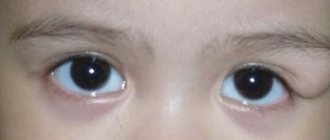
It is not difficult to notice the development of keratopathy, because when it appears, clouding of the pupil occurs. Visual acuity also decreases, and it is as if a veil is cast over the eyes, through which a person can barely distinguish the outlines of well-known objects.
What other symptoms most often bother the patient:
- Severe pain of a different nature. It can be a nagging discomfort, cramping or acute pain.
- Serious visual impairment up to its complete loss.
- Sensation of a foreign body in the eyes.
- Swelling or increased tearing may occur.
In almost all cases, symptoms develop gradually. After the first signs appear, about a month passes, and the person’s well-being noticeably deteriorates, visual acuity decreases almost to zero.
A dangerous inflammatory disease is iridocyclitis.
Untreated conjunctivitis can give rise to the development of the disease
The right approach to vision correction – which lenses for the eyes are best to choose.
Laser therapy helps cure the problem without a transplant
The main method of diagnosis is visual examination and questioning of the patient. If a person complains of pain and decreased visual acuity, and the doctor notices clouding of the pupil, then the diagnosis is more than clear.
However, to confirm it, it is still better to conduct research using special equipment. Most often, doctors resort to the following research methods:
- biomicroscopy;
- tonometry;
- ophthalmoscopy;
- visometry.
The point form is perfectly determined using biomicroscopy. If we are talking about ribbon keratoscopy, the doctor may resort to determining the level of uric acid and calcium.
Only after a comprehensive diagnosis and the correct verdict can we proceed to discuss treatment options.
Ophthalmoscopy
In what cases is the use of Catarax eye drops indicated, read the article.
Visometry
When mites become a real problem - eye demodicosis symptoms and treatment.
Prevention measures
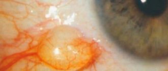
A conjunctival cyst is not dangerous to vision if it is detected and treated in a timely manner . In addition to preventive examinations by an ophthalmologist, the following measures will help to avoid the appearance of cysts and the development of their complications:
- compliance with hygiene rules, including when using contact lenses and cosmetics;
- attention to all doctor’s instructions during the rehabilitation period after operations for other ophthalmological pathologies;
- reducing, if possible, the impact of irritating factors on the eyes in everyday life or professional activities;
- control of the general condition of the body, correction of immunity;
- avoiding injuries and bruises to the organs of vision.
A conjunctival cyst is not dangerous - it is only important to carry out timely diagnosis and treatment . You should not hesitate to visit an ophthalmologist or be afraid of surgical intervention - the risk of possible postoperative complications is many times lower in comparison with the consequences of neglected cystic cavities.
Share the article on social media. networks:
Classification
The disease has a number of pathomorphological signs that determine the stages of progression of the disease. Let's look at the main types of actinic keratosis:
- Hypertrophic. Nuclear cells appear in the skin, producing light and dark keratin.
- Pigmentary. A large number of melanin cells accumulate in the basal layer of the epidermis, giving the integument a dark brown color.
- Lichenoid. A lymphocytic infiltrate forms in the basal cells of the skin, spreading to the upper layers of the dermis.
- Proliferative. Foci of hyperkeratosis appear against the background of colloid dystrophy with the deepening of epidermal cells into the layers of the integument.
- Atrophic. The stratum corneum becomes thinner and cracks appear.
- Acantholytic. Tumor-like processes arise in the dermis, growing upward to the outer layers of the skin.
- Buoenoid. The initial stage of development of the oncological process.
Treatment options
The most effective method of treatment is a corneal transplant or prosthetics. However, not all patients are ready to take such extreme measures. That is why the following conservative treatment methods are popular:
- the use of medications that eliminate the sensation of the presence of a foreign body and combat dry eye syndrome;
- exposure using laser therapy;
- corneal cell prosthetics;
- transplantation of a part of the lens - surgery;
- installation of special contact lenses capable of transmitting oxygen;
- in the case of a band-shaped form, calcium deposits are removed.
If a person is diagnosed with a climatic form of keratopathy, a change of place of residence may be required. Doctors also recommend wearing special glasses that reduce the negative impact of the environment.
Some experts recommend resorting to the use of folk remedies. For example, you can drop Siberian fir oil into your eyes before bed. This remedy effectively eliminates corneal clouding. However, you should resort to traditional methods of treatment only after consulting a doctor, using them as an option for additional therapy.
Visual inspection is the main diagnostic method
Necessity or needless torment? – Injections in the eyes for retinal dystrophy.
ICD 10: Item D31 Benign neoplasm of the eye and its adnexa
Composition of the item
- D31.0 Benign neoplasm of the conjunctiva
- D31.1 Benign neoplasm of the cornea
- D31.9 Benign neoplasm of the eye, unspecified part
- D31.2 Benign neoplasm of the retina
- D31.3 Benign neoplasm of the choroid
- D31.4 Benign neoplasm of the ciliary body
- D31.5 Benign neoplasm of the lacrimal gland and duct
- D31.6 Benign neoplasm of the orbit, unspecified part
Treatment
The method of therapy is selected by a dermatologist individually for each patient based on the disease picture and the results of the studies. Conservative therapy, removal of affected skin areas, as well as the additional use of traditional medicine recipes are allowed.
Hardware therapy methods
It is impossible to predict the further development of keratosis on the skin, therefore, in order to prevent malignancy of the formation, it is recommended not to treat it, but to remove it.
The following methods are allowed:
- Cryodestruction. A targeted area of skin is exposed to liquid nitrogen, resulting in controlled tissue death. After the procedure, a scar remains at the site of the keratotic spot. There is a risk of skin hyperpigmentation in the affected area.
- Radio wave, laser, electrical removal. The affected area is excised using one of the listed methods, which also leaves a scar after healing.
Medicines
A conservative approach to the treatment of solar keratosis is the use of pharmacological drugs. Thus, local chemotherapy products, for example, formulations with fluorouracil, can be used to remove the formation. Their application to the skin destroys diseased cells, blocking their functions.
It is acceptable to use other means:
- Imiquimod cream to stimulate the protective function of the skin. Its use in the early stages of the disease leads to the body’s independent rejection of damaged cells.
- Non-steroidal anti-inflammatory drugs, including 3% Diclofenac gel.
Folk remedies
Home methods of alternative medicine are used as auxiliary methods. They are aimed at softening the skin and increasing their level of protection from solar radiation.
The most commonly used recipes are:
- Cream with celandine. The dried leaves need to be crushed and the powder mixed with melted pork fat in a ratio of 1 to 3. The resulting ointment, after hardening, is applied to the affected areas three times a day.
- Therapeutic oil rubs. To soften tissues, it is recommended to treat keratotic areas twice a day with a cotton pad soaked in sea buckthorn or fir oil.
- Before applying the ointment to thick skin, you need to take a hot bath with soda. To do this, fill a container with water at the maximum permissible temperature and dissolve several tablespoons of baking soda in it. Steaming duration is 10 minutes.
- For internal use, tea is prepared from burdock roots. The crushed plant component is brewed with hot water in the proportion of 4 tablespoons per liter of liquid. After infusing in a thermos for 5 hours, the composition is filtered. During the day you need to gradually drink 0.5 liters of the drink.
The main dangers of diagnosis
The main danger posed by such a disease is the likelihood of complete loss of vision. This forecast is relevant in cases where a person does not pay attention to the progress of the problem and allows it to develop.
Due to clouding of the cornea, visual acuity is noticeably reduced, which makes it impossible to drive a car or other vehicles. Symptoms of keratopathy can cause pain and pain with prolonged tension of the eye muscles. Consequently, it becomes impossible to work at the computer or read for a long time. That is why it is necessary to immediately pay attention to emerging symptoms and eliminate them using medical techniques.
Normalizes metabolic processes and moisturizes the sclera - Cationorm eye drops instructions.
Loss of vision is the main danger when keratopathy occurs
Treatment of cataracts must begin with proven remedies. Read the instructions for using Katachrom eye drops here.
Diagnosis of the disease
A general conclusion about the presence of a neoplasm can be made based on a visual examination and collection of patient complaints, but then a number of examinations may be needed:
- ophthalmoscopy - in order to identify concomitant changes in the fundus; with a cyst, distortion of the shape of the optic nerve head can be observed;
- viziometry – testing visual acuity;
- tonometry - measurement of intraocular pressure, which tends to increase in the presence of pathological cavities;
- biomicroscopy - examination under magnification of the conjunctiva, cornea, lens, iris using a slit lamp - allows you to assess the condition of the tissues and diagnose the spread of the cyst to the cornea;
- examination of a histological specimen (tissue sample obtained during cyst removal) - helps to detect inflammation and tissue degeneration, as well as determine the type of tumor and the prognosis for relapse.
Based on the studies, drug or surgical treatment may be prescribed. Some cysts only require observation and may resolve on their own.
Diagnostics
Examination of a patient with bullous form of keratopathy involves an external examination and the use of special diagnostic methods. Visually, the ophthalmologist determines persistent clouding of the anterior part of the eyes, often in combination with hyperemia of the orbital conjunctiva. The complex of ophthalmological examinations includes: • Biomicroscopy of the eye.
At the initial stage, local swelling of the endothelial layer and single folds on the decementum membrane are detected.
2 is characterized by persistent limited swelling, diffuse dispersion of pigment, and multiple folds. At the next stage, in addition to the above-described manifestations, injection of conjunctival vessels, signs of superficial keratitis, erosive defects, and increased neovascularization occur. At 4, the swelling spreads to all layers. The final stage is characterized by vascular opacities of varying density. The cornea is replaced by dense scar tissue with focal ulcerations. • Keratopachymetry.
Signs of local or diffuse edema are determined.
The thickness of the cornea in the central part varies from 600 to 1500 microns. • Gonioscopy.
At the fifth stage, obliteration of the anterior chamber angle is detected, which is caused by the organization of exudate and the deposition of pigment granules.
Iridocorneal adhesions are visualized. The aqueous humor is transparent and contains vitreous fibrils. • Visometry.
Visual acuity is often in the range of 0.1-0.3 diopters.
In severe cases, only light perception is preserved. • Ophthalmoscopy.
Ophthalmoscopic examination is possible only at stages 1-3 of the disease.
The reflex obtained from the fundus of the eye is pink, less often gray. Characteristic signs of retinal detachment. • Ultrasound of the eye.
Posterior synechiae and pupillary exudative film are visualized at stage 3.
It is used in all patients at 5 due to fusion of the pupillary opening. Vitreous opacity or destruction may be detected. • Non-contact tonometry.
With total damage, an increase in IOP is observed, which is caused by a violation of the outflow of intraocular fluid.