Causes of the disease
Heredity is one of the main factors of the disease.
A cyst can appear in the womb, early or teenage years. Chronic illnesses, abuse of tobacco products, and various alcoholic beverages carry the greatest risk to the fetus. And they lead to the appearance of dermoids in the eyes. Another cause of the neoplasm can be trauma, wounds, mechanical damage, surgery, or removal of the pterygium. According to ophthalmologists, a parasitic and inflammatory process can lead to the disease. There are cases when the disease occurs without any prerequisites.
Impaired fluid outflow from the glands and inflammatory processes can be caused by low-quality decorative cosmetics. It contains a lot of oils that cause clogged pores.
Hypothermia also causes cyst formation. It is often found on the lower eyelid, but can also appear on the upper eyelid. The pathology is not related to the skin. If you press on it, it will be able to move freely. The eyelid turns red. And the sizes can vary from a few millimeters to 1 cm.
Causes
Most often, formation is formed during the following conditions or diseases:
- excessive secretion production by the sebaceous glands;
- Gastrointestinal pathologies – inflammatory conditions of the stomach, esophagus, intestines, pancreas;
- the spread of a bacterial infection that clogs the gland canal;
- frequent inflammatory diseases of the eyes and eyelids (blepharitis, conjunctivitis, keratitis);
- penetration of a microscopic mite causing inflammation in the meibomian glands;
- violation of hygiene rules, especially when wearing contact lenses;
- contact with the eyes of a foreign object that causes an inflammatory condition or an infectious process;
- allergies to seasonal flowering of plants, household factors, when severe inflammation and redness of the eyes occurs;
- penetration of viruses (herpes, adenovirus);
- the use of low-quality cosmetics or their expiration date.
These reasons do not always lead to the appearance of a cyst. They are only negative factors that increase the possibility of its formation.
What is a dermoid cyst?
Dermoid is a benign neoplasm, a tumor in the form of a cyst, which forms as a result of pathologies of embryogenesis and blastogenesis.
The cyst is lined with keratinizing thin squamous epithelium, like the dermis, has a fibrous capsule and includes by-products of the skin. Dermoid cysts have cavity structures, which, during development in the embryonic period, are filled with various contents, such as particles of teeth, glands, skin, and fatty deposits. The most common location of a cystic formation is the outer or inner area of the orbit of the eye.
Reasons for development
The occurrence of dermoid is associated with a disruption in intrauterine development. During the formation of the skin membranes, a portion of the ectoderm is detached from other zones, and an abnormal fusion of dermal tissue occurs. In certain cases, hormonal imbalances are a factor in the appearance of cysts. Many scientists believe that dermoids can also occur due to a genetic predisposition on the maternal side.
This disease can be caused by irradiation of the fetus.
The tumor is considered an abnormal formation. It appears from detached and displaced islands of the epidermis at 6-7 weeks of fetal formation. The main causes of the disease include:
- the effect of radiation on the fetus;
- long-term infectious diseases;
- use of drugs that cause teratogenic effects.
Symptoms of the disease
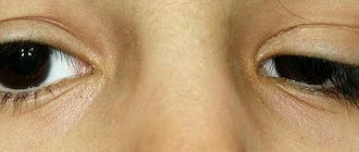
Small dermoid cysts develop asymptomatically. Increasing education is very risky for the life and well-being of a child. A small formation in the orbit of the eye may be hidden on the back of the eye and cause minor discomfort. Large tumors can move the eye forward or to the side, which leads to limiting the physical activity of the eye. As the cyst enlarges, it can be identified visually; a small bump will be visible through the skin or cornea, depending on its location. Common symptoms may include the following:
- elevated temperature;
- processes of inflammation or decay;
- drooping eyelid;
- optic nerve degeneration;
- general malaise.
In appearance, such a tumor is a round formation on the eye. Distinctive features of a dermoid cyst:
- yellow tint of the neoplasm;
- flat or convex (spherical) shape;
- germination of fine white hairs.
Causes
The reasons for the occurrence of these neoplasms are not fully understood, but it is believed that hormonal changes in the body (puberty, pregnancy, menopause) or traumatic exposure are factors in the beginning of the growth of dermoids.
On the eyelid, a dermoid cyst can form in the embryonic period, however, it is not always noticeable at the time of birth. Over time, the dermoid can grow to the size of a large pea or even a walnut. The internal cavities of dermoid cysts (single- or multi-chamber) contain a sebaceous mass mixed with hair or keratinized skin flakes. At the same time, depending on the contents of the cysts, they are divided into dense and soft.
Types of pathology: brief description
Since there are many causes, experts consider several types of the disease:
- Congenital cyst. It appears in infants a few months after birth. The iris is stratified due to penetration of the corneal epithelium.
- Traumatic. Occurs after physical trauma or unsuccessful eye surgery.
- Glaucoma (exudative). It can form due to glaucoma or taking medications that contract the smooth muscles of the eye.
- Dermoid occurs due to disturbances in the development of epithelial cells. This neoplasm contains particles of skin and hair. It is also called teratoma.
- Spontaneous appears without specific reasons. In appearance it is like a small bead. This type includes serous and pearly cysts.
Neoplasms often appear on the connective membrane of the eye, but can also appear on the eyelid and extend to the temple. The cyst occurs without clearly defined symptoms. It does not cause any unpleasant sensations to a person. Often specialists diagnose it in later stages. For examination, special ophthalmic lenses and mirrors are used.
Cyst on the eye: eyelid, removal, treatment, causes, folk remedies, symptoms
A dermoid cyst or dermoid is a tumor that has the shape of a cyst with a wall of connective tissue. A dermoid cyst is an organoid teratoma consisting of connective tissue. A characteristic feature of such formations is that they are smooth on the outside (placed in an oval capsule) and rough on the inside.
The inner layer is similar in structure to the skin and consists of periderm and stratified epithelium, with sweat and sebaceous glands, hair and fatty inclusions.
As a rule, dermoids occur in people of different ages and are located on the temporal region, the upper or inner edge of the orbit, on the eyelids, neck, scalp, in the oral cavity, in the area of the manubrium of the sternum, etc.
Types of eye cysts
The disease is classified according to causes and clinical symptoms:
- Congenital cavity - occurs due to dissection of the iris. It can develop within several years after birth, but appears at a young age before 7 years.
- Spontaneous cyst on the eyelid - serous (in terms of fluid content) and pearly (resembles a white dense ball).
The causes are unknown, cavities appear at any age. It is known as a lower eyelid cyst because it most often appears on the lower eyelid. Serous tumor of the upper eyelid. - The traumatic type (or corneal cyst) is formed due to the impact of epithelial cells on the eye organ. For example, an eyelash can get into the mucous membrane of the eye and gradually grow.
- Exudative - develops due to eye diseases (for example, glaucoma).
- Cyst on the conjunctiva of the eye - there are two types: implantation (after surgical treatment) and retention (due to stagnation of lymph and purulent fluid).
Tumor on the conjunctiva of the eye organ. - Stromal - the cavity appears unexpectedly, can move throughout the eye, and then disappear. Causes significant discomfort.
- Dermoid cyst of the eye - is formed due to fragments of epithelial cells of the hair, skin and nail plates.
- A Moll cyst develops due to blockage of the sweat glands of the eye.
Most often, an eye cyst is conjunctival, as it occurs on the conjunctiva.
The most effective drugs
If the tumor is small, it is necessary to use antibacterial drugs to treat a Moll cyst on the eyelid, a photo of which is not provided for ethical reasons.
- Antiviral ointments for external use: Acyclovir, Hydrocortisone and yellow mercury ointment. The dosage will be selected taking into account the general condition of the patient.
- Antiseptic liquids: iodine, brilliant green, Fucoricin.
- Antiherpetic drugs for oral administration.
- Novocaine blockades for severe pain.
- Anti-inflammatory drops that relieve swelling.
General information about eyelid dermoid in children
By their nature, dermoid formations, including those localized in the eyelid area of a child, are among the benign neoplasms of the fibro-epithelial type. In other words, it is a mixture of epithelial (integumentary) tissues with fibrous, connective tissue fibers.
In addition, dermoids belong to the class of teratomas - congenital, embryonic neoplasms that consist of certain types of connective tissue fibers .
These tumors have a specific structure - they are smooth and even, dense on the outside, but on the inside they have an uneven, rough structure and can be filled with various foreign tissues that are not typical for a given tumor location.
Although this is initially a benign formation, under certain conditions, and not in all patients, it can transform into a malignant tumor.
On the outside, the dermoid cyst is covered with a dense and completely smooth capsule, has an oval regular shape, its size ranges from the size of a pea to the size of a walnut. The cavity of a dermoid cyst can be single, isolated, or it can be a multi-chamber formation filled with caseous masses (similar to sebum) with an admixture of skin flakes and hair.
Causes
The development of a cyst on the eye is provoked by a number of factors, mainly eye diseases:
- Congenital abnormalities (for example, delamination of iris pigment).
- Eye infections (conjunctivitis).
- Diseases of the eyelids (stye).
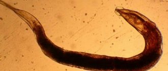
- Worm infestation.
- Inflammation of the sclera.
- Long-term course of treatment, including surgery to restore vision.
- Mechanical damage.
- Constant friction due to contact lenses.
In rare cases, decorative cosmetics and false eyelashes can provoke the unwanted development of a benign cavity.
The tumor also appears as a postoperative complication. Some causes of eye cysts are due to external influences (for example, dust or sand).
Risk group
The following categories of patients are most susceptible to the disease;
- often suffering from inflammatory eye diseases;
- suffering from metabolic disorders, hormonal imbalances, and gastrointestinal pathologies;
- infected with eyelash mites;
- daily neglect of hygiene rules;
- suffering from allergic reactions to negative environmental factors or food consumed.
Such patients should periodically visit an allergist, ophthalmologist, or dermatologist to detect pathology in the early stages of development.
Vision can be restored without surgery
Source: https://alternativa-mc.ru/lechenie/dermoidnaya-kista-veka.html
Symptoms
This pathology, like other benign formations, may not manifest itself for a long time. A dermoid cyst on the eyelid can be detected upon careful examination as a slightly elongated or rounded elastic “ball” localized under the skin. Symptoms of formation usually appear when it becomes inflamed, suppurates, increases in size, and begins to put pressure on nearby tissues and organs.
A cyst on the eyelid is characterized by the following:
- Usually has a round shape;
- Elastic and dense to the touch;
- Painless on palpation;
- Does not adhere to the skin;
- The skin over the dermoid cyst on the eyelid is of a normal color, without ulcerations, rashes, etc.;
- It does not increase in size for a long time.
The clinical manifestations of a dermoid cyst on the eyelid are usually determined by its size and the age of the patient. Dermoids, which are small in size, do not affect health and do not cause damage to the eye structures. With small sizes, this is just a cosmetic defect. However, as the formation begins to grow, it begins to interfere with clear vision. In addition, the risk of its inflammation and malignancy (the appearance of malignant properties) increases, which becomes the reason for its mandatory removal.
Symptoms of the disease
|
|
The neoplasm may have a red-brown, transparent, whitish-blue tint. Once these symptoms appear, you should consult a doctor. A timely diagnosed cyst is a guarantee of quick treatment.
Return to contents
Symptoms
The early stages of development of the pathology pass without visible symptoms. The only thing that can worry a person is the feeling of a small knot under the fingers while examining the eyelid. In many cases, the formation resolves on its own after 2-3 weeks, without affecting your health in any way.
When self-resorption does not occur, the formation begins to actively grow. We are already talking about the fact that the cyst becomes similar in size to a pea, it is easy to visualize. During this period, the first unpleasant symptoms appear:
- Vision decreases. At first it is difficult and then impossible for a person to focus his gaze on one object, a point.
- The horizons become limited.
- “Black dots and flies are flying before my eyes.”
- Tearfulness increases.
- Discomfort and pain occur during blinking and even at rest.
- There is a sensation of a foreign body in the eye.
The cyst can now be clearly seen. It acquires a red-brown, white, blue tint. Can be completely transparent.
The appearance of alarming symptoms should be a reason to contact an ophthalmologist. Timely detection of a cyst is the key to successful, quick therapy and preservation of eye health.
Diagnosis of the most common DISEASES OF THE EYELIDS
What can a general practitioner advise patients with chronic blepharitis? How to recognize malignant neoplasms of the eyelids? How to manage patients with entropion and ectropion?
In medical practice, eyelid diseases are often encountered, which a general practitioner can treat independently. Conventionally, they are divided into three large groups: inflammatory and infectious diseases, benign and malignant tumors and eyelid deformities.
- Inflammatory and infectious diseases
External stye.
Stye is an acute staphylococcal infection of the hair follicle and adjacent glands. Accompanied by painful swelling at the leading edge of the eyelid, which gradually resolves. Styes go away on their own, and antibiotics and surgery are rarely needed.
| Figure 1 (left). External styes go away on their own |
| Figure 2 (center). In case of recurrent meibomian cysts, meibomian gland cancer and BCC should be excluded |
| Figure 3 (right). Fatty cysts of the eyelids do not differ from similar types of cysts of any other location and must be removed in the same way |
Meibomian cyst.
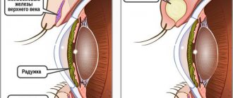
The cause of the development of a meibomian cyst, or chalazion, is a blockage of the duct of the meibomian gland, the fatty gland of the cartilaginous plate of the eyelid. Each eyelid has about 30 glands that open at its posterior edge. The secretion of these glands serves to lubricate the edges of the eyelids and constitutes the outer lipid layer of the tear film.
The cyst contains sebum and becomes inflamed, forming walls of granulomatous tissue. It is a slowly growing painless tumor on the eyelid. A large cyst can put pressure on the cornea and cause astigmatic vision impairment. A chalazion is usually round in shape, hard to the touch and has the appearance of a granuloma when the eyelid is everted. If relapses occur, a biopsy should be performed to rule out meibomian gland carcinoma and basal cell carcinoma. Most cysts go away without treatment, but if they persist, they are excised and scraped out under local anesthesia, then antibiotic ointment is applied and the eye is tightly sealed with a bandage for several hours.
Internal stye.
It is an acute staphylococcal infection of the meibomian gland. The contents of such a lesion are released on the posterior edge of the eyelid, so this type of disease more often requires surgical intervention than external barley.
Other cysts.
Moll cysts are clear swellings found on the anterior margin of the eyelids. Zeiss cysts are not transparent, but are otherwise similar to Moll cysts. Their contents are aspirated using a needle or catheter.
Fatty cysts of the eyelids do not differ from similar cysts of any other location and also need to be removed.
Blepharitis.
Blepharitis is a chronic disease of the eyelid margin, often affecting the conjunctiva. The cause of this disease is not fully understood, but staphylococcal infection and seborrhea play an important role in the pathogenesis.
Seborrheic blepharitis indicates dysfunction of the Zeiss glands and often accompanies seborrheic dermatitis.
Dysfunction of the meibomian glands leads to the development of posterior blepharitis, in which lipid secretion increases, causing burning pain and blurred vision. This condition is often accompanied by anterior blepharitis.
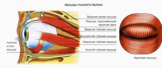
| Figure 4. Eyelid: vertical section |
Blepharitis is a common disease, although often missed by doctors, since the clinical signs are too subtle, despite the complaints of patients whose condition, as a rule, worsens in the morning.
Staph infection affects the base of the eyelashes, causing chronic irritation, itching and soreness, moderate photophobia and weeping. The blood vessels at the edges of the eyelids dilate. Clusters of dry flakes at the base of the eyelashes are called lace collars. After removal of such collars, small ulcers often remain on the edge of the eyelid, which tend to deepen and hypertrophy.
Sometimes the eyelashes curl inward, causing a foreign body sensation and damage to the cornea. Hypersensitivity to staphylococcal exotoxin can lead to inflammation of the inferior tarsal conjunctiva and punctate corneal erosions, usually affecting the lower third of the cornea.
Seborrhea can be oily or dry. It is believed that the excess neutral lipids produced in this condition are broken down by Corynebacterium acnes into irritating fatty acids.
Symptoms and signs of seborrheic blepharitis are similar to those of staphylococcal blepharitis, but less pronounced. The eyelids seem greasy and the eyelashes stick together. Soft scales are easily removed.
In posterior blepharitis, oil globules can sometimes be seen on the posterior edge of the eyelid, and the conjunctiva is moderately injected.
| Figure 5 (top left). Blepharitis |
| Figure 6 (top center). Molluscum contagiosum |
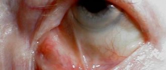
| Figure 7 (top right). Dermoid cyst |
| Figure 8 (top). Basal cell carcinoma |
Styes, meibomian cysts and marginal keratitis often develop against the background of blepharitis.
Treatment of blepharitis consists of constant monitoring of the patient’s condition and minimizing the inconvenience that arises from it, since a cure is not possible. The mainstay of treatment is eyelid care, which includes removing crusts and toxic substances from the edges of the eyelids.
To do this, take a flannel or cotton ball soaked in a weak solution of baby shampoo or soda. This procedure is carried out twice a day, and less often when the condition stabilizes (as necessary).
If there are signs of a staphylococcal infection, antibacterial ointment is applied to the edges of the eyelids.
P
For severe exacerbations, when the diagnosis is beyond doubt, 0.5% eye drops with prednisolone and neomycin or 0.5% hydrocortisone eye ointment can be prescribed. They quickly relieve discomfort and inflammation, but should not be used long-term due to the risk of developing iatrogenic glaucoma and cataracts.
Dry eyes are characteristic of staphylococcal blepharitis, and in this case artificial tears help. It is also important to treat other areas of seborrheic dermatitis, especially the scalp and eyebrows. Difficulties may occur in the treatment of posterior blepharitis, so it is better to refer such patients to the ophthalmology department. It may be necessary to re-examine the glands, which is best done using a slit lamp.
Benign tumors.
Molluscum contagiosum, a viral infection most often found in children, can also affect the eyelids. Desquamation of virus-laden cells causes follicular conjunctivitis and, rarely, superficial keratitis. The lesions are painless and look like pearls with a depression in the center. Treatment consists of squeezing or cauterization, but uncomplicated lesions should not be touched; they go away on their own.
Common benign skin tumors, including warts, squamous papillomas, dermoid and fatty cysts, actinic keratoses and seborrheic keratoses, also affect the eyelids. Less common are keratoacanthomas and cutaneous horn.
The principles of treatment are the same as for tumors of other locations, but any lesion requiring excision that crosses the edge of the eyelid should be referred to an ophthalmologist.
Hemangiomas.
Hemangiomas often occur in children, most often in the first six months of life. These flat and red lesions disappear spontaneously by age five, requiring no treatment unless they obstruct the visual axis. If in doubt, send your child to the eye department.
Xantellasma
- this is the accumulation of lipids in the medial part of both eyelids, they are slightly raised above the skin level and have a yellowish color; as a rule, they indicate an increase in blood lipid levels. They can be excised, but usually they appear again.
- Malignant tumors
Basal cell carcinomas (BCCs) are the most common malignant tumors of the eyelids. As a rule, they affect the lower eyelids and medial corner of the eye. The typical age of patients is 60 years and older.
| Figure 9. BCC: The appearance of BCC varies from a pearl-like nodule to a typical “gnawed out” ulcer. The sclerosing type is a flat formation with poorly defined boundaries |
The appearance varies from a pearl-like nodule to a typical “gnawed” ulcer. The sclerosing type is a flat formation with poorly defined boundaries
These tumors do not metastasize, but are prone to local invasion (especially those located in the medial corner of the eye). The method of choice in the treatment of such tumors is their removal, involving 3 mm of the eyelid margin, followed by histological examination.
Squamous cell carcinomas (SCC) account for only 5% of the total number of malignant eyelid tumors. They appear as an ulcerated area, nodule, or papilloma. They are characterized by faster growth than BCC and the ability to metastasize to the preauricular and submandibular lymph nodes.
Treatment is the same as for BCC, but a larger area is excised.
Sebaceous gland carcinoma is a rare tumor of the meibomian glands. It is a separate nodule of a slightly yellowish color; it can easily be mistaken for a relapse of chalazion. Multifocal tumors resemble chronic blepharitis with areas of inflammation along the eyelid margin.
This tumor metastasizes, with a five-year mortality rate of 30%, perhaps in part due to diagnostic difficulties. Treatment is surgical, the same as for PCI.
- Deformations of eyelids and eyelashes
| Figure 10 (top). Squamous cell carcinoma accounts for only 5% of malignant eyelid tumors |
Ectropion.
It is an inversion of the lower eyelid, leading to weeping and chronic inflammation of the conjunctiva. Gradually the conjunctiva hypertrophies and thickens.
A common form of involutional ectropion, in which the muscles and tendons weaken and the horizontal dimension of the lower eyelid lengthens. Due to the fact that the eyelid is not adjacent to the eyeball, there is an excessive amount of tears and damage to the cornea with the development of keratitis and a foreign body sensation.
Treatment depends on the degree of eyelid weakness. Mild weakness in the medial part of the eyelid is corrected by applying a thermal cautery to the conjunctiva. In more severe cases, eyelid shortening is required. Other causes of ectropion include scarring, both congenital and caused by facial paralysis.
| Figure 11 (bottom). Entropion |
Entropion.
Entropion is an inward turning of the eyelid, causing discomfort, photophobia, and weeping.
The most common form is involutional, or senile, entropion. The eyelid turns inward as its cartilaginous plate thins and atrophies, as a result of which the upper edge bends more than the lower one. The fatty layer of the palpebral fissure also becomes thinner, and enophthalmos is often observed, aggravating the patient’s condition.
Temporary relief comes from attaching the eyelid to the cheek with bandages or removing the eyelashes to prevent damage to the cornea. However, the radical treatment method is surgery.
Trichiasis.
Eyelashes that curl inward and scratch the cornea cause discomfort, photophobia and weeping, as in entropion. Treatment consists of epilation by electrolysis or cryotherapy at least every three months. The latter can lead to depigmentation of the eyelid margin, especially noticeable in dark-skinned patients.
Districhiasis.
In this condition, the patient has a second row of eyelashes on the inner edge of the eyelid. They can be congenital or appear as a result of scarring of the eyelids. Treatment is the same as for trichiasis.
Note!
- For recurrent meibomian gland cysts, it is necessary to rule out meibomian gland carcinoma using a biopsy.
- Treatment of blepharitis consists of strict monitoring of the patient’s condition and minimizing the inconvenience that arises from it. Therapeutic measures are based on eyelid care; if there are signs of staphylococcal infection, antibacterial ointment is applied to the edges of the eyelids. For severe exacerbations, prednisolone and neomycin drops quickly relieve discomfort and inflammation, but they should not be used for a long time
- The most common eyelid tumors are basal cell carcinomas. As a rule, they affect the lower eyelids and medial corner of the eye. The typical age of patients is 60 years and older. These tumors do not metastasize, but are prone to local invasion, especially those located in the medial corner of the eye
- Treatment of ectropion depends on the weakness of the eyelid. Slight weakness of the middle part of the eyelid is corrected by applying a thermal cautery to the conjunctiva. Medial conjunctival repair involves removing a diamond-shaped area of tissue just below the reshaped area. In more severe cases, eyelid shortening is required
- With entropion, temporary relief comes from attaching the eyelid to the cheek with bandages or removing the eyelashes. The radical method of treatment is surgery
Dermoid eyelids: symptoms, signs, diagnosis and treatment
The symptoms of a dermoid cyst, regardless of its location, are characterized by an implicit nature and practically do not manifest themselves, since they develop slowly.
Clinical manifestations occur when the size of the tumor exceeds 5-10 cm and begins to put pressure on nearby organs, the growth becomes inflamed, and suppuration occurs.
In some cases, symptoms visually manifest as a cosmetic defect (hair cyst on the eye, neoplasm on the head). In most cases, the growth is discovered accidentally or during an exacerbation.
Manifestations depend on the location of the formation:
- Symptoms of a large ovarian dermoid cyst put pressure on neighboring organs, which causes constant aching pain in the lower abdomen, tension in the peritoneum, and enlargement of the abdomen. There is a disturbance in the functioning of the intestines (constipation, diarrhea), a disorder of urinary processes (frequent urge, pain in the lower abdomen during urination). A complicated neoplasm with suppuration provokes severe pain in the woman’s ovarian area and an increase in temperature. The danger of ovarian dermoid formations lies in the possibility of their malignancy, therefore these formations require mandatory surgical intervention;
- Large growths in the pararectal area compress the rectum, resulting in pain and difficulty during bowel movements. Due to pressure on the lumen of the rectum, the feces become flat and take on a ribbon-like shape;
- Large dermoids in the middle sections of the chest cavity (mediastinum) begin to put pressure on the trachea, lungs, and pericardium. The result is difficulty breathing, regularly occurring tachycardia, cough, and bluish skin.
Causes
The appearance of a dermoid is associated with a malfunction in the intrauterine development of the embryo. During the formation of skin layers, part of the ectoderm is detached from other areas and incorrect fusion of dermal tissue occurs. The cause of a cyst can be hormonal imbalances, but these are extremely rare cases.
Some scientists believe that dermoids can also appear as a result of genetic predisposition on the maternal side.
A dermoid cyst appears in a child from birth. Due to its small size, the neoplasm cannot be seen immediately, but only after a period of time. Great discomfort is caused by the visual presence of a tumor, which can reach the size of a walnut . Upon examination, the dermoid has the following characteristics:
- Dense texture when palpated;
- Painless palpation;
- Round or flat shape;
- The skin over the cyst is without redness or swelling;
- There is no adhesion to the skin.
Dermoid can form anywhere:
- Eyes;
- Neck area;
- Brow ridge;
- On the child's head;
- bridge of the nose;
- Behind the ear;
- Coccyx;
- The groin of a child.
If a dense enlargement is detected in a child, you should contact your pediatrician.
Causes of dermoid cysts
The neoplasm owes its formation to disturbances that occur during embryonic development. The main causes of dermoid cysts include:
- Hormonal factor. The development of a neoplasm can be triggered by hormonal changes during puberty and menopause;
- Consequences of trauma or contusion of the peritoneum.
Ovarian dermoid cyst - teratoma is a dense hollow formation, the contents of which consist of keratinized elements, particles of hair, bones, sebaceous and fatty inclusions. The main cause of the tumor is embryonic anomalies. The development of formation to visible sizes occurs at stages of age-related hormonal changes: menopause, puberty.
In most cases, a dermoid cyst of the right ovary is formed. This occurs due to the more active functioning of the right organ. Hormonal imbalances affect the right-sided ovary more strongly. A dermoid cyst of the left ovary is rare, and the teratoma formed on the organ does not reach large sizes. Usually the largest volume of the tumor is 6 cm.
Ovarian teratomas must be removed, since one of the consequences of this type of tumor is infertility.
A dermoid cyst on the eyebrow is a congenital pathology. This neoplasm is manifested by external deformation of facial tissues and is detected at an early age. The development of education is asymptomatic, but manifests itself visually.
The structure of the growth is dense with clear boundaries, painless and mobile. Pain syndrome occurs with the development of inflammation.
When the tumor suppurates, the surrounding skin becomes painful, the temperature rises, and attacks of headache, nausea and weakness may occur.
The formation must be removed surgically, since a hollow node can deform bone tissue and have a negative effect on the nasopharynx and brain.
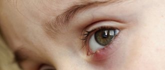
A dermoid cyst of the eye is a benign cavity formation, in most cases of a congenital nature. The growth is localized in the area of the upper eyelid, in the cornea, on the apple, and sclera.
The development of the formation is asymptomatic, but its increasing volumes can affect the reduction in the size of the eye and provoke disturbances in visual perception that cannot be corrected with lenses or glasses.
The formation is a dense capsule filled with contents from particles of the epidermis and hair follicles. Hair is often visible on the surface of the growth, which is why this formation is called a “hairy cyst.”
A dermoid cyst on the head is a hollow growth containing elements of hair, dermis, and keratinized particles. Not characterized by clear localization.
It can form on the lips, eyelids, eyeballs, back of the head and any area of the scalp, bridge of the nose, ears, nasolabial folds, oral cavity, nasopharynx.
At the initial stage of development, it is asymptomatic, and its development is clearly visualized.
Treatment is possible exclusively through surgery. Removal of a tumor is not performed in children under 5 years of age, since general anesthesia is used during the operation. The exception is cases when the neoplasm threatens the health and life of the child.
Stages of development of eyelid dermoid in a child, classification
Based on what contents the dermoid cyst growing on the child’s eyelid will be filled from the inside, the dermoid can be divided into two types:
- Solid form
- Soft shape.
In terms of its main stages of formation, this dermoid is no fundamentally different from any other benign elements.
They grow, practically without damaging the surrounding tissues, and by moving them apart, the increase in size occurs extremely slowly, but the growth of the cyst does not stop at all throughout the child’s life.
Without timely elimination of the dermoid, there is a risk of serious complications, which can be caused both by the cystic formation itself and by the reaction of surrounding tissues. The cavity may suppurate, as well as transform into a squamous cell form of a cancerous tumor.
Source: https://bugmk.ru/bolezni-glaz/dermoidnaya-kista-veka-2.html
Treatment of eyelid cyst
If everything is left to chance, this may lead to partial loss of vision and distortion of the lens (astigmatism). In medicine, several treatment methods are used: medication, surgery and laser. With the parasitic form, specialists practice an integrated approach. An examination with mirrors and a survey will help diagnose the origin of the cyst and determine treatment therapy.
Drug therapy
It is carried out in case of a neoplasm that appears when an infection enters the human body. Often such cysts resolve on their own. The patient is prescribed anti-inflammatory drugs, antipyretics, emulsions and drops to strengthen the immune system. For example, Sofradex, Albucid, Prednisol, Dexamethasone, Ophthalmoferon.
| Important! Medicines should only be taken after consulting a doctor! |
The duration of therapy is 14 days. Medicines may have side effects and contraindications. There is no need to self-medicate.
Surgical removal
Surgery provides a 100% guarantee of cure. This method is used if the cyst grows rapidly, causes discomfort to the patient, or is dermoid. There is no need to go to the hospital for surgery. A few hours after the operation, which lasts about 30 minutes, the patient is sent home.
Laser cyst removal
The rays have antibacterial and anti-inflammatory properties. If the procedure is performed correctly, then the possibility of relapse is reduced to zero. It is performed under local anesthesia.
Return to contents
Folk remedies
This method of treatment should be discussed with a doctor. This will help avoid complications and waste of time. The most common recipes:
- Chamomile decoction. The tea bag is brewed with boiling water, then strained. Using gauze, lotions are made. Chamomile has anti-inflammatory properties and a bactericidal effect.
- A mixture of honey and rose water is rubbed into the growth or an application is made.
- A compress is made from a decoction of guava leaves. A cotton pad is soaked in it and applied to the affected area for 10-15 minutes. This will help relieve redness and pain.
- A piece of fabric is soaked in a decoction of acacia leaves and applied to the eyelid 3 times a day for 10 minutes.
- Eyebright decoction is also used for compresses.
Certified specialists are categorical about traditional methods. They can cause an allergic reaction and itching, and infect the tumor. Symptoms of episcleritis may also occur.
Features of therapy for cysts
When diagnosing a cyst, doctors, as a rule, are in no hurry to prescribe a special course of therapy, preferring to wait. But if the tumor does not resolve on its own within 2-3 weeks, then the patient is prescribed a complex of treatment.
It will depend on how long ago the pathology developed, the size of the formation, as well as the presence of concomitant pathological processes.
Ignoring the symptoms of the disease can lead to serious complications, including: deformation of the lens of the eye, decreased visual acuity, and deterioration in the patient’s quality of life.
Various methods can be used to treat cysts, from the use of medications to surgical treatment. For maximum therapeutic effect, an integrated approach can be used.
Effective Treatments
Conservative method
Therapy for dermoid on the eyeballs depends on the location of the tumor. At the beginning of treatment measures, the size of the formation and the occurrence of inflammatory processes in it are established. If there are inflammatory reactions, physiotherapy is not prescribed, due to the fact that this process can cause a violation of the integrity of the cyst and the formation of an abscess. In this case, antibacterial therapy is performed. If the size is relatively small and the growth is not accompanied by infection, the following is prescribed:
One type of treatment is eyelid massage.
- hydrocortisone ointments;
- eye drops;
- UHF;
- eyelid massage;
- warming up;
- compresses.
Surgery
Elimination of dermoid tumors using classical surgical methods is considered the most reliable. The cyst membrane is directly removed, and this reduces the possibility of the disease returning. The process of eliminating the tumor begins with the introduction of anesthetics into the area of the cyst for the purpose of pain relief, after which the ophthalmologist-surgeon opens the growth and eliminates the contents together with the surrounding tissues. The procedure ends with the application of a suture and a disinfected bandage. After surgery, special anti-inflammatory creams or eye drops are used.
Laser removal
Laser removal of dermoid formations is considered the most popular method for removing many types of cysts. This method has many advantages:
Most often, a dermoid cyst is removed by laser surgery.
- The operation is carried out with great precision, healthy cells are not damaged.
- The laser acts on the dermoid formation and destroys it.
- Insignificant degree of complications after surgery.
- Minimal contact with the conjunctivoid membrane, which prevents infection.
It is worth noting that not all cysts can be removed with medication or laser. This directly depends on the size and location of the formation.
Treatment
Treatment of a dermoid cyst on the eyelid is possible only surgically; there are no other options. The fact is that the causes of these tumors have not been fully studied, so there is no therapeutic treatment or prevention for them.
During the operation, excision of the cyst is performed within the boundaries of healthy tissue; excision of nearby areas is performed somewhat less frequently (to prevent possible complications). When removing a cyst on the eyelid, both local and general anesthesia can be used, which depends on the nature of the formation and the age of the patient.
For children under seven years of age, such operations are performed under general anesthesia; for older patients, under local anesthesia.
The operation is performed on an outpatient basis, and its duration does not exceed a quarter of an hour. The intervention leaves no traces or scars; after it is performed, the patient goes home the same day.
By contacting the Moscow Eye Clinic, each patient can be sure that some of the best Russian specialists will be responsible for the results of treatment. The high reputation of the clinic and thousands of grateful patients will certainly add to your confidence in the right choice. The most modern equipment for the diagnosis and treatment of eye diseases and an individual approach to the problems of each patient are a guarantee of high treatment results at the Moscow Eye Clinic. We provide diagnostics and treatment for children over 4 years of age and adults.
Should I be concerned about malignant oncology?
This question is relevant for those who are faced with the treatment of benign neoplasms. The likelihood that the cyst will become malignant is high. Therefore, a timely diagnosed disease guarantees recovery. The cyst occurs in the choroid of the eye, so it goes unnoticed for a long time. The neoplasm affects:
- iris (the most common is a cyst of the neuroepithelial diaphragm);
- choroid - a pigmented layer responsible for the blood supply to the retina and the front of the eye;
- the ciliary body, which is responsible for focusing the eye on near and far objects.
Delayed therapy can result in:
- Lymphoma, which affects the middle of the eyeball, its tissues and tear ducts.
- Sebaceous carcinoma, similar to a stye or chalazion.
- Basal cell carcinoma - this pathology is prone to relapse, despite its complete removal.
- Melanoma.
- Squamous cell carcinoma, which is prone to metastases.
- Merkel's cancer is a cancer that grows quickly and has purple-colored fleshy masses.
Return to contents
Diagnosis of the disease
To make an accurate diagnosis of a cyst that has formed, it is worth going through a number of procedures:
- External examination of the skin of the eyelids and face.
- Visiometry - this study reveals a person’s visual acuity, which may be reduced due to certain diseases.
- Perimetry.
- Analgizemetry - allows you to determine the sensitivity of the cornea.
- Biomicroscopy of the eye.
- Examination of the eyes for the presence of transparency of their environment.
- Ophthalmoscopy.
Chalazion treatment
If we talk about Moll cysts, then you will have to take scrapings from the affected skin to determine antibodies to the virus. A general blood test is also taken to determine whether inflammation is present in this disease. When diagnosing a dermoid cyst, computed tomography or MRI is considered an effective method; they visualize in detail the required area with the cyst.
Prevention of cysts on the eyelid
Following a few simple rules will help you recover quickly:
|
|
It is forbidden to scratch the tumor or open it yourself. A light superficial massage will help eliminate swelling.
Forecasts and methods of prevention
There are no direct methods for preventing conjunctival cysts. But the risk of its formation, like any other ophthalmological pathology, can be significantly reduced if you do not forget about visual hygiene, do not cause infectious diseases if they arise, and avoid eye injury. Even in the absence of complaints, it is recommended to undergo an examination by an ophthalmologist once a year.
Compliance with basic hygiene rules and regular visits to an ophthalmologist is the best prevention of neoplasms in the eyes
If there is a hereditary tendency to formations in the eye, you should be examined by an ophthalmologist at least twice a year. If a dermoid has been detected and removed, then in the next two to three months you should especially carefully monitor eye hygiene and avoid excessive stress on them. You should not spend too much time at the computer, TV, or get too carried away with reading, and in sunny weather, be sure to wear sunglasses.
If the cyst was identified and operated on in a timely manner, the prognosis is favorable; this pathology will not have any effect on visual acuity or quality of life. If the formation has reached a large size, complications may include a decrease in visual acuity and an increase in intraocular pressure.
Summary: a conjunctival cyst is not a health-threatening ophthalmological pathology. Often it does not make itself felt and does not bother the patient; sometimes it occurs only periodically and resolves on its own. But more often, the cyst begins to grow, turns into a noticeable cosmetic defect, impairs visual acuity and causes unpleasant symptoms. For this reason, it makes sense to immediately consult your doctor after detecting a suspicious tumor and determine further actions. Under unfavorable circumstances and lack of therapy, a conjunctival cyst can degenerate into a malignant tumor.
Types of conjunctival cysts
| Type of neoplasm | Distinctive features |
| Primary | It develops as a result of separation of the area of the iris of the eye, which protrudes beyond the mucous membrane. |
| Implantation | It is provoked by external influences, often by blows, including through a closed eyelid, as well as surgical interventions on the eye. |
| Spontaneous | It is not possible to establish the reasons for the development. Cysts, however, can form on any part of the conjunctiva. |
| Conjunctival retention cyst | It looks like a small vial with thin walls, the cavity of which contains a clear liquid. It has no symptoms and sometimes disappears on its own. |
| Dermoid cyst of the conjunctiva | It looks like a yellowish neoplasm with a heterogeneous structure, dense consistency and smooth surface. It is the most dangerous species, prone to malignancy. |
| Exudative | It is a solid neoplasm that develops as a result of previous glaucoma or long-term use of pharmacological drugs. |
Dermoid cyst in a child: causes, symptoms, treatment, surgery
A dermoid cyst in a child is a congenital benign pathology. The disease develops as a result of a malfunction in the intrauterine development of the fetus. Consists of epidermis, sebaceous glands, dermis particles and hair follicles. The cyst can grow to a very impressive size, which will disrupt the normal functioning of the organs.
The tumor can be localized on different parts of the body: near the inside of the child’s eye socket, on the lower part of the neck, on the head (hair area), brow ridge and bridge of the nose.
Prevention of conjunctival cysts
This neoplasm is not dangerous if it is detected early and treated adequately. Preventive measures that will help avoid its occurrence are as follows:
- regular preventive examinations with an ophthalmologist;
- compliance with hygiene rules when using contact lenses;
- using high-quality eye cosmetics that have not expired;
- strict adherence to the doctor’s recommendations after ophthalmic surgery;
- minimizing the impact of irritating factors on the eyes;
- complete nutrition, providing the body with all the necessary vitamins and microelements;
- minimizing the risk of eye injury.
Ask a question
Entropion (turning of the eyelids)
This is an incorrect position of the upper, lower or both eyelids, in which the free edge of the eyelid is shifted inward towards the eyeball.
There are 2 types of entropion:
- Congenital:
- hereditary etiology: the genetic basis for the development of entropion has not been established. There is a breed predisposition: Chow Chow, Shar Pei, St. Bernard, Leonberger, Bernese Mountain Dog, Cocker Spaniel, English Bulldog, Labrador Retriever, Mastiffs;
- the presence of skin folds, lack of tone of the skin of the eyelids, the size of the eyeball, the length of the palpebral fissure - all these are predisposing factors to the development of entropion;
- Acquired:
- Spastic (resulting in soreness in the eye area)
- Scarring (from injury or chronic inflammation in the area)
The clinical manifestations of this pathology are very diverse - from asymptomatic disease to more severe clinical manifestations (blepharospasm, epiphora, corneal diseases)
Diagnosis: complete ophthalmological examination + fluorescein staining of the cornea.
Treatment: surgical eyelid surgery
Complications
If a cyst occurred on the lower or upper eyelid, and it was removed in time or a course of drug therapy was prescribed, then the main concern that should be avoided is a relapse. Another risk associated with the appearance of a tumor is its malignancy. The percentage of malignancy is quite high; only timely diagnosis and properly selected treatment will help to avoid this.
In an advanced state, a benign formation often becomes the cause of visual impairment. Even after the removal of the cyst, it is not always possible to completely restore vision. This once again speaks to the need for timely diagnosis and therapy.