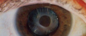
Symblepharon is a medical term that means that the patient has undergone a process of fusion of the conjunctiva of the eyelids with the conjunctiva of the eyeballs. Doctors distinguish 2 types of this disease. In the first case, complete fusion of the eyeball with the entire surface of the eyelid occurs.
In the second variant of the development of the disease, a partial connection of organs is recorded; for example, there may be separate bridges between the eyelid and the eyeball, or adhesions cover separate areas of the eyelids. This type of disease is divided into 2 types:
- Anterior lesion. When diagnosing this disease, the doctor may place a probe under symblepharon.
- Posterior lesion. In this case, the patient’s anterior zone of the eyelids is completely free, but the areas of the arches are fused.
Below we will describe the causes of the development of the disease, symptoms, prevention of the disease, methods of examining the patient and his treatment.
Symptoms of the disease
The main symptoms of ankyloblepharon include:
- Patients experience fusion of the eyelids. Mainly occurs from the temple to the bridge of the nose. There are cases of fusion of the edges of the eyelids from the bridge of the nose to the outside of the eyeball.
- If the pathology is not treated, symptoms of vision impairment appear.
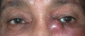
- Sometimes patients note pain in the eyes, lacrimation, pain and moderate itching, swelling of the conjunctiva.
- Congenital ankyloblepharon is combined with concomitant defects: cleft of the hard palate, unilateral lip deformation, microphthalmia. The listed signs indicate a congenital malformation. If a child is born with an eyelid defect, other symptoms of the abnormality should be looked for.
The manifestations of the acquired disease directly depend on the severity of the patient’s condition and the timeliness of treatment.
If the patient has suffered a serious eye injury, there is an overgrowth of dense connective tissue that fuses the eyelids together.
Causes of appearance and diagnosis
Causes of ankyloblepharon include:
- Hereditary predisposition plays a major role in the development of the disease.
- Hay-Wells syndrome belongs to a group of congenital dysplasias. A newborn child has a cleft lip, a cleft palate, fused rudimentary eyelids, and dystrophy of the nail plates. Microblepharon is one of the manifestations of the anomaly.
- Popliteal pterygium syndrome presents with similar symptoms as Hay-Wells syndrome. The pathology is characterized by the presence of connective tissue strands on the eyelids and in the oral cavity. It is difficult for the baby to open his mouth and eyes.
- A complicated form of diphtheria can lead to ankyloblepharon.
- Burns from caustic substances and gas vapors provoke cicatricial changes in eye tissue.
- Ulcers in the eye area after infectious pathologies can scar and cause fusion of the eyelids.
- Chronic ophthalmological pathology leads to complications (keratitis, conjunctivitis).
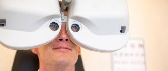
Ankyloblepharon can be suspected at the examination stage. The doctor checks visual acuity, fields, and function of the oculomotor nerve. The doctor measures intracranial pressure and examines the fundus. Performs biomicroscopy using a slit lamp.
Additionally, he prescribes general clinical tests and CT scans of the orbits. To exclude concomitant ailments, the patient is referred for echography of the soft structures of the eye.
There are cases of complete overgrowth of the eyeball. Then an ultrasound and MRI of both orbits are required.
Disease prevention
Among the main measures to prevent the pathological condition are:
- Heredity cannot be influenced. Expectant mothers who suffered from ankyloblepharon in childhood should try to reduce the risk of developing the pathology in the child. During pregnancy, it is necessary to lead a healthy lifestyle (eliminate bad habits and unhealthy foods). The mother's diet should be filled with vitamins and minerals. A correct attitude towards your own health reduces the risk of developing ankyloblepharon. There is a chance to give birth to a healthy baby.
- Careful handling of various alkalis, acids, and devices prevents traumatic damage to the organ of vision. People who work with welding must wear eye protection.
- If chronic ophthalmological diseases are detected, treatment should be started immediately. Timely visits to an ophthalmologist, diagnosis and proper treatment prevent serious complications of the pathology.
After a traumatic eye injury, you should see a doctor. The doctor will be able to identify the development of cicatricial deformities and fusion of the eyelids. Early surgery prevents vision problems.
Factors influencing the occurrence of the disease
For the development of this disease, a person must have corresponding pathological conditions. These include the following conditions:
- The patient suffered chemical or thermal burns to the organs of vision.
- A person has been diagnosed with mechanical injuries to the eye and its appendages.
- The cause of the disease may be the development of an inflammatory process of the conjunctiva. Symblepharon usually develops as a complication after the onset of symptoms of trachoma, diphtheria, and pemphigus.
- The disease may appear after eyelid surgery.
- The appearance of symptoms of symblepharon is possible if a person has developed chronic blepharitis or signs of cicatricial pemphigus appear in the eyes.
- The described disease can manifest itself during atopic conjunctivitis or epidemic-type keratoconjunctivitis.
- The disease can occur due to radiation, for example, if a person has been in the affected area for a long time or works with radioactive substances.
Long-term use of drops against glaucoma or a low-grade inflammatory process on the eyelids can lead to the appearance of symblepharon. Doctors often encounter the appearance of this disease in patients with Stevens-Johnson syndrome.
As scientists have found, symblepharon develops in any condition that leads to a violation of the integrity of the conjunctival membranes of the eyeballs and eyelids. In this case, fusion of the wound surfaces of the contacting structures often begins. Patients with this diagnosis are sent to a special hospital for treatment of eye ailments.
Etiology and pathogenesis
Causes of symblepharon:
- complications after illness.
- thermal and chemical burns;
- eye injury;
- trauma to the organs of vision;
- chronic course of infectious conditions of the conjunctiva;
- radiation poisoning (for example, when working in hazardous conditions);
- long-term use of antiglaucoma drugs.
Attention! The article provides informational information. To obtain advice, you must contact a specialist.
A remedy for pupil constriction (miosis) – Pilocarpine eye drops.
Burns are the most common cause of the disease.
Symblepharon may cause insufficient or inadequate treatment of the following diseases:
- pemphigus;
- trachoma;
- diphtheria;
- pemphigoid;
- Stevens-Jones disease.
Consequences of the pathology:
- inability to completely close the eyelids (lagophthalmos);
- drying of the mucous membrane of the eye;
- the appearance of cloudy and ulcerated areas on the cornea;
- cosmetic defects;
- keratitis;
- thorn;
- trichiasis
If injuries, wounds or burns to the eye occur, you must immediately contact an ophthalmologist to prescribe treatment. In the absence of timely assistance, the development of ocular symblepharon occurs much more often and progresses faster.
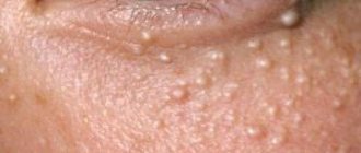
This article will help you find out whether pinguecula of the eye is dangerous.
The problem may lead to a cataract
A drug with antibacterial and anti-inflammatory effects - instructions for Sofradex eye drops.
Classification of the disease
Based on the severity of the pathology, ophthalmologists distinguish the following forms of lagophthalmos:
- Mild degree. The disease leads to a slight widening of the palpebral fissure and drooping of the edge of the lower eyelid. In this case, the patient retains the ability to almost completely close his eyes, but during sleep the palpebral fissure does not close;
- Average degree. The pathology is characterized by pronounced symptoms and a decrease in the folding of the skin. The patient has to make a lot of effort to completely close the eyelids. During sleep, the affected eye remains open;
- Severe degree. Lagophthalmos results in the patient being unable to completely close the eyes. As a result, the cornea and conjunctiva dry out, which contributes to the development of inflammatory and dystrophic processes.
Symptoms
The disease is determined visually and based on the patient’s sensations:
- eye asymmetry;
- entropion of the eyelid;
- the presence of jumpers on the mucosa;
- change in eyelash growth;
- burning and pain;
- lacrimation;
- dry eyes;
- swelling of the conjunctiva.
It is not always possible to make an accurate diagnosis based on visual examination alone. To obtain a complete picture of the disease, the ophthalmologist must conduct a series of studies.
Why eyes fester is described in detail in the article.
Entropion of the eyelid is a visual symptom
A dangerous degenerative disease of the retina is retinoschisis.
Symblepharon in cats
The cause of the development of symblepharon in cats in most cases is infection with the herpes virus, which affects the surface of the conjunctiva and cornea, forming a pathological condition identical to burns. The process causes inflammation and adhesion of tissue structures, as a result of which the conjunctiva floats onto the cornea. Pathology also occurs as a consequence of thermal or chemical burns, injuries to the cornea or conjunctiva. Symblepharon in cats can be a congenital pathology or a complication of a corneal ulcer.
In a pet with symblepharon, a film forms on the eye or becomes cloudy. In more severe cases, when the pathology affects most of the eye, a decrease in vision is recorded.
The main signs of the disease are redness and swelling of the conjunctiva, the presence of exudate, mostly purulent. A cat may scratch its sore eye with its paw and squint. In some cases, increased lacrimation may occur as a result of fusion of the nasolacrimal ducts. If symptoms of the disease are detected, you should contact a veterinary clinic. A veterinary ophthalmologist makes a diagnosis based on a visual examination. If the need arises, microbiological swabs are taken to analyze for feline herpes virus, an ultrasound of the eye and an electroretinogram are performed. The treatment strategy is chosen depending on the severity of the disease. In particularly complex and advanced cases, microsurgical intervention is performed.
With partial symblepharon, the veterinarian takes the course of the disease under control. This type of pathology does not require surgical intervention, since it is prone to relapse.
Diagnostics
Methods for examining patients with suspected symblepharon:
- visual inspection;
- visometry (recording the results of a visual acuity test using specialized tables);
- tonometry (determination of intraocular pressure);
- perimetry (visual field examination);
- biomicroscopy (using a slit lamp);
- Norma test (to determine the condition of the lacrimal membrane);
- Schirmer's test.
As a result of diagnosis, not only the presence of the disease is determined, but also its type. Symblepharon, depending on the severity and location of adhesions, has several types.
- Partial – with a small area of pathology development: posterior and anterior (a probe is inserted).
- Complete – with general fusion.
In any case, tissue scarring occurs, which can only be prevented by surgery.
If signs of symblepharon develop on other organs, the patient will be scheduled for consultation with an appropriate specialist.
A description of methods for diagnosing and treating retrobulbar optic neuritis can be found at the link.
To monitor the course of the disease, it is important to conduct periodic tonometry
Vitamin therapy for the visual organ - Riboflavin eye drops.
Diagnostic measures
If the development of lagophthalmos is suspected, the ophthalmologist performs an external examination. To establish the severity of the pathological process, the patient is asked to close his eyelids. In the early stages, a special ophthalmological examination is required, which involves the following procedures:
- biomicroscopy. The procedure allows you to determine the presence of damage to the cornea and conjunctiva. If inflammatory or infectious processes are identified during the procedure, then surgical treatment of lagophthalmos is possible after compensation of pathologies;
- visometry. Determination of visual acuity is a mandatory stage of diagnosis, since pathology leads to the development of severe visual dysfunction;
- Ultrasound in B-mode. The study is carried out if the development of endo- or panophthalmitis is suspected. Against the background of inflammatory diseases, the procedure is contraindicated. In such situations, optical coherence tomography is performed.
If lagophthalmos develops against the background of paralysis, the patient needs additional consultation with a neurologist. For pathologies of the thyroid gland, treatment by an endocrinologist is necessary.
Treatment
Treatment of the disease is carried out surgically. There are several options for intervention.
- For small areas of adhesions, dissection of adhesions followed by suturing is used. It is possible to use specialized prostheses and therapeutic lenses
- In case of extensive development of pathology, mucous membrane is transplanted from adjacent areas or the patient’s lips. To do this, you need a piece of fabric with a leg. Subsequently, a course of antibiotics is prescribed (1 week), the use of ointments and eye drops with glucocorticosteroids. Rehabilitation takes about 1 month.
Before surgery, the patient undergoes standard preparation. Blood and urine tests are required.
The effect after surgery is observed immediately. In some cases, several interventions followed by plastic surgery are necessary.
Detailed information describing the causes of photophobia of the eyes can be read here.
Untreated pemphigoid can lead to the onset of the disease.
When every day is important and can cost complete loss of vision - treatment of retinal detachment.
Dissection of adhesions
For the treatment of eye symblepharon, the use of proteolytic enzymes is common. They are able to prevent and destroy existing adhesions. They are administered to the patient by injection or electrophoresis. Contraindications:
- presence of ulcerated areas;
- scleral defects;
- the period immediately after injury or burn (10-14 days).
The administration of proteolytic enzymes can only be prescribed by an ophthalmologist in situations where the area of fusion is small.
A proven drug to combat bacterial infections. – Instructions for using Signicef eye drops are presented here.
The introduction of proteolytic enzymes will help at the initial stage of development of the problem
Forecast
To evaluate the effectiveness of treatment, the following parameters are used:
- area and depth of fusions;
- maintaining the ability to rotate the eye;
- changes in visual acuity;
- the presence of ulcers and opacities.
After surgery, the motor activity of the eyeball is restored, and the field of vision increases. In severe cases, keratoplasty and keratoprosthesis are used. The prognosis is more favorable with partial tissue fusion.
The effectiveness of treatment depends on the availability of timely qualified assistance. Only a specialist can make a timely diagnosis and prescribe a surgical procedure. Symblepharon progresses rapidly and intervention is required immediately.
An important factor in assessing the success of treatment is the mobility of the eyeball
What is symblepharon?
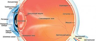
With symblepharon, the inner (conjunctival) part of the eyelid fuses with the mucous membrane of the eyeball .
If the eyelid fuses completely with the eye, we are talking about the full form of the disease . Sometimes the eyelid is connected to the mucous membrane of the eye by separate bridges. In this case, partial symblepharon .
Reference! Technically, this pathology is one of the types of complications characteristic of infectious diseases, burns and other violations of the integrity of the eye membrane.
If you do not take into account burns, which can be obtained at any age, and consider symblepharon specifically as a complication, the risk of its occurrence arises already at the age of thirty years and older.
Prevention
Preventive measures include:
- compliance with eye hygiene rules;
- protection when working with hazardous chemicals and welding machines;
- compliance with technical safety rules;
- providing timely assistance for burns, wounds and eye injuries;
- adequate treatment of infectious eye diseases.
In case of serious eye injuries, the patient should be under constant supervision of the treating ophthalmologist.
What does personal hygiene include?
Symblepharon occurs due to wounds and burns of the eye and as a consequence of the course of infectious diseases. It can be complete or partial. The only effective treatment is surgery.
The prognosis is favorable only in the presence of bridges, without widespread tissue fusion. The main preventive measures are timely assistance in case of eye injury and adequate treatment of pathological conditions. We also recommend that you read our article which will tell you what retinal dystrophy is.
Causes of symblepharon
Symblepharon can occur as a result of a chemical or thermal burn, as well as against the background of certain forms of conjunctivitis, which are characterized by a severe course. Fusion of the conjunctiva of the eyelid with the conjunctiva of the eyeball is a symptom of some systemic and ophthalmological diseases (Stevens-Johnson disease or cicatricial pemphigoid of the eye). The pathology may also be an anomaly of embryonic development. The reasons for the appearance of symblepharon include:
- chronic inflammatory diseases of the cornea;
- chronic bullous dermatosis of the mucous membranes of benign or allergic etiology;
- surgical intervention;
- epidemic keratoconjunctivitis;
- atopic conjunctivitis;
- radiation exposure or hazardous working conditions.