Definition of disease
The vitreous body occupies about eighty percent of the entire eyeball and has a jelly-like structure. In the back of the eye there are vital blood vessels, and if their integrity is disrupted, bleeding occurs. Blood can also get inside the eyeball from abnormal vessels that do not correspond to the anatomical norm. When the disease occurs, most often only one eye is affected.
Structure of the eye
Symptoms and classification
There are no nerve fibers in the internal environments of the organs of vision; when they are damaged, there are no standard clinical manifestations: discomfort, pain, itching. The only sign of the disease is a sharp drop in the level of vision, up to absolute blindness.
Clinical features of the disease make it possible to divide hemophthalmos:
- Total type - when filled with biological fluid more than three quarters. Similar problems arise after trauma to the visual organs. Symptoms are manifested by an almost complete absence of vision, with preservation of sensitivity to light. The patient cannot distinguish objects located in the field of view or freely navigate in space;
- In subtotal - the internal cavity is filled with blood from one third to three quarters. It is provoked by diabetic damage to the blood vessels of the fiber. The patient sees with the affected eye vague outlines of things and silhouettes of people;
- Partial – when less than a third of the vitreous glass is filled with blood. It is one of the frequently recorded types of hemorrhage; it occurs under the influence of diabetes mellitus, retinal detachment, and hypertension. Characterized by flashing black dots, reddish stripes in front of the eyes or nebula.
This form affects both organs as an exception; mainly one eyeball is affected. The main goal of therapy is observation without specific treatment; partial forms disappear spontaneously.
If primary symptomatic manifestations of hemorrhage in the scleral area appear, the patient should immediately seek medical help. Timely identification of the source of the deviation will allow you to prescribe an adequate course of therapy and prevent the formation of complications.
Causes
The causes of hemorrhage into the eye from ordinary and newly formed vessels vary.
From normal vessels:
- Traumatic injury.
- Vitreous detachment. Most often it occurs against the background of age-related changes in sixty to eighty years.
- Antiomatosis of the retina. Hemorrhage occurs from dilated vessels.
- Surgical operations.
- Terson syndrome. The cause is increased intracranial pressure.
From abnormal vessels:
- Diabetic damage to the organs of vision.
- Retinopathy.
- Retinal dystrophy.
- Intraocular tumors and neoplasms.
Types and differences of hemorrhages in the eye
In young people, any bleeding in the eye is associated with an accidental blow or fight. Older people will most likely think about a jump in blood pressure and will begin to strongly advise urgently going to see a therapist or at least using a blood pressure monitor.
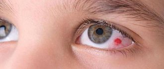
Hyposphagma
This is the entry of blood from the vessels under the conjunctiva. In the diagnosis made by the doctor in the medical history, such blood spillage in the sclera is also referred to as “subconjunctival hemorrhage in the eye.”
People will make the diagnosis “a vessel has burst,” which will also be correct, because such an accumulation of blood is caused by ruptured capillaries, from where the blood enters that part of the white of the eye that is located around the iris.
Possible reasons
- Traumatic impact to the eye; its long and strong friction; a sharp jump in atmospheric pressure (working in tanks, hyperbaric chambers, sealed enclosed spaces, etc.), a blow to the cornea with a foreign object (a large insect at high speed), exposure to aggressive chemical reagents.
- Poor blood clotting caused by congenital or acquired hemophilia, uncontrolled and excessive use of blood thinners in the form of aspirin, dipyridamole, heparin, ticlid, etc.
- A surge in pressure during vascular spasms, childbirth with its attempts, constipation, sharp, “heartfelt” sneezing, blowing a very stuffy nose, an attempt to lift an unbearable weight, suffocation, vomiting, convulsive cough.
- Eye infections.
- Consequences of eye surgery.
- Fragility of blood vessels caused by vitamin deficiency with deficiency of vitamins K and C; diabetes, atherosclerosis, systemic connective tissue lesions (vasculitis, lupus).
Hyposphagma, when viewed as a cosmetic defect, is considered the most harmless and quickly passing disease. Compared to subcutaneous hematomas (bruises), it does not change in color, but simply turns pale, gradually dissolving.
The feeling of an obstacle in the eye in the presence of such a hemorrhage can be attributed, rather, to psychological inconvenience, as an obstacle it is perceived more at the level of self-hypnosis.
Thus, the disappearance of the blood stain inside the sclera occurs without drug intervention.
We advise you to read: How an eyesore forms in a person’s eyes and how to treat it.
Although the resorption of the red spot can be accelerated with potassium iodine eye drops. And if you detect the beginning of the formation of a bruise in the sclera, and it continues to expand, then stop this process by instilling the popular drip products “Vizin”, “Naphthyzin for the eyes”, “Octilia”, etc.
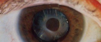
Hyphema
The location of the hemorrhage is the compartment between the iris (which gives our eyes their unique color) and the convex outer lens of the clear cornea. This place is called the anterior chamber of the eye.
In the normal state, the chamber is filled with a transparent substrate of complex composition. Hyphema is the leakage of blood into this chamber.
The accumulation of such blood looks like a segment in the lower part of the iris, and the larger the hemorrhage, the larger the area this segment occupies.
The anterior chamber, as a result of extensive hemorrhage, may even be completely filled, giving the iris, whatever color it was previously, a pure bloody color.
Why do eyes turn red, and how to prevent diseases of the organs of vision?
The cause of the pathology is always a rupture of the vessel. What are the most common causes of breakups?
Injuries
Types of injuries:
- Penetrating - with eye damage starting from the cornea and further inward, forming a through channel between the internal cavity of the eyeball and the external environment. The most common injuries are caused by piercing sharp objects. Less often - from blunt blows, but then the blow must be strong.
- Non-penetrating. With such an impact, the eye may remain intact, but the impact results in consequences reminiscent of a contusion, up to and including dynamic destruction of the internal structures of the eye. This type of injury is almost always caused by blunt force trauma. May be accompanied by a concussion.
- Trauma also includes all surgical manipulations of the eyes.
Eyeball diseases
Diseases of the eyeballs associated with the appearance and growth of defective blood vessels in the eye (neovascularization). New vessels always have some structural defects, which makes their walls more fragile than normal ones. When these walls rupture with minimal impact, hemorrhage occurs into the cavity between the cornea and the iris.
Neovascularization occurs from:
- Diabetic angiopathy in diabetes
- Defects of the venous network in the retina and the beginning of its detachment
- Tumors in the eye and inflammation of intraocular structures.
Diagnostics
The obviousness of the signs of the disease does not raise doubts about the diagnosis, and the doctor can only prescribe treatment, based on the very fact of hematofilling of the anterior chamber and the amount of blood in it. The diagnosis is made based on simple steps:
- Examination of an eye with hyphema
- Measuring pressure inside the eyes with a special tonometer
- Determination of visual acuity percentage
- Examination of the eye using a hardware method (using an ophthalmological microscope).
Before treating hyphema, it is necessary to accurately determine the causes of its appearance and eliminate the external factors that caused such a pathology, such as:
- Cancellation of blood-thinning drugs (if they were prescribed)
- Vitamin deficiency, if present
- Ophthalmic infections
- Bad habits, especially heavy smoking
However, if the hyphema occupies up to 1/3 of the iris segment, its resorption is only a matter of time. To speed up this process, use 3% drops of potassium iodide and medications designed to reduce intraocular pressure (Azopt, Timolol, Latanoprost).
If the course of the hyphema is complicated, surgical intervention is indicated.
Hemophthalmos
The vitreous body is normally a gel of ideal transparency, in which the light flux passing through the aperture of the pupil and focused by the lens must freely reach the retina in the posterior hemisphere of the eye. Any foreign inclusions, including blood, interfere with the passage of light, causing sensations of an obstacle of varying degrees of density and opacity in front of the eyes.
Cases of hemorrhage into the vitreous body, or hemophthalmos, are most often associated with painful inflammation of the retina. Or rather, with ruptures of the microvessels covering this inner hemisphere under the photosensitive elements.
Causes
Vascular ruptures are possible from:
- Concussion impact (a strong blow to the head with symptoms of a concussion) against the background of intracranial pressure caused by such impact, which goes far beyond normal limits.
- Rupture of the membranes of the eye from a penetrating wound
- Blood clots in the retinal vessels
- Retinal vascular abnormalities
- Hematopathologies associated with low coagulation
- hypertensive crises
- Diabetes
- Oncological problems of the eye.
- An abrupt increase in pressure in the sternum from coughing, labor, with strong vomiting spasms, extreme strength loads, sneezing).
The use of potassium iodide leads to rapid results in the treatment of bacterial and fungal infections and resulting mechanical damage to the eye, eliminating hemophthalmos.
There are no nerve receptors in the glassy gel, so a person with hemophthalmos does not experience pain. Hemophthalmos can manifest itself only in the form of decreased vision, sometimes to complete blindness.
Kinds
These hemorrhages are:
- Total, when the internal volume of the eye is filled with blood by more than ¾. They almost always occur as a result of traumatic effects on the eye. Characterized by blindness with the presence of light perception, but without distinguishing the details of the surrounding world.
- Subtotal, with a volume of blood filling from 1/3 to ¾. The most common cause is diabetic vascular damage. Vision in this case is the discrimination of silhouettes and general contours of objects.
- Partial, with a lesion volume of less than 1/3. It is observed in cases of the onset of retinal detachment, with arterial hypertension or progressive diabetes. Manifestations – black dots, a red streak or a red veil in the eyes.
Treatment
When the first symptoms appear, you should immediately visit an ophthalmologist for a thorough diagnosis, which includes:
- Initial examination;
- Determination of visual acuity;
- Fundus examination;
- Angiography;
- Tomography (including using a computer);
- Gonioscopy and biomicroscopy.
After determining the nature and severity of the problem, the ophthalmologist prescribes treatment.
By medication
Mild vitreous hemorrhage resolves with drug treatment. The patient is prescribed drugs that have a positive effect on the state of the blood (Vikasol), as well as intravenous administration of sodium chloride and local administration of calcium chloride. In addition, dexamethasone solution is prescribed as a corticosteroid drug.
Vikasol is used to improve blood condition
During the treatment period, it is important to take food and additional medications containing large amounts of calcium. This is important to prevent relapses and complications.
Surgically
One of the most commonly prescribed treatments for bleeding is laser coagulation. This procedure can eliminate the problem of hemorrhage from abnormal vessels, as well as prevent relapses and possible complications of the disease.
The most radical surgical treatment for the disease is vitrectomy, which is the removal of the affected area of the vitreous. This method is shown:
- For retinopathy;
- If the condition worsens two to three months after the onset of the disease;
- In case of traumatic injury;
- With retinal detachment;
- If there are no positive results from treatment with other methods.
Performing vitrectomy
Folk remedies
Traditional methods of treating diseases of this type are not usually used as alternatives to professional ones. They are effective as a complement to the main methods.
The most useful in this case:
- Chicory root. For thirty grams of product you need to take a glass of boiling water and leave it for ten minutes. Take half a glass three times a day.
- Aloe juice. Take a teaspoon orally after meals.
- Blueberry juice. Mix one part of the product with two parts of water and use for instillation.
Traditional recipes can be actively used during the recovery period after treatment.
Symptoms of vitreous hemorrhage
The clinical picture is due to several signs characteristic of this disease.
Loss of vision
Depending on the volume of blood trapped in the vitreous body, loss of visual function occurs:
- partial (hemorrhage occupies 1/3 of the vitreum area);
- subtotal (up to 75% of the vitreous structure is affected);
- total (with blood spread more than 75%).
In the first case, visual impairment is accompanied by a loss of clarity of visible objects, the appearance of “floating circles”, a “veil” before the eyes, and visibility in a reddish tint.
The subtotal type of pathology is characterized by significant loss of vision, while a total diagnosis indicates complete blindness.
Typically, ocular hemorrhage occurs in only one eye. Quite rarely, the disease affects both eyes, unless due to traumatic causes.
The pathology is characterized by a slight improvement in vision after sleeping with the head raised, due to the displacement of the blood stain to the lower part of the eye.
Visualization.
Even minor hemorrhage is easy to notice.
Painless.
The vitreous body does not contain nerve endings, so hemorrhage does not cause pain. However, if the pathology is caused by mechanical damage (trauma), pain is possible.
Symptoms of vitreous bleeding may intensify if the patient has not consulted a doctor for a long time or the pathological process progresses. Small blood clots dissolve on their own, but very slowly, and can lead to serious complications. Therefore, if a disease is detected, you should immediately visit an ophthalmologist.
Prevention
There are no special preventive methods for diseases of this type. To prevent it, it is recommended to maintain a healthy lifestyle and monitor your general health. If there are provoking diseases, their treatment is necessary. In addition, after forty years, systematic measurement of intracranial pressure is desirable.
It is also useful to avoid the possibility of injury and undergo regular diagnostics by an ophthalmologist to check for problems with the organs of vision.
Eye drops: list of names
This article will tell you what aphakia of the eye is.
All about the drug Atropine https://eyesdocs.ru/medicinaoperacii/lekarstva/glaznye-kapli-na-osnove-atropina.html
How to prevent and prognosis?
Prevention of hemorrhage into the vitreous cavity does not have clear rules. Ophthalmologists recommend regularly monitoring your health. For patients suffering from diabetes, it is important to maintain normal glucose levels. These people are at risk for many diseases, in particular ophthalmological ones, so regular examinations are required - at least 2 times a year. For patients over 40 years of age, it is important to keep blood pressure levels normal and undergo an annual examination with an ophthalmologist to determine intraocular pressure. The main measures in prevention are considered to be measures to maintain the normal condition of blood vessels.
The prognosis depends on the stage of development of the pathology. Partial hemorrhage, which does not exceed 12%, in most cases resolves on its own and does not pose a great danger. Damage to a quarter of the vitreous complicates the treatment process and increases the risk of retinal detachment. The total type of hemophthalmos involves irreversible blindness. In 95% of such patients, disability awaits.