Medical statistics show that 20% of people with eye pathologies turn to an ophthalmologist due to the rapid development of marginal blepharitis. What is blepharoconjunctivitis and its further treatment?
- 5.1 Traditional medicine
Forms of the disease
With conjunctivitis, there is a burning sensation and pain in the eyes, lacrimation, but when the eyelids are included in this process, blepharoconjunctivitis is formed. The clinical picture is a set of symptoms characterizing blepharitis (eyelid hyperemia) and conjunctivitis. When these symptoms come together, treatment becomes quite difficult. The risk zone includes both children and adults.
The disease has several forms:
- Acute, characterized by the appearance in a short time.
- Subacute. It mainly affects children. Appears gradually, in the initial period in the corners of the eye sockets.
- Chronic . The infection is of a sluggish nature, aggravated by periods. Sometimes without symptoms.
Varieties
- Allergic blepharoconjunctivitis. Caused by individual increased sensitivity of the body to a specific allergen.
- Bacterial (epidemic). It is provoked by staphylococcus.
- Seborrheic (dermatic). The background for development is severe seborrheic dermatitis or rosacea.
- Demodectic blepharoconjunctivitis. It is caused by the parasitic demodex mite.
In hot climates, a person can become infected with the Koch-Wicks bacillus. Blepharoconjunctivitis, provoked by the Koch-Wicks bacillus, is of an acute epidemic nature. It spreads through the unwashed hands of a sick person, infected things and objects. It is characterized by outbreaks of the disease in hot climates, especially among children. Blenorrheal blepharoconjunctivitis occurs in infants infected with gonococcus during childbirth by a mother with untreated gonorrhea. Blepharoconjunctivitis, caused by the Morax-Axenfeld bacillus, is subacute in nature, chronic and localized in the corners of the eyes.
Causes
Among inflammatory diseases of the organ of vision, conjunctivitis (at least 67%) and blepharitis (approximately 23%) are especially common.
According to statistics, up to 50% of all outpatients who see an ophthalmologist have inflammatory eye diseases of an infectious and allergenic nature. Moreover, the main part of them are patients with red eye syndrome.
In terms of the total number of diseases, demodectic blepharitis and blepharoconjunctivitis account, according to various sources, from 39 to 88%.
The most common route of infection of the conjunctiva is from the outside, and autogenous infections are not uncommon. Factors predisposing to the onset of the disease also include microtrauma of the conjunctiva, previous infections, myopia, astigmatism, weakened immunity due to hypothermia/overheating.
Blepharoconjunctivitis is transmitted by contact, as it has a viral or adenoviral etiology, less often - allergenic. The causative agents of blepharoconjunctivitis are usually microorganisms of a coccal nature (streptococci, gonococci, pneumococci, staphylococci, as well as Koch-Wicks or Morax-Axenfeld bacilli), or microscopic mites of the genus Demodex.
Causes and types
Blepharoconjunctivitis has several types:
- Bacterial. The causative agent is Staphylococcus aureus. A large amount of purulent discharge appears from the eyes. When one organ is affected, most often the second is involved in the disease process if the infection cannot be eliminated immediately.
- Scaly (seborrheic). Scales similar to dandruff appear on the edges of the eyelids.
- Allergic . Reaction to interaction with any allergens.
- Meibomian blepharoconjunctivitis. Characteristically, there is a secretion from the meibomian glands that glues the eyelids together and has an opaque appearance.
- Demodectic. It is a consequence of infection with demodectic mites.
- Viral. The main cause is the presence of herpes or adenovirus infection. It may appear after or during a viral illness. It can be observed in childhood.
Factors that contribute to the appearance and development of blepharoconjunctivitis:
- weakened immune system;
- unbalanced diet (consumption of foods low in vitamins);
- diseases of the gastrointestinal tract, ENT organs;
- allergy;
- anemia;
- myopia, eye injuries;
- the presence of worms;
- recurring viral infections;
- long-term use of antibacterial drugs.
Symptoms and signs
Regardless of what caused the development of blepharoconjunctivitis, patients have:
- severe swelling of the eyelids;
- redness of the conjunctiva and adjacent tissues;
- photophobia;
- itching;
- lacrimation;
- the formation of purulent mucous discharge of varying thickness.
When the pathology is bacterial in nature, people are annoyed by:
- redness of the eyes;
- formation of a “collar” of acne at the roots of the eyelashes;
- sticking of the eyelids, especially in the morning, due to the secretion of thick purulent mucous secretion and its drying;
- loss of eyelashes in clumps.
With seborrheic appearance, the following come to the fore:
- swelling and redness of eye tissue;
- burning sensation;
- profuse lacrimation.
The intensity of the symptoms of the disease may increase after eating spicy foods.
With viral, allergic and demodectic blepharoconjunctivitis, patients suffer more from itching and profuse discharge.
Risk factors
The most common provocateurs of blepharoconjunctivitis are:
- weakened immunity;
- poor nutrition;
- vitamin deficiency;
- gastrointestinal diseases;
- myopia, other pathologies;
- anemia;
- eye injuries;
- neglect of personal hygiene rules;
- viruses, such as ARVI, influenza;
- fungi;
- infection with worms;
- caries;
- proximity of allergens;
- diseases of the ear, nose and throat, such as tonsillitis;
- long-term use of antibiotics.
Rarely does any single cause lead to the development of the disease; as a rule, blepharoconjunctivitis requires a combination of a whole range of factors. Blepharoconjunctivitis is an acquired disease from which even a newborn baby is not immune: during childbirth there is a risk of eye infection.
Reasons for development
Most often, blepharoconjunctivitis occurs as a result of infection from the external environment. Less commonly, the disease occurs against the background of autoimmune pathologies.
Provoking factors may be:
- conjunctival injuries;
- penetration of a foreign object into the organs of vision;
- infectious diseases;
- refractive errors of the eyes, including myopia, astigmatism, hypermetropia;
- decreased immune system as a result of colds, hypothermia, surgical interventions, or previous serious illnesses;
- poor nutrition, lack of vitamins;
- diseases of the digestive tract;
- dental pathologies;
- helminthic infestation;
- prolonged use of antibiotics;
- ENT diseases, for example, tonsillitis;
- insufficient personal hygiene;
- fungal infections;
- contact with an allergen.
Usually, human infection occurs through interaction with an infected patient, and less often during surgical interventions on the organs of vision if sterility is not maintained.
Symptoms
Manifestations of the disease vary so much that a person cannot independently determine what type of illness he has. Therefore, if you have the slightest concern in the eye area that lasts for more than three days, you need to seek qualified help from a specialist (ophthalmologist). Symptoms of blepharoconjunctivitis are determined depending on its type. The common signs that unite each type of disease are:
- swelling of the eyelids;
- lacrimation;
- reduced eye shape;
- red eyes, photophobia;
- thickening of blood vessels;
- eye fatigue.
Each blephoroconjunctivitis has its own characteristics, regardless of the general symptoms:
- The bacterial species is characterized by the appearance of ulcers at the base of the eyelids and abundant discharge of pus, with slight redness.
- Seborrheic – characterized by constant strong burning and redness (of the whites and conjunctiva) in the eyes, the edges of the eyelids peel.
- Allergic – manifests itself in the presence of an allergen. Patients experience severe watery eyes, swollen eyelids, and itching.
- Viral - accompanied by severe redness, lacrimation, and body hyperthermia may be observed.
- With meibovian blepharoconjunctivitis, an accumulation of secreted opaque secretion is added in the corners of the eyes, with demodectic blepharoconjunctivitis - scales located between the eyelashes.
Read in a separate article: Is it possible to wash with conjunctivitis, swim in a sauna and pool?
Pathogenic factors (causes)
To date, there is no single, statistically reliable idea of the role and correlation of the main causes of blepharoconjunctivitis. In most sources, blepharoconjunctivitis is divided according to etiopathogenetic criteria into three types.
Infectious, in which different classes of pathogens are characterized by specific features of the clinical picture:
- viral inflammations, as a rule, begin acutely, against the background of the general symptoms of ARVI, and are manifested by photophobia, burning, foreign body sensation, hyperemia, pain, lacrimation;
- The distinctive features of bacterial infections are abundant mucopurulent discharge, beginning in one eye with possible involvement of the other, swelling, redness, and in severe cases, ulceration of the eyelids, conjunctiva, and cornea;
- similar in nature, but, as a rule, less pronounced and much more persistent symptoms are inherent in fungal infections; typical is the formation of various kinds of films, “gluing” mucous accumulations, nodules and erosions on the skin of the eyelids.
Allergic reactions usually develop as part of a more general, systemic reaction, and are characterized by irresistible itching (especially along the edge of the eyelids), lacrimation, severe swelling of the eyelids, and violent accompanying symptoms from the nasopharynx.
Seborrheic (dermatic) are caused by functional disorders in the eyelash follicles and sebaceous glands; manifested by peeling, accumulation of dandruff flakes and scales on the eyelids and eyelashes, itching, a feeling of rough dryness, and a tendency to secondary infection when scratching.
A number of provoking and potentiating factors are also described:
- ametropia (refractive errors) to a strong degree in case of refusal of optical correction, which causes constant eye strain;
- exhaustion of the body, weakened immune system, anemia, hypovitaminosis;
- endocrine and other disorders in which the biochemical composition of the secretion of the eye glands may change;
- “pink” acne (a common cause and companion of the so-called “blepharitis rosacea”);
- the presence of multiple microtraumas and damage to the conjunctiva, which creates conditions for secondary infection. Such trauma can be associated with constant mechanical, physical or chemical irritation (dust, ultraviolet burns, aggressive fumes, etc.), as well as with the activity of a microscopic skin parasite - the Demodex mite.
The last factor should be discussed in more detail.
What is the result?
You will strengthen your immunity. The inflammatory process is localized. The pain, itching and burning that tormented you will disappear. Redness and swelling will disappear. Metabolism in tissues, muscles and the cornea of the eyes will improve. Your body will begin a powerful process of regeneration and restoration. Immunity will increase. The risk of vision-threatening complications will be eliminated.
Powerful and effective treatment with the Vizulon device will quickly eliminate the clinical manifestations of the disease, localize the foci of pathology, and reduce the dosage of medications. This will allow you to live a full life and forget about your illness forever.
Your body will quickly cope with the inflammatory process and return to its normal functioning. Unpleasant symptoms of inflammation will disappear, appearance will improve and cosmetic defects will be eliminated. And most importantly, you do not have to worry about the serious consequences of the disease associated with deterioration or loss of visual functions.
Symptoms of the disease
What are the symptoms of blepharoconjunctivitis? Patients often complain of itching and burning in the orbital area. Sometimes a person develops a fear of light, a feeling of sand in the eyes. Tearing immediately begins if the patient looks at the rays of the sun or a bright lamp.
Redness and swelling appear around the eyes. Visual acuity decreases if part of the cornea is inflamed. In the presence of a bacterial pathology, patients suffer due to the periodic formation of pus with an unpleasant odor.
Watery discharge is present when the pathology is of viral origin. Patients complain of severe pain in the eyes. Nearby lymph nodes often become inflamed.
The allergic type is accompanied by bilateral damage to the eyeball. A viscous secretion is also released. But the lymph nodes do not become inflamed. You cannot wear contact lenses if you have this condition. This intensifies the described manifestations and increases irritability of the mucous membrane of the visual organ.
Treatment
Treatment of blepharoconjunctivitis is carried out locally. For viral blepharoconjunctivitis, drugs are prescribed in the form of Actipol, Poludan drops, Acyclovir tablets, which have an antiviral effect. The dosage and duration of use is determined by the patient's condition. The bacterial species requires treatment with antibiotics (Tetracycline, Ciprofloxacin), Metronidazole ointment and drops containing Dexamethosone. Treatment of eyelashes and eyelids with alcohol-based solutions is used for demodectic disease. Eye protection medications (Oftagel) are instilled in advance. To prevent infection from affecting your face, you should consult a dermatologist.
For all blepharoconjunctivitis, anti-inflammatory drugs are used. Drops have proven themselves to be effective in restoring damaged eye tissues. (Taufon, Taurine) The drug Systane Ultra can relieve dry eyes.
In combination with drug therapy, it is possible to use traditional medicine recipes, among which we can particularly highlight the use of herbal infusions (yarrow, calendula, and chamomile) for washing the eyes. You can use regular tea leaves for this purpose. A new gauze pad should be used to rinse each eye.
Ways to get rid of the disease
Before using any medications, you should consult your ophthalmologist. Only a specialist can prescribe correct, competent treatment. The choice of medications depends on the type of blepharoconjunctivitis, since first of all it is necessary to influence the etiological factor.
It should be remembered that blepharoconjunctivitis is contagious, so it is important to adhere to good hygiene. In addition, you should regularly clean your eyes using a cotton pad soaked in an antibacterial solution.
Treatment for bacterial inflammation
The cause of the disease is bacteria, so the main role in treatment is given to antibacterial drugs - ointments and drops. Local application, that is, directly to the lesion.
- Levofloxacin (solution). Instill into the eye every 2 hours for 2 days; in subsequent days of illness, the drug should be used once every 4 hours.
- Levomycetin (drops). Apply 3 times a day. The average course of treatment is 10 days.
- Erythromycin (ointment). Place behind the eyelid 2-3 times a day. The course is quite long - 1–2 months.
- Tetracycline eye ointment. Use 3-4 times a day, no more than two weeks (depending on the degree of damage).
Medicines for ticks
To combat demodectic blepharoconjunctivitis, the following medications are used:
- 1% mercury ointment (antiseptic). The dosage regimen should be selected by a specialist, as it depends on many factors.
- 10% sulfapyridazine sodium (solution). Instill in the morning and evening. The course of treatment is 30 days.
- Glycodem (cream). Apply only to eyelashes and eyelids. Use with caution, avoid contact with the mucous membrane of the eyes.
Treatment of allergic blepharoconjunctivitis
The main condition of therapy is the identification and destruction of the allergen. Medications include antihistamine tablets (oral) and antiallergic drugs for topical use in ophthalmology:
- Opatanol (drops). Apply 2 times a day with an interval of 8 hours. If necessary, treatment lasts up to 4 months.
- Lecrolin (drops). Use 4 times a day until symptoms disappear.
- Diazolin or Suprastin (H1-histamine receptor blockers). Take 1-3 times a day. Therapy is considered complete after the symptoms of the disease disappear.
Fighting viral eye infections
For this form of the disease, the main group of drugs are antiviral:
- Bonafton (0.05% ointment). Apply behind the eyelids 3-4 times a day. To enhance the effect, it is used in combination with the tablet form.
- Okoferon is available in powder form for preparing a solution. Buried every 2 hours for no more than 7–10 days.
- Acyclovir (ointment). Placed in the lower conjunctival sac 5–6 times a day. Treatment continues until symptoms disappear completely.
General therapy
To relieve swelling of the eyelids, corticosteroid medications are used:
- Hydrocortisone ointment;
- Dexamethasone drops.
To improve the separation of tear fluid, use vasoconstrictor nasal drops:
- Naphthyzin;
- Tizin.
Adequately prescribed therapy will get rid of the problem in 7–10 days
If a patient complains of dry eyes, the following will help moisturize and protect the cornea:
- Ophtolic;
- Hilo Chest of Drawers.
Use as needed, but no more than 4 times a day.
To speed up the healing process, ophthalmologists recommend the use of non-steroidal anti-inflammatory drugs:
- Diklo-F (solution);
- Indocollier.
The dose and duration of use are selected individually by the doctor.
Medications: drops, ointments - photo gallery
Acyclovir - antiviral eye ointment, placed behind the lower eyelid Levomycetin - antibacterial drops Opatanol is used for allergic blepharoconjunctivitis
Hydrocortisone ointment relieves swelling of the eyelids
Oftolik moisturizes and protects the cornea Indocollir accelerates the healing process Glycodem is used to combat demodectic blepharoconjunctivitis
Therapy for blepharoconjunctivitis
It is necessary to treat the disease in accordance with the etiology and characteristics of its course. An effective treatment regimen for blepharoconjunctivitis is prescribed by a qualified ophthalmologist. If necessary, consultation with an infectious disease specialist, dermatologist, or allergist is carried out. Self-medication is unacceptable. During therapy, it is important to eat properly and follow a daily routine.
Removal of secretions from the conjunctival cavity is carried out by washing the eyes with antiseptic and disinfectant liquids.
For bacterial forms of pathology, erythromycin ointment can be effective. Basically, the treatment of blepharoconjunctivitis is carried out using drip solutions and liniments for local action. The table shows drops and ointments used for blepharoconjunctivitis of various etiologies:
| View | Group of drugs | Name of drugs |
| Bacterial | Antibacterial | "Tobrex" |
| Dibiomycin ointment | ||
| Erythromycin ointment | ||
| "Sintomycin" | ||
| Tetracycline ointment | ||
| Sulfanilamide | "Albucid" | |
| "Sodium sulfate" | ||
| Viral | Antiviral | "Poludan" |
| "Aktipol" | ||
| "Oftan" | ||
| "Trifluridine" | ||
| Seborrheic | Antifungal | "Fluconazole" |
| "Ketoconazole" | ||
| "Metrogil" | ||
| Allergic | Antihistamine | "Allergodil" |
| "Fenistil" | ||
| "Eden" | ||
| "Sodium cromoglycate" | ||
| "Olopatadine hydrochloride" |
Solcoseryl will help restore damaged corneas. For any type of blepharoconjunctivitis, patients are recommended to take vitamins A, B, C. If the cornea is damaged, medications are prescribed that stimulate its regeneration, including: Solcoseryl, methyluracil ointment. It is contraindicated to apply an aseptic dressing, as secondary keratitis may result.
Treatment with folk remedies
You can supplement the main therapy with traditional medicine. Herbal decoctions that have bactericidal and antiseptic effects are used. Treatment with folk remedies should be carried out with the permission of the treating specialist. Effective recipes for infusions that can be used to wash your eyes:
- Mix chamomile, calendula, sage, eucalyptus leaves (1 tbsp each).
- Brew the mixture with boiling water (200 ml).
- Leave for 10-15 minutes.
- Cool the liquid.
- Filter.
Another way:
Rose petals can be infused in boiling water and the resulting solution can be used to wash the visual organs.
- Collect 1 tbsp. l. red tea rose petals.
- Pour boiling water (1 cup) overnight.
- Rinse your eyes with the resulting infusion in the morning.
For those suffering from chronic blepharoconjunctivitis, thyme infusion is recommended:
- Dry grass (1 tbsp) pour water (200 ml).
- Let it sit for a day.
Treatment of acute and chronic forms of the disease
Etiopathogenetic therapy is focused on eliminating the pathogenic factor as completely as possible. For viral infections, the basis of treatment is measures to strengthen the immune system (drugs containing interferon or stimulating its secretion); for bacterial infections, antibiotics of various spectrums of action are used, in various pharmaceutical forms and dosages (determined by the specificity and sensitivity of the pathogen, prescribed exclusively by a doctor, taken and discontinued only under medical supervision); for fungal infections - antimycotic ointments, drops, etc.
Allergic inflammation requires suppression of sensitivity to the allergen (if it cannot be radically excluded from the living space), i.e. prescribing a desensitizing and antihistamine regimen, often with the inclusion of hormonal anti-inflammatory drugs.
Additionally, according to indications, various moisturizing, soothing, anti-inflammatory drugs can be prescribed, incl. decoctions of chamomile, calendula, sage, tea.
Blepharoconjunctivitis of dermatitis, fungal, demodectic, rosacea origin are, as already emphasized, therapeutically resistant and easily chronic diseases. Of primary importance in therapy are local antiseptic and antiparasitic procedures - washing, wiping, ointments (for example, based on ether, zinc, dimexide, etc.). For each specific case, the therapeutic strategy, usually combined (including immunostimulation and elimination of secondary infectious factors), is selected individually; treatment is usually long-term, with variable therapeutic success, i.e. the likelihood of relapses and exacerbations is very high.
Taking into account the above, the most effective treatment for blepharoconjunctivitis is its prevention: timely elimination of foci of infection and exacerbations of somatic or endocrine pathology, compliance with sanitary, hygienic and ergonomic standards, improving the diet, giving up destructive habits and self-medication. At the first signs of inflammation of the eyelids or conjunctiva, you should urgently see a qualified ophthalmologist, because even quite reasonable advice from a pharmacist from the nearest pharmacy or the use of folk remedies in practice often results in serious consequences.
Prevention
Methods for preventing the disease come down to following simple hygiene rules:
- do not touch your eyes with dirty hands, wash them often and thoroughly;
- do not use other people’s personal hygiene items;
- carry out timely replacement and treatment of contact lenses;
- avoid injury to the organs of vision;
- If infectious diseases occur, treat them promptly;
- Maintain cleanliness and order in the living area.
Blepharoconjunctivitis is an insidious disease which frequent relapses are possible, therefore, at the first symptoms, you must immediately consult a doctor and strictly follow his recommendations. The disease can only be defeated through the combined efforts of the patient and the doctor.
Development of the disease
Primary ocular HSV infection
Primary herpes infection is characterized by unilateral damage to both the eyelids and the conjunctiva of the eye. The disease is accompanied by blistering rashes on the conjunctival membrane in combination with enlarged parotid lymph nodes. To make a diagnosis, the presence of herpetic vesicles on the skin or along the edge of the eyelids is important.
Ocular HSV infection is characterized by blistering rashes, inflammation of eyelash follicles, swelling and redness of the eyelids
Repeated ocular HSV infection
A new herpes infection can affect any tissue related to the organ of vision, including:
- eyelids;
- conjunctiva;
- cornea;
- iris;
- trabecular meshwork;
- retina
The most common clinically diagnosed eye diseases with HSV are:
- Blepharoconjunctivitis.
- Epithelial keratitis.
- Stromal keratitis.
- Iridocyclitis.
The most mild forms of eye damage due to herpetic infection are blepharogerpes and conjunctivitis (more often they are combined with each other). It is quite difficult to determine whether this is a primary disease or reactivation of the virus. But with untimely or incorrect treatment, a transition to more severe forms with damage to the deep layers of the eye is possible.
Herpetic blepharoconjunctivitis is characterized by a blistering rash on the eyelids, swelling, and redness.
How inflammation of the conjunctiva manifests itself - video
Herpetic keratitis
Herpetic keratitis is classified as follows:
- Epithelial keratitis. This is an inflammation of the superficial parts of the cornea of the eye caused by the herpes virus. The following types are distinguished: Vesicular keratitis, when small blisters form on the surface of the cornea.
- Arborescent keratitis is so called due to the similarity in shape of the corneal lesion to a tree branch. The lesions may merge into several branching tree-like ulcerations with spherical thickenings.
- Geographical keratitis is a complication of the dendritic type, usually occurs during treatment with glucocorticosteroids and is characterized by the formation of deep ulcers.
- Non-necrotizing:
- interstitial herpetic keratitis - characterized by the appearance of single or multiple whitish opacities of the deep parts of the cornea and the absence of ulcerations;
Types of herpetic keratitis in the photo
Geographic epithelial keratitis (fluorescein stain)
Tree herpetic keratitis (fluorescein staining)
Herpetic disciform keratitis
Herpetic necrotizing keratitis
Quite often, different forms of herpetic keratitis can be present simultaneously.
Necrotizing stromal herpetic keratitis - video
The result of the spread of infection to the deeper parts of the eyeball may be the development of inflammation of the ciliary body and iris (iridocyclitis). The disease can also occur independently.
Change in iris color characteristic of iridocyclitis
What will happen if left untreated?
In most cases, the development of blepharoconjunctivitis leads to severe complications that pose a real threat to your health and visual acuity, up to and including complete loss of visual function. Among these complications:
- Trichiasis (abnormal eyelash growth)
- Scar deformation of the eyelids
- Chalazion (chronic inflammation of the periciliary gland)
- Keratitis
- Loss of vision
If you do not take urgent measures or use ineffective treatment, then powerful pathological processes will inevitably start in your body, which will lead to serious disorders of visual function. The progressive inflammatory process will not only cause discomfort and pain, but will also jeopardize your vision.
Complications of blepharoconjunctivitis can cause partial or complete loss of visual functions, lead to irreversible consequences and forever deprive you of the usual joys of life. Timely treatment will help the body quickly cope with inflammation and begin the recovery process.
Classification
There are blepharoconjunctivitis:
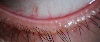
- Epidemic, divided into acute and chronic. It is characterized by:
- redness of the eyes without any significant changes in the eyelids;
- purulent mucous “collars” at the base of the eyelashes;
With epidemic blepharoconjunctivitis, pus appears on the eyelashes
- small erosions along the ciliary edge;
- eyelash loss.
If the pathogen is viral in nature, then blepharoconjunctivitis begins acutely with lacrimation and a rash of small blisters on the inner fold of the conjunctiva.
- Seborrheic blepharoconjunctivitis develops as a consequence of seborrhea (increased production of sebum by the sebaceous glands with the formation of dandruff) or rosacea (small pink acne with purulent heads). Its symptoms:
- hyperemia of the conjunctiva and sclera of the eyes;
- an unpleasant burning sensation and “sand in the eyes.”
Seborrheic blepharoconjunctivitis can be recognized by the hyperemic sclera of the eyes
- The allergic type of disease has various manifestations, its main symptom is its occurrence in connection with the action of some allergen. Symptoms may become more intense after eating spicy, spicy foods.
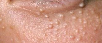
- Demodectic. Its characteristic symptoms:
- severe itching of the eyes, especially painful in the morning;
- sticky discharge with epithelial scales accumulating on the eyelashes.
Types of blepharoconjunctivitis
There are different types of the disease, which are characterized by certain symptoms:
Seborrheic
It begins with seborrheic dermatitis from which the patient suffers. The patient has oily skin, eyebrows, eyelashes, epithelium under the hair are affected, and in men, the growth areas of the mustache and beard are affected. Rosacea may be diagnosed, in which a bright pink rash is visible. The main symptoms are severe hyperemia, itching and an unpleasant burning sensation.
Demodectic
This type of disease appears when demodex mites come into contact with the skin. Microorganisms are found in hair follicles, and with low human immunity and favorable conditions, they begin to multiply quickly. The area around the eyes is affected, epithelial scales accumulate near the eyelashes and crusts form. Patients complain mainly of itching and pain in the eyes.
Viral
The disease can be viral in nature and manifest itself after the patient has suffered from acute respiratory viral infection. In such a situation, a person experiences redness of the eyelids and severe inflammation of the conjunctiva. Sometimes the disease provokes infection of the human body with dangerous herpes viruses or chlamydia.
Other types
Epidemic blepharoconjunctivitis (its other name is “bacterial”) is provoked by staphylococci. Sometimes the disease begins to develop after the Koch-Wicks bacillus enters the body, which is spread by insects.
Patients experience symptoms of blepharitis and severe redness of the mucous membrane. Yellowish discharge is noted, pus covers and sticks together the eyelids, and a so-called “collar” is formed in the eyelash area. The secretion gradually dries out, and the presence of crusts leads to eyelash loss.
If the body reacts sharply to contact with pollen, fluff or certain foods, allergic blepharoconjunctivitis may develop with hyperemia of the sclera and eyelids, burning in the eyes and severe itching
If the entry of microbes into the human body is recorded, such types of blepharoconjunctivitis are diagnosed as: diphtheria, pneumococcal, diplobacillary, etc.
The disease may be of autoimmune origin (drug-induced, hay fever, tuberculous-allergic blepharoconjunctivitis, etc.)
Complications
Blepharoconjunctivitis is not a harmless pathology.
In some cases, the disease can lead to adverse consequences.
Possible complications:
- secondary ectropion of the century,
- development of keratitis, chorioretinitis,
- dacryocystitis,
- dry eye syndrome,
- cicatricial entropion,
- orbital phlegmon,
- extremely rarely - thrombosis of the sigmoid sinus.
Remember! To prevent such diseases, it is necessary to take a responsible approach to the treatment of the disease and strictly adhere to medical recommendations.
Diagnostics
To make a correct diagnosis, an ophthalmologist conducts laboratory and instrumental research methods .
First, the doctor collects a thorough medical history, trying to find out the cause of the disease, and conducts a visual examination. Then he proceeds to diagnose:
- Visometry. With blepharoconjunctivitis, most often visual acuity is slightly or not at all impaired.
- Biomicroscopy. The doctor detects swelling and redness of the conjunctival vessels and skin of the eyelids. Pathological discharge accumulates along the peripheral edge.
- Fluorescein instillation test. This method allows you to detect violations of the integrity of the tear film.
- A swab-scraping from the conjunctiva followed by bacterial culture. With the help of diagnostics it is possible to identify the etiology of the disease.
Reference! If necessary, the ophthalmologist can prescribe a consultation with specialized specialists - an allergist, a dermatologist, an ENT doctor.
With their help, therapy is selected aimed at completely eliminating the cause of the disease.