Phosphenes in ophthalmology
Phosphenes appear as spots, formations, or colored figures of varying configurations, sizes, and colors .
It is worth noting! Such visual images can be visualized with different intensities and frequencies and appear in different quantities, differing in brightness and shape.
Such phenomena, even if they change with great frequency and appear constantly, are never repeated in configuration , each time being unique optical phenomena.
Mostly such formations have soft pastel colors and can be yellow, green, orange and blue, while different people may have one or another color as their main color.
This phenomenon does not happen in people who are blind from birth, but if blindness is acquired, by irritating certain parts of the brain, visual images can appear.
In sighted people, this disorder can manifest itself both with closed and open eyes.
In the second case, phosphenes are superimposed on the contours of visible objects and can take on their outline and similar color.
Prevention of keratoconus
To minimize the occurrence of keratoconus, young people first of all need to regularly visit an ophthalmologist and follow all his recommendations. If inflammatory processes are detected in the organs of vision, prompt measures should be taken to eliminate them.
While reading, working at the computer, or watching TV, it is necessary to control eye strain
To prevent excessive strain, it is necessary to ensure sufficient lighting of the place where work or activities are carried out that require concentration and attention to the eyes.
You should not neglect protective equipment under circumstances that can harm your eyes: dusty air, cold wind, bright light.
Proper nutrition and a lifestyle without bad habits will benefit the entire body and the eyes in particular.
It is important to take prompt measures when allergic processes occur and maintain eye hygiene by washing them with decoctions of healing plants
All of the above recommendations will help minimize the risk of keratoconus.
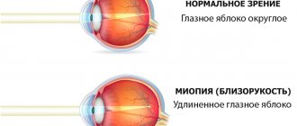
Keratoconus is a disease in which the cornea thins and the eyeball loses its shape. This contributes to the development of various visual impairments. The pathology has been known since the 18th century. Modern correction methods can slow down the progression of the disease and maintain clear vision.
Reasons for appearance
Keep in mind! The appearance of phosphenes is not an ophthalmological pathology, the causes of which lie in physical diseases of the eyes.
The source of such phenomena is the brain, but in general, such manifestations may have the following pathological causes :
- a sharp increase in intraocular pressure;
- traumatic brain injuries;
- hemorrhages in the eyeballs;
- eye injuries;
- sudden turns of the head;
- intoxication of various origins;
- oxygen starvation (including carbon monoxide poisoning).
Often such visual images are seen by people whose activities involve long periods of work at the computer .
In such cases, a few minutes of rest are enough for the visions to disappear.
Often this disorder appears in people over 40-45 years of age , since the tissues of the organs of vision and brain are subject to dystrophic, atrophic and sclerotic processes, as a result of which such optical disorders often occur.
Causes
Phosphenes cannot be considered an ophthalmological disorder, since they appear as symptoms of the development of eye diseases. As a rule, the appearance of phosphenes occurs in the brain. It is also worth highlighting violations that can provoke the appearance of images:
- traumatic brain injury;
- hemorrhages in the eyeballs;
- mechanical eye injuries;
- very sharp turn of the head;
- intoxication with toxic substances;
- insufficient oxygen in the room.
Also, visual images can appear as symptoms of certain diseases not related to ophthalmology. Such diseases include:
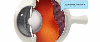
- disorders associated with the vitreous humor of the eyes;
- retinal detachment;
- presence of migraines;
- problems with the cardiovascular system;
- diabetes;
- inflammatory process in the blood vessels of the eyes.
Types and symptoms of the disorder
Note! The appearance of phosphenes may be accompanied by the following symptoms (or some of them):
- eye fatigue;
- pain in the head , eyebrow area and shoulders ;
- frequent blinking;
- difficulties when trying to focus the gaze on certain objects and doubling of objects;
- feeling of the presence of foreign bodies and “sand” in the eyes;
- increased sensitivity to light and uncontrolled tearing;
- redness of the conjunctival membrane.
Symptoms can vary and appear more or less intense - it depends on the reasons for the appearance of the images.
Such optical images can most often appear (lines, dots, circles, ovals, spots).
Symptoms
Symptoms in which phosphenes appear usually indicate a certain disorder in the body. Most often, such images indicate problems with the organs of vision. Phosphenes appear along with headaches and painful sensations in the eyes.
In the presence of hypertension, visual images may occur when there is a sharp change in blood pressure readings. This is most often encountered by patients whose blood pressure increases spontaneously.
Some eye diseases can cause severe dizziness with loss of balance. The same symptoms appear in the presence of a brain tumor. If symptoms appear that may accompany several pathologies, the patient may be referred for examination to other specialists.
Also, along with the visual organs, the following may appear:
- eye fatigue;
- soreness in the area above the eyebrows and in the shoulders;
- increased eye blinking;
- problems with focusing attention on individual objects;
- doubling of objects;
- sensation of foreign objects in the eyes;
- feeling of sand under the eyelids;
- photosensitivity and increased lacrimation;
- redness of the eye membranes.
Artificially induced phosphenes
There is no practical sense in intentionally causing visual images to appear.
But in a healthy person, such phenomena can be easily caused by external stimuli, for example, electromagnetic fields .
Thus, phosphenes are often observed among workers of power plants and nuclear power plants.
The artificial appearance of this optical disorder can be provoked by introducing electrodes into certain areas of the cerebral cortex .
You can independently create such an optical illusion by pressing your fingers on the drooping eyelids of your eyes.
Diseases
The appearance of visual images can be a symptom of a number of diseases, and not in all cases the pathology is related to the organs of vision. Among these:
- pathological processes of the vitreous body;
- retinal detachment;
- migraine;
- diseases of the cardiovascular system;
- diabetes;
- inflammation of the choroid;
- malignant brain tumors.
The cause of retinal detachment can be not only ophthalmological diseases, but also serious physical activity, injury or a stressful situation.
Possible concomitant diseases
Need to know! Such phenomena may be a secondary sign of some ophthalmological diseases associated with disturbances in the structures of the visual organs.
But in such cases, additional symptoms appear in the form of headaches and eye pains, increased blood pressure, dizziness, and loss of balance.
Typically, in such cases, choroiditis, retinitis, retinal rupture or and pathological processes in the vitreous body .
Diseases in other areas include the following pathologies, accompanied by the described phenomenon:
- the appearance of tumors and neoplasms of various origins in the brain area;
- migraine;
- hypertension;
- diabetes;
- pathologies of the vascular system.
Causes
It is already known that phosphenes are not directly related to the organ of vision and are projected directly into the cerebral cortex. There are a number of external and internal reasons that can provoke this phenomenon. Such reasons include the following violations:
- skull injuries;
- blow to the eye area;
- some diseases;
- ocular hemorrhage;
- sudden increase in intraocular pressure.
Phosphenes can also occur due to sudden turns of the head, loss of balance and, as a result, poisoning of the body with any toxin. Post-alcohol syndrome is almost always accompanied by the appearance of clearly distinguishable optical phantoms. Difficulty breathing due to the appearance of even a small amount of carbon monoxide in the air is accompanied by the appearance of bright color figures. This symptom is a harbinger of loss of consciousness, so it is necessary to leave the contaminated area as quickly as possible. In some cases, phosphenes can also appear in the light. In this case, they are superimposed on the visible image, which causes optical illusions accompanied by dizziness and loss of spatial orientation. Very often, the appearance of phosphenes is preceded by some kind of pathology in the body, but this is not necessary.
Optical phantoms in the eye can occur from prolonged computer use, prolonged reading, or exposure to bright light. In this case, the appearance of phosphenes is associated with eye fatigue and after a certain rest they disappear. A cause for concern may be a situation where light figures appear too often and for no apparent reason. It should be borne in mind that in people over 40 years of age, phosphenes appear as a consequence of the aging process of the body in general and the visual organs in particular.
Symptoms of eye fatigue
Phosphenes may indicate a serious illness. If they occur unexpectedly, you should consult a doctor.
Diagnostics
It is not possible for specialists to diagnose the appearance of hallucinations, and a doctor can only learn about the appearance of a disorder from the words of the patient , who describes the intensity and type of formations.
Important! With such symptoms, the ophthalmologist should always prescribe certain diagnostic procedures that will identify concomitant diseases:
- angiography (x-ray examination of blood vessels);
- ophthalmoscopy;
- measurement of intraocular pressure;
- Ultrasound of the organs of vision;
- determination of visual acuity;
- for medical reasons - MRI and CT.
Sometimes it is not possible to determine the exact cause of the images even after these procedures, and in such cases the patient needs to undergo blood tests and undergo a complete examination of the body .
Diagnosis and treatment
It is almost impossible to diagnose phosphenes. The doctor can only form an approximate opinion about what the patient will tell him and prescribe a series of procedures to identify any ailment:
- angiography (vascular examination);
- measurement of intraocular pressure;
- ophthalmoscopy;
- Ultrasound of the eyes;
- visual acuity test;
- MRI.
If these procedures do not help identify the causes of phosphenes, then other tests are done. Next, you will be treated, perhaps not by an ophthalmologist, but by another medical specialist.
As already noted, phosphenes are not a disease, but only a manifestation of some ailment. In this regard, you will have to fight the underlying pathology, after which the phosphenes will disappear.
MagazinLinz.ru team
Treatment and prevention
There is no symptomatic treatment for the appearance of phosphenes, since this is only an indirect manifestation of certain pathologies, with the treatment of which the symptom disappears.
Treatment is always selected individually based on the diagnosis , the patient’s age and the manifestation of other symptoms.
In some cases , the presence of an ophthalmologist alone is not enough, and cardiologists, neurologists and angiologists (specialists in vascular diseases) may be involved in the treatment.
If the reason lies in ophthalmological pathologies , are prescribed for instillation into the eyes.
If there is no effect, surgery may be prescribed .
Remember! Throughout the course of treatment, vitamin complexes are used as supportive and strengthening therapy.
Every fifth case of visual hallucinations associated with ophthalmological disorders is a sign of maturing or progressive retinal detachment or disorders in the vitreous body.
In such situations, only surgery will help, which must be performed as early as possible.
Prevention of the appearance of phosphenes has no specific features and consists only of maintaining a healthy lifestyle and, if possible, avoiding junk food, alcohol and tobacco.
Treatment
Before you begin to treat visual manifestations, you need to understand that they are just a concomitant symptom indicating the development of a particular pathology. Based on this, it is important to find the root cause, which must be treated first.
In each case, therapeutic therapy is prescribed individually, focusing on the underlying disease, its severity and the patient’s age category.
In cases where the underlying cause is not an ophthalmological disease, other specialists participate in the treatment process along with the ophthalmologist. Since phosphenes can be a symptom of any disease, consultation with a neurologist, cardiologist and other specialists in vascular pathologies may be necessary. Therapeutic therapy is carried out under the supervision of an ophthalmologist and other necessary specialists.
For eye disorders with an infectious nature, the ophthalmologist may prescribe the use of antimicrobial drugs for topical use and in the form of other eye drops. If phosphenes appear due to eye injuries, treatment using a laser or surgery is possible if conservative treatment methods are insufficiently effective.
Most often, the need for surgical intervention may arise when there is a risk of retinal detachment or other pathological processes associated with the vitreous. Also, as part of general therapy, it is possible to prescribe vitamin complexes and other preparations containing the necessary microelements that are needed to maintain the patient’s immunity.
fosfenos
Phosphenes are visual sensations due to which a person can perceive very small points of light , about the size of the tip of a pin, or visual
In most cases, phosphenes are harmless, but if they recur, you should pay attention as they may be a symptom of a more serious condition .
Phosphenes are the result of irritation in the retina, which, when it occurs, creates an impulse that is perceived as light. light spots in the eyes are associated with fly flies, retinal detachment, migraines or other conditions.
To meet the definition of a phosphene it is sufficient to say that those rays, flashes or very small spots of light which appear in theamp or visual after closing the eyes very forcefully, rubbing the eyelids or receiving a blow to the face.
Rays of light in a central or visual environment usually appear and disappear very quickly and for no apparent reason.
Additionally, when a person states that they see light in their eyes, it is important to determine whether it is phosphene, photopsia, or glare.
In general,
bright lights occur due to the presence of light that disturbs and interferes with the body or visually. In phosphenes there is no presence of external light that can justify stains.
photopsia, unlike phosphenes, is the sensation of seeing spots of light without external light stimuli.
Most light flashes or phosphenes occur when the vitreous gels or changes and exerts a pull on the retina , causing it to react with a stimulus that is perceived as light.
When we rub our eyes or close our eyes tightly , this stimulus is also created when pressure is applied to the retina.
However, although most of the time the presence of phosphenes in the lungs or vision is not a warning sign, it is important to be very careful if it becomes something regular, as it could be a sign of more serious medical conditions.
The meaning and differences between phosphenes and photographic films are often confused.
This misconception is common because both conditions are very similar , although they have large differences and have different consequences.
La difference between phosphenes and photographic films is that phosphenes they are similar rays or spots of light caused by mechanical stimulation while on photographic films syphilis or flashes appear or visual without external light stimuli, therefore phosphenes can be seen with eyes closed .
When eye phosphenes become a recurring discomfort, it is important to visit an ophthalmologist to check your eye health and rule out a number of diseases associated with these flashes of light.
Las- medical causes of phosphenes are:
When phosphenes or light spots are accompanied by dark floaters in the camp or visually, we can say that these are floaters . There are several causes that cause myodesopsy , from less common ones such as retinal detachment , or uveitis to the most common cataract surgery .
Usually
the treatment is simple, just do laser vitrolysis or, in more serious cases, vitrectomy .
El retinal detachment occurs when the vitreous hooks to the retina in such a way that when detached, it can be pulled out by applying such force that it comes to separate part of the retina .
El retinal detachment associated with phosphenes can occur if we rub our eyes very hard since what we do is press the vitreous body against the retina and subsequently release it .
The appearance of phosphenes in the camp or visual Similarly, the product is the traction exerted by the vitreous body on the retinal tissue .
An ocular migraine is
a severe headache that may begin with episodes of aura where a person may see spots of light on the amplitude or visual .
In this case, the phosphenes are caused by spasms in the blood vessels of the brain .
Whenever a person has light spots on their eyes or visual appearance that is recurring or persistent, you should immediately go to an ophthalmologist for a complete review of your visual health.
Las- the main signs why you should see a doctor:
- Begin to see spots or rays of light in the camp or visual that they have not seen before and that do not disappear.
- Increase the amount of phosphenes or light rays in the amplifier or visual
- See flashes of light along with spots or opaque dots.
- See a dark area in the camp, or a visual or curtain of sorts that interferes with your vision.
- See rays of light after suffering a fall or blow .
Most ocular phosphenes do not require treatment because they do not pose a risk to visual health and go away on their own.
In some cases, when light rays are abundant and impair vision, you may have a vitreous clearing, more commonly known as a vitrectomy .
However, it is important to carefully analyze the need to perform this type of surgical cleaning.
When light spots cause migraines, it is necessary for a person, in addition to visiting an ophthalmologist , to visit a neurologist to prevent headaches and thus avoid the appearance of phosphenes in theamp or visual
Source: https://areaoftalmologica.com/ru/retina/fosfenos/
Phosphenes in the eyes - what are they, causes, symptoms, types, diagnosis, treatment - About the eyes
In order for human organs to function fully, blood circulation must be normal.
Blood delivers oxygen and essential nutrients. The process must also function in the eye area, otherwise the development of diseases cannot be avoided. Vascular eye problems worry many people. Due to inattention to oneself, many pathologies go unnoticed. To avoid complications, you should visit the hospital promptly and undergo medical examinations.
Burst capillaries in the eyes
How our eyes work
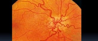
Among the circulatory system, vessels occupy an important place; they deliver nutrition to the eyeball, optic nerves, and retina. Disruption of their work can lead to a disruption in blood supply. The organs of vision will not be able to function fully, this will lead not only to a decrease in visual acuity, but also to complete blindness.
This article talks about the structure of the artery and vein in more detail.
The veins that facilitate the return transportation of metabolic products are slightly different in structure. There are no valves to prevent blood from flowing backwards.
Important! The venous blood flow passes near the brain. Any inflammation or infection in the eyes can be transmitted to the brain.
The arteries of the eye are divided into several branches: the central artery provides adequate nutrition.
Additional ones come from it:
- ciliary;
- muscular;
- lacrimal
Both arteries and veins in the eyes are located next to each other, which is why they have the same name.
Common pathologies
Damaged blood vessels in the eyes can lead to the development of many unpleasant and dangerous consequences.
There are the following vascular diseases of the organ of vision:
- Thrombosis - forms in the central optic artery or in the branches extending from it. As a result of cholesterol deposits on the walls of the arteries, they become clogged. High blood coagulation can contribute to its development.
- Ischemic neuropathy occurs due to impaired blood circulation, as well as when the innervation of the arteries of the fundus is impaired.
- Chorioretinitis is an inflammatory process that affects the vascular branches of the eye.
- An aneurysm is another pathology of the arteries of the fundus, during which their lumen is limited and the walls protrude. If blood coagulation is low, severe bleeding occurs.
The photo below is an example of some diseases
Common pathologies
Among the numerous problems, diseases of the vascular tract of the eye account for approximately 5%.
The most common pathologies are the following:
| Pathology | Characteristic |
| The pathology is characterized by the absence of the iris. The pupil is dilated to its maximum, only blackness is present. | |
| An eye defect can be acquired or congenital. The pupil takes the shape of a pear, and fundus defects may appear. | |
| There may be two or more pupils, one is large, the others are smaller. Visual acuity decreases. | |
| The location of the pupil is eccentric, vision decreases, and strabismus may develop. | |
| The form of the anomaly is harmless and is often found in children. Dense membranes in the center of the lens affect visual acuity. |
Diseases of the eye vessels are dangerous and require immediate intervention from medical personnel.
Characteristic symptoms
If the eye vessels hurt and some pathologies occur, a person may notice the following symptoms:
- visual acuity decreases;
- the radius of the visible area decreases several times;
- objects may fall out of sight;
- burning sensation in the eyes;
- acute pain in the affected area;
- sensation of a foreign body and sand in the eyes.
The problem is that different diseases can have a common clinical picture, which makes it difficult to make a diagnosis.
Important! Symptoms may indicate not only damage to blood vessels in the eyes, but also to the lens, retina, etc. Only a doctor can make an accurate diagnosis.
The vessels of the fundus of the eye may change in hypertension. Blood pressure may increase, and it is thanks to this that problems can be identified in a timely manner.
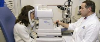
Diagnostics
In order to identify how the pathology has affected the blood vessels of the eyes and correctly prescribe treatment, the patient must undergo a full examination.
Diagnostic measures used for this may be the following:
- Ophthalmoscopy - examines the fundus of the eye, the membrane of blood vessels, and the retina. An ophthalmoscope is used to carry out the procedure. The beam of light is directed to the right place, and the doctor can see everything he needs. There are several methods for performing ophthalmoscopy, each of them involves the use of different mirrors.
- Fluorescein angiography. A contrast liquid is injected into the vascular system, and the specialist analyzes the condition of large and small vessels, the retina and the fundus of the eye. The method is often used to identify the choroid.
- Doppler - ultrasound diagnostics, allows you to evaluate the nature of blood flow in the vessels, speed, coagulation, etc. During the study, ischemic changes can be detected.
- Rheography - unlike the method listed above, you can calculate the amount of blood that enters the eyeball over a certain period of time.
As a result of the research methods carried out, a diagnosis can be made and treatment can be prescribed. This is important, since many pathologies can lead to irreversible consequences.
Treatment
In order to treat diseases affecting the eye vessels, the following medications and methods may be prescribed:
- Eye drops, for example, Albucid, Floxal. The effect is local, drops are used to treat the retina and membrane. It is possible to relieve inflammatory processes and stop the further spread of infection.
- Eye ointment – Acyclovir, Tetracycline. The drug is applied behind the lower eyelid or on its surface. Just like drops, it has a local effect; symptoms can be relieved in a few days.
- Tablets – medications in the form of tablets can be used to treat vascular diseases. The composition of the blood improves, coagulation decreases, and the formation of blood clots is prevented.
- Treatment by surgery. An operation is performed only if neither drops nor medications give the desired effect. The procedure is performed by a specialist in ophthalmic-oncology clinics.
Since many vascular diseases occur deep at the bottom of the eyeball, treatment becomes difficult. Drugs with local action are ineffective, so it is recommended to combine them with other methods.
Laser therapy
A new and effective treatment method. Suitable for vascular deformation and disruption. Patients are given local anesthesia using drops; the procedure lasts no more than half an hour. The patient does not experience pain and does not require further follow-up with a specialist.
The laser beam acts directly on defective areas of blood vessels. Can be used for prevention.
Surgery is performed to treat the following diseases:
- age-related changes in the retina;
- angiomatosis;
- retinal dystrophy;
- hypertensive retinopathy;
- retinal tear.
Laser coagulation can be carried out after surgery, so the results can be consolidated.
If the patient has serious pathologies, such as diabetes, there may be a relapse after laser coagulation. Overall, this is an effective and simple method of therapy. The percentage of complications is low, the rehabilitation period is fast.
Laser beam directed at the pupil
Vitamins for eyes
Modern lifestyle makes the eyes vulnerable to the development of many pathologies. There are many provoking factors: working at a computer, poor lighting, stress, etc. In order to maintain the health of the visual organs, it is recommended to drink vitamin complexes several times a year (see Vitamins for strengthening the blood vessels of the eyes - who needs them?).
Complivit Oftalmo
The main advantage of vitamins is that they have a positive effect on all eye functions. Contains many vitamins and minerals. The deficiency of nutrients is replenished, the walls of blood vessels are strengthened, and regeneration processes are accelerated.
Vitrum Vision
Contains everything you need for your visual organs. The eyes are supplied with everything they need, visual acuity increases, and blood circulation improves. The development of many diseases can be prevented. It is not recommended to take it together with other multivitamins; if necessary, you should consult your doctor.
Blueberry Forte with lutein
Everyone knows about the beneficial properties of blueberries. It contains all the substances that activate recovery processes. The permeability of cell membranes is restored, and the walls of blood vessels are strengthened.
Among the shortcomings, one can highlight only intolerance to the components included in the composition.
Star eyebright
The vitamin complex is designed for connoisseurs of the power of nature. The plant has a positive effect on tissue regeneration and is suitable for people who spend a lot of time at the computer. Blood circulation improves and blood vessels are strengthened.
Biorhythm Vision
The organs of vision are constantly under tension during the day, so they must fully rest at night. When under load, an additional complex of beneficial substances for the eyes is required. Vitamins maintain visual acuity, strengthen the walls of blood vessels, and prevent the development of many diseases.
Omega-3
Many people mistakenly believe that the drug is only suitable for the heart, but this opinion is erroneous. The eyes need support not only under heavy loads, but also with age. With the help of Omega-3, atherosclerotic processes can be prevented, and all conditions for cleansing will be created. Instructions for use are included in the package.
Proper nutrition
To maintain health, it is necessary to maintain proper nutrition.
The following products should be present in the diet:
- Carrot juice - the eyes will be saturated with vitamin A, twilight vision will improve. In order for it to be better absorbed by the body, it is necessary to mix it with milk in a 1:1 ratio. The cocktail must be drunk every day for one month.
- Parsley – removes toxins, relieves inflammation from the optic nerve. In winter, dried herbs can be used to maintain eye health.
- Blueberry. It is useful in any form, both frozen and fresh. Preparations with blueberries are not as effective as the natural product.
- Apricot – contains potassium, which is good for the eyes.
The price of such products is low, and many people grow them in their gardens.
Prevention
It is easier to prevent the development of diseases than to fight them later. Moreover, treating vascular eye pathologies is difficult and time consuming.
In order to prevent their development, you must follow simple rules:
- Control your blood cholesterol levels. It settles on the walls of blood vessels, plaques form, which in turn lead to thrombosis. To prevent this from happening, its level should be controlled. For this purpose tests are taken.
- Take vitamin complexes several times a year. With their help, the arteries are maintained in good shape and do not become thinner. Blood composition improves.
- Follow a regime when you have eye strain. Spending time at the computer, TV, or book should alternate with rest. You need to take a break every hour. Spend this time doing eye exercises.
Source: https://4hospital.ru/veki/fosfeny-v-glazah-chto-eto-prichiny-simptomy-vidy-diagnostika-lechenie.html
Phosphenes in the eyes: what are they, causes, symptoms, types, diagnosis, treatment
Often a person in the dark or with his eyes closed sees specific colored formations or phosphenes that differ in shape, color, and brightness. Despite the fact of numerous requests from patients and complex medical research, the essence of such manifestations is not fully determined.
Astronauts were the first to talk about such formations. One of these representatives turned out to be L. Armstrong. NASA took the news very seriously and research activities began. It was soon discovered that the three characteristic parameters of formations (brightness, size and shape) are always variable.
What are phosphenes?
Phosphenes
– visual sensations of bright points and figures, manifested in conditions of absence of light.
People who are blind from birth do not see such optical phantoms, however, in people with acquired blindness, a similar sensation can be caused by irritating the brain.
Phosphenes are not classified as an ophthalmological pathology, and the mechanism of their manifestation lies in the development of physiological eye disease. The source of this manifestation is the brain, however, the cause of such a pathology can be one of the following phenomena:
- receiving a traumatic brain injury;
- bruising of the eyeballs;
- mechanical damage to the organ of vision;
- sudden turn of the head;
- poisoning with various toxic substances;
- lack of oxygen.
The appearance of visual images can occur in people whose work involves spending a long time at the computer. To eliminate the undesirable phenomenon, it is enough to give your eyes a rest for a couple of minutes.
Often this deviation can be observed in people in the age category of 40-45 years, and this is associated with ongoing dystrophic, atrophic and sclerotic processes in the human visual organs and brain.
Phosphenes in the eyes are manifested by the following symptoms:
- feeling of visual fatigue;
- frequent blinking;
- heaviness in the eyelids;
- sensation of grains of sand getting into the eyes;
- Red eyes;
- a seeming veil before the eyes;
- excessive tearing;
- significant photosensitivity;
- splitting of visible objects;
- difficulty focusing on an object;
- headache;
- pain in the area of the shoulder joints and the superciliary part.
The manifestation of concomitant symptoms is associated with the development of a related disease, which, as a rule, is directly related to the organ of vision.
The diagnosis of this deviation is conditional, since the specialist does not have the opportunity to see hallucinations, but can judge their intensity and types of formation only from the patient’s words.
The task of diagnosis is to determine concomitant diseases. For these purposes, the following procedures may be required:
- fundus examination;
- radiography of blood vessels;
- determination of intraocular pressure;
- Ultrasound of the organ of vision;
- visual acuity test;
- MRI
- CT.
There are clinical cases in which, after carrying out the above procedures, it is not possible to determine the specific cause of visual illusions, then a blood test and a full examination of the main body systems should be performed.
Diseases
The appearance of visual images can be a symptom of a number of diseases, and not in all cases the pathology is related to the organs of vision. Among these:
- pathological processes of the vitreous body;
- retinal detachment;
- migraine;
- diseases of the cardiovascular system;
- diabetes;
- inflammation of the choroid;
- malignant brain tumors.
The cause of retinal detachment can be not only ophthalmological diseases, but also serious physical activity, injury or a stressful situation.
The prescribed therapy depends on the specific diagnosis, individual characteristics of the body, age category and other symptoms. The course of treatment is carried out under the supervision of not only an ophthalmologist, but also with the participation of a neurologist, cardiologist, and angiologist.
If the cause of the visual images is an ophthalmological pathology, then a solution is prescribed for eye drops. If there is no improvement, surgical intervention is an option, since this may indicate an impending or progressive retinal detachment or a pathological process of the vitreous body.
Any prescribed course of therapy must be accompanied by the use of vitamin complexes for general strengthening of the body.
Thus, visual hallucinations appear due to the development of physiological pathologies of the eye. The process is characterized by a fairly clear clinical picture. Specialists of various profiles take part in the treatment of pathologies.
5 out of 5:
/ 2
Please rate the article:
Source: https://103doc.ru/fosfeny-v-glazah-chto-eto-prichiny-simptomy-vidy-diagnostika-lechenie.html
Phosphenes or strange stuff in the eyes.
Hi all.
About a year ago I was diagnosed with keratoconus
.
Keratoconus
(from ancient Greek κέρας - “horn” and κῶνος - “cone”) - a degenerative non-inflammatory disease of the eye, in which the cornea thins and takes on a conical shape.
Well, let's start in order.
Until I was 19-20 years old, I had 100% perfect vision, I didn’t complain, I lived and didn’t grieve.
When I was 20, during the summer holidays, I noticed that my eyes were watering quite often, especially the left one. I went to a regular optician, where they told me that I had astigmatism in the early stages. What the hell?
Astigmatism
translated from Latin - absence of a (focal) point. Astigmatism occurs due to the irregular (non-spherical) shape of the cornea (less commonly, the lens).
I didn’t attach any importance to it then, I was young and stupid. In principle, he was not interested in his health. They prescribed me glasses, which I did not wear because they were uncomfortable and awkward. I still saw well, didn’t complain, studied and moved on with my life.
At the age of 22-23 I decided to go to the gym. There was success, I gained good weight, my working weight was growing, I was very proud and happy about it.
He got married at 24 and was a gym fanatic. Programs, diet, weight gain and weight.
When I was 25, I bought a car and noticed that at night I couldn’t see very well behind the wheel. My fault was the glare from oncoming headlights. I went to the hospital, to the ophthalmologist for “night glasses.” The ophthalmologist upset me... Suspicion of keratoconus and an urgent request to stop driving altogether.
Thanks to my wife, she kicked me into going for an examination at a specialized eye hospital. I myself was very careless about my health.
After a bunch of procedures and tests, the diagnosis was confirmed. Keratoconus, and quite advanced at that. BUT, they reassured me and said that I could still do one procedure - cross-linking
. Its essence is that special agents are dripped onto the cornea.
solution and heated with ultraviolet light. A frame is formed on the cornea, which prevents deformation of the cornea. The procedure is not cheap, it cost about 200,000 tenge, it was done by a doctor from Moscow (I live in Almaty).
In general, we also had and still have specialists, but this one was strongly recommended to me by the center itself.
The operation, or even better, the procedure, was scheduled for April. And I had the tests in February. After waiting a couple of months, I came to the hospital on time for an appointment with this miracle doctor. But the miracle did not happen.
After looking at my sore eye, she made a verdict: there is no way to do cross-linking. It turns out that there are indications - the thickness of the cornea should be at least 400 microns. For me it has become very thin, in some places up to 200 microns.
There is only one way out - corneal transplantation.
Due to stupidity and ignorance, I was not upset. I thought, what’s the big deal, they’ll take it and attach it like a lens. I made a mistake.
It turns out it doesn't exist
organic corneal transplants. And if there is, it’s definitely not here and only in theory.
We do everything the old fashioned way - they take a corpse, cut out the cornea and change it for you.
We went to the doctor who was recommended at the clinic.
She is a surgeon with many years of practice. He treats complex cases throughout Kazakhstan. If necessary, I will send you the contact information in a private message.
As it turned out, the process of destruction of the cornea is quite a long process, BUT, constant training with weights significantly accelerated the process.
She said that classes should be stopped immediately, try not to bend over, only if you squat. Do not lift heavy objects, as this causes eye pressure.
From physical education - only walking.
After looking at me, she confirmed the indications - only transplantation. And urgent.
Well, there’s nothing to do, we have to do it. But, as it turned out, everything is not very simple. In Almaty, and probably in general in KZ, there is no center where finished fabrics are stored. You have to wait, and it’s unclear how long. Some people wait years for suitable fabrics. I was very lucky. After 3 months, the call came - there was a suitable fabric. I urgently need to undergo surgery.
Taking a change of clothes and shoes with me, I went to the operation. I called work beforehand and said that I would be gone for about a couple of months.
The operation itself takes place under local anesthesia and takes about an hour. First, the eye is anesthetized, then a part of the cornea is cut out according to a special template. The donor's cornea is also cut out using the same template.
Sew by hand (the seam looks like it was sewn with a machine). A hard lens is put on and sent to the ward to rest. You need to spend at least 5 days in the hospital, during which special drops are given every 2 hours.
The operation itself is practically painless, except for the injection in the eye. But the first days are the most difficult. Especially the first night. I think I know what phantom headaches are. Neither painkiller injections, nor pills, nor sleeping pills helped.
My head, especially the left side, was simply splitting. In the morning I fell asleep and was ready to sleep until lunch, but we are in the hospital, and there is a regime there. We got up at 7 am to put in the first drops.
To be honest, it was very scary to open my eyes and look at myself in the mirror.
The left eye was swollen and had some mucus inside. I didn’t wash it because it was boring. This went on until 9 am.
At 9 am, an appointment with the surgeon who performed the operation. She removed the lens, washed the eye and gave an injection and dropped drops. In general, injections were given for 10 days, 2 times a day. Painful, infectious. The entire eye and eyelid were punctured.
As I said earlier, the most difficult are the first days. Because it is on these days that it is decided whether the new tissue will take root. Very often, especially in young people with normal health, foreign bodies are rejected.
“Thank you” to our immunity for this. So after such operations it is necessary to greatly reduce immunity. To do this, I was given some kind of injections, which were also painful and had some side effects, there was severe itching.
So 2 days passed, they dripped and pricked me). The eye was just as swollen, I could hardly see anything with it. I lay and thought all day.
On the 3rd day, at the appointment, the doctor said that the cornea was not taking root... There was severe rejection, and most likely I would have to have a second operation.
To be honest, I fell apart.
Well, imagine for yourself - he lived for himself, did not grieve. At least I saw it somehow, and perhaps I would have seen it for just as long, but maybe not. And then they cut off a piece, sewed another one - but there was no use!
I lay there and thought about what to do. My wife and parents were very supportive. I had a serious conversation with my father, the essence of which was not whine, but do it, you have a family, children.
There was nothing to do, I wrapped the snot around my fist and talked again with the doctor, more substantively.
As it turned out, repeated surgery does not guarantee 100% that it will be successful. And 3 and 4 and 5. One patient had surgery for the 3rd time and things weren’t going well for her.
The main thing I understood is that even if I don’t have surgery and the cornea doesn’t heal, it will become cloudy. Vision, of course, will be almost zero.
BUT, there is still a chance that everything will be fine, that the drops that I apply every 2-3 hours + regimen will bear fruit and the transplant will take root.
I decided that I had nothing to lose - any time, if it takes root - good, no - I need to do it again, which I was not yet ready for either mentally or physically (in terms of money). Therefore, I asked to be discharged home, houses and walls help. And I lie in the hospital all day, tormenting myself with different thoughts. And you can come for injections every day.
The doctor, of course, was against it, but I was very persistent)
The point is, six months have passed. The cornea has healed, and now vision is about 30% in the left eye. The doctor is surprised by this fact, since there have been no such cases in her practice.
If the swelling does not subside after a couple of days, then complete rejection usually occurs. But adherence to the regime and attitude did their job - slowly, vision is restored.
In May I will go to remove the stitches, then another six months and I will be able to go for laser correction.
I had a GCL cut into my right eye – now I wear it and can’t imagine how I used to live without it. In a couple of years I will go to have my right eye done. Here, too, it’s just a transfer. Although, who knows, maybe by this time there will be a different type of treatment.
I hope that in a couple of years I will be able to return to the gym. Of course, you won’t be able to lift very much, but benching acceptable weights is fine.
Thanks to the doctor, who knows what would have happened if she had not had the operation. And she did it very professionally.
To all those who have vision problems, run to the doctors! The problem can most often be solved in its infancy. It’s very scary to see the faces of people who are told that they most likely won’t be able to see... and during the days that I was in the hospital, I saw several such cases while sitting during procedures.
1. Male, under 50. Driver. He came with a complaint that there was a “curtain” on his eye. I wore glasses all my life, and every year I simply prescribed them at a nearby optician with thicker lenses.
We looked and it turned out we needed a lens transplant. It seems like a small thing, but it’s an ordinary procedure. Done. We looked through this lens and it turned out to be a detached retina. And this is very serious.
The result is disability and loss of vision in one eye.
2. Boy, 13 years old. Problems started when he was 8 years old. They weren’t really examined. Now I have problems with eye pressure, which gives me severe pain in my head.
3. My case - I could get by with lenses, and not for constant wear, but, for example, only night ones. But we are all so confident in ourselves and our health.
In general, the story turned out to be a little crumpled; I wrote it from memory. If you have any questions, I will be happy to answer
Do not be ill.
PS: I will not disclose the price for the operation, since it is all very individual. I was more or less lucky, the amount was recoverable + almost all of it was restored under insurance
Source: //pikabu.ru/story/fosfenyi_ili_neponyatnaya_shnyaga_v_glazakh_3369596
Let's evaluate weight, operating temperature and sun resistance
The sun damages the most plastic products, that is, PVC hoses. The plastic becomes cloudy and rough. When purchasing, look for a UV protection indicator on the label. Often the color you like best is chosen. At the same time, algae grows in transparent products, which impairs flow and complicates the operation of devices connected to the hose, for example, sprinklers. The most practical ones are brightly colored, they are visible on the grass, and are more difficult to step on or damage when mowing the lawn or aerating the soil.
The weight of the hose depends on the material, diameter, wall thickness and length. Maximum length – maximum weight. When purchasing, consider how much area you will irrigate and who will carry the hoses around the garden. The most common weight parameters for multilayer PVC are as follows: with a 15 m coil - up to 8 kg; with a 20m coil - up to 10kg; 25m - up to 13kg. It turns out that on average, a meter of an inch hose is half a kilogram with a coil. The weight will be less with a smaller cross-section, for example, with a diameter of half an inch - 0.2 kg, with a diameter of 3/4 - 0.3 kg. A meter of high-quality thick rubber hose with an inch diameter, black, from Russian manufacturers, weighs one and a half kilograms.
Most often, the operating temperature range for watering hoses is minus 20 to plus 60 degrees Celsius. This is more than enough to use them at any time of the year, except winter. Please note that if you use the hose in cold weather with sub-zero temperatures, the rubber and plastic will bend much less easily. To prevent your inventory from spoiling, roll it up before it gets cold and hide it indoors.
Causes of hyperemia of the conjunctiva of the eye
Causes of hyperemia of the conjunctiva of the eye
Below, the obaglaza.ru project has collected common causes of redness of the conjunctiva.
Conjunctival infection
Inflammation of the ocular mucosa also spreads to neighboring organs, that is, the eyelids. Often a person notices swelling and a local increase in temperature in the area affected by inflammation. If you carefully move the eyelid away, you can notice hyperemia of the conjunctiva and on the mucous fold.
The common disease conjunctivitis manifests itself differently depending on its course. Thus, acute processes are characterized by diffuse redness of the mucous membrane, and chronic processes are characterized by redness in the area of the cartilage of the eyelids.
The transition of the folding of the membrane to the eyeball is painted bright scarlet. In this place, protrusion of small vessels is also observed, which easily move around the eye along with the mucous membrane.
Often, a patient with conjunctivitis complains of the imaginary presence of sand in the eyes and itching, in addition to conjunctival hyperemia. There are unpleasant sensations in bright sunlight.
Ogaglaza.ru warns you against unwanted actions that aggravate the disease. This is self-medication and constant touching of the affected area.
Ciliary infection
Complications of conjunctivitis include iritis and, rarely, iridocyclitis. The clinical picture of the disease differs from classic conjunctivitis. So, on the eyeball you will not see inflamed, red capillaries; instead, the eye will turn purple and the iris will turn reddish. The reason for this manifestation is the deep location of the vascular layer of the eye, which becomes inflamed.
Mixed infection
This is a simultaneous lesion of both the conjunctiva and the deep structures of the eye. Occurs during acute inflammatory changes. The route of transmission of infection is through the blood vessels that supply the eye; the medical term for this route is hematogenous. If the disease has penetrated to the ciliary body, then there is a slight blurring of the contours of the iris, purulent (hypopyon) and inflammatory exudate. Conjunctival hyperemia is also possible.
During an examination of the conjunctiva, a specialist can see enlarged lymphatic vessels, into the dilated ducts of which blood cells penetrate. This phenomenon is called hemorrhagic lymphangiectasia.
Don’t worry, such cases are extremely rare and account for tenths of a percent according to medical practice, the results of which Obaglaza.ru specially studied for you.
Examination of conjunctival vessels with special instruments under enormous magnification often leads to the detection of a “sludge phenomenon”. This is a process in which red blood cells stick tightly to each other, clogging the lumens of the capillaries and stopping the normal blood supply to the organ.