Often, angiopathy of the retina or retina is not an independent disease, but develops against the background of other internal ailments that occur in the body with complications. This vascular pathology often affects middle-aged people, but is sometimes diagnosed in children. If angiopathy is not treated promptly, irreversible consequences begin to progress in the organs of vision, the most severe of which is complete loss of vision.
This pathology does not have an ICD code, since it is not an independent disease, but progresses against the background of other internal disorders.
Symptoms
With functional phlebopathy, there are no external anatomical abnormalities, but the patient’s subjective sensations make one suspect pathology. Patients have no external signs of varicose veins. Symptoms of the disease occur against a background of satisfactory health and appear more often in women. With functional phlebopathy you may be concerned about:
- Night cramps;
- Swelling and pastiness that occurs in the evening;
- Fullness and heaviness in the legs;
- Severe fatigue when standing in a static position.
Why is it dangerous?
With a long course of the disease and the absence of necessary treatment, phlebopathy can cause complete loss of vision. Thrombosis of the vessels of the eye with subsequent formation of an embolus is a factor in the development of heart attacks or strokes due to blockage of smaller vessels. In addition, when one organ of vision is involved in the process, the second one necessarily suffers over time, since they are interconnected.
Phlebopathy provokes constant hemorrhages in the retina.
Types of phlebopathy
Phlebopathy includes several types of disease:
- Hormone dependent, which is caused by taking hormonal contraceptive medications.
- Orthostatic phlebopathy occurs as a result of prolonged standing or sitting, as this develops congestion in the lower extremities.
- Phlebopathy during pregnancy is caused by increased intra-abdominal pressure due to fetal growth. As a result, the normal outflow of blood from the venous bed to the heart is disrupted.
- Hereditary phlebopathy is genetic in nature and is inherited.
Prevention
In order to prevent the development of phlebopathy, it is necessary to follow some preventive measures. First of all, you should lead an active lifestyle and not remain motionless for a long time. It is recommended to take a walk in the fresh air every day, and this is especially useful during pregnancy.
A good way to prevent phlebopathy is a contrast shower, which is recommended to be taken in the morning. You can strengthen your ankle muscles and immunity through swimming or water aerobics.
Hormone dependent
In case of long-term use of hormonal contraceptive pills, the venous network undergoes a number of changes that affect the vascular wall:
- Against the background of an increased concentration of estrogens, the size of the inner and middle layers of the wall increases. In this case, endothelial cells undergo degeneration.
- Progesterone affects elastin and collagen, causing their degeneration.
- The combined administration of estrogen and progesterone drugs leads to the destruction of the venous wall and an increase in the lumen of blood vessels. This is due to the loss of the muscle layer and phlebosclerosis, in which the growth of connective tissue occurs.
A few months after starting to take the drugs, you can notice the first symptoms of phlebopathy. In this case, there is pain and heaviness in the calf muscles, swelling and pastiness of the legs and feet, spider veins and bruises form in places of physical contact.
Diagnostics
To diagnose retinal phlebopathy, a variety of methods are used that can identify vascular anomalies and determine the degree of vision changes. The examination begins with standard ophthalmological procedures:
- visometry
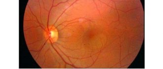
, or assessment of visual acuity and quality using special tables; - ophthalmoscopy
, or examination of the fundus using a special device through a pupil dilated with drugs; - refractometry
, or measurement of the refractive and focusing functions of the eye.
Additionally, the doctor may measure intraocular pressure.
To detect changes in venous outflow, instrumental studies are used:
- angiography of the vessels of the fundus
- an x-ray examination, during which the doctor visualizes areas of dilated veins and recognizes areas of tissue atrophy in the images; - angiography of cerebral vessels
in cases of suspected cerebral origin of vascular insufficiency; - To T or MRI of the vessels of the head
, in which the doctor visualizes the veins of the eyeball and brain, determines the degree of change in the tissues adjacent to the pathological vessels, etc.
Additionally, laboratory tests are carried out aimed at identifying possible causes of vascular changes:
- blood tests for hormones;
- blood tests for cholesterol;
- blood chemistry;
- blood and urine testing for sugar.
After identifying the true causes of vision problems, the doctor can begin to draw up a treatment plan.
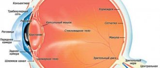
Retinal phlebopathy
With diabetic angioretinopathy, minor bleeding from the vessels of the retina often occurs, and varicose veins also form. This disease can result in blindness. Phlebopathy in the initial stages is characterized by the appearance of vascular tortuosity, an increase in the diameter of the veins, and stagnation of blood in the vessels in the fundus area. The caliber of the retinal vessels becomes heterogeneous, microaneurysms are formed (expansion of the diameter of the vessels).
As a result of stagnation of blood in the vascular bed against the background of phlebopathy, metabolism in the vitreous body is disrupted, and the blood can be destroyed. Due to the accumulation of breakdown products (urea, lactic acid), the liquid media of the eyeball become more acidic. Against the background of such metabolic disorders, the processes of varicose veins of the retina intensify. Fundus vessels can grow into the vitreous substance, leading to hemorrhages and a subsequent decrease in visual function up to complete blindness.
The prognosis for phlebopathy due to diabetes mellitus is quite favorable, but subject to timely and high-quality treatment. With rational insulin therapy, blindness can be avoided in 85-90% of cases. At a young age, these numbers are less rosy. For 20% of patients who have lost vision due to diabetes, survival does not exceed five years.
Retinal inflammation
Inflammation of the retina of the eyeball (retinitis) can occur as a unilateral or bilateral process. The causes of retinitis are varied: infectious diseases of a bacterial and viral nature (including tuberculosis, syphilis, AIDS, herpes), toxic eye damage due to other pathologies, allergic diseases, chronic heart and kidney diseases.
Manifestations of retinitis can be different and depend on in which parts of the retina the pathological process is localized. Common symptoms for any form of retinitis include changes in visual fields (scotoma) and a decrease in its acuity. Scotomas are focal loss of visual fields; their size and location directly depend on the location and size of the inflammatory focus. If retinitis is untimely or incompletely treated, persistent vision deterioration occurs, which is especially noticeable in poor lighting conditions.
Treatment of retinitis should be aimed at identifying and eliminating the cause that caused the inflammation of the retina. The most effective is an integrated approach, including drug treatment, injections of anti-inflammatory and antibacterial drugs, and laser coagulation of retinal vessels.
Read more about the treatment of retinal inflammation >>>
Causes
Phlebopathy is a consequence of a violation of the structure and function of the vein wall. This changes its elasticity and permeability. Usually the veins in the lower extremities undergo transformation. With age, the condition of the muscle layer in the blood vessels is disrupted. This process can be aggravated by congenital and acquired pathologies. Among the causes of acquired phlebopathy are:
- Features of professional activities that involve long-term static loads on the lower limbs (surgeons, hairdressers, cooks).
- Impaired blood outflow from the venous bed due to increased intra-abdominal pressure during pregnancy, in the presence of scars, adhesions, or a tumor process.
- Excessive physical activity.
- Various fluctuations in hormonal levels in women due to the use of hormonal drugs, menopause, pregnancy, and menstrual irregularities.
For what reasons can illness occur?
The appearance of symptoms of phlebopathy is promoted by venous stagnation of blood, and this, in turn, is caused by the following reasons:
- prolonged load on the venous outflow of blood (increased blood outflow or its decrease), that is, the risk of developing phlebopathy is directly related to a person’s prolonged sedentary work or if a person spends the entire working day “on his feet”;
- failure in the human hormonal system, this most often concerns pregnant women or women during menopause;
- hereditary predisposition to the disease. According to studies, phlebopathy is observed in 50% of patients who are at risk due to heredity.
Thus, factors contributing to the development of phlebopathy include :
- professional activity;
- sedentary lifestyle;
- compression of leg veins during pregnancy;
- impaired blood flow due to a pelvic tumor;
- changes in hormonal levels;
- excessive physical activity.
These reasons can be combined with each other, so it cannot be said that the presence of the disease in a patient is caused by one of these reasons. Typically, phlebopathy develops as a result of the interaction of several causes.
Treatment
Due to the fact that phlebopathy is based on thinning of the muscular layer of the vein wall, it is usually not possible to correct the disease surgically. In this regard, the main method of treating phlebopathy is the prescription of medications, as well as the use of elastic compression. In the latter case, special therapeutic knitwear is used, which varies in degree of compression. Such underwear can also be used for prevention, for example, during pregnancy.
Venotonic effects are used for drug therapy for phlebopathy. For hormone-dependent forms of pathology, Antistax has proven itself well. Local remedies with heparin also have a positive effect on the condition of the legs (eliminate pain and swelling). These include Lyoton gel, which helps improve microcirculation of lymph and blood, eliminates dry skin, relieves pain and itching.
What treatment is prescribed?
Drugs
If retinal angiopathy is diagnosed in a timely manner, a conservative treatment regimen is prescribed, the main objectives of which are to normalize blood pressure, strengthen blood vessels, and reduce the risk of developing intraocular bleeding. The following groups of drugs are used:
Often, with such a pathology, the patient is prescribed Aisotin drops.
- vasodilators;
- vitamin eye drops to strengthen the retina;
- drugs that improve blood microcirculation.
Effective drops for retinal angiopathy are:
- "Emoxipin";
- "Taufon";
- "Quinax";
- "Isotin."
Therapy with folk remedies
Alternative medicine also helps fight pathology, but before you start treatment with folk remedies, you first need to consult a doctor. To improve the nutrition of vascular structures, it is recommended to consume more fiber, which is found in vegetables, fruits, and herbs. For angiopathy, the following remedy will benefit:
- Grind fresh parsley leaves in a blender and squeeze out the juice.
- Drink 1 tbsp daily. juice in the morning before breakfast.
Physiotherapy
Physiotherapeutic procedures prescribed by a doctor will help normalize blood circulation and nutrition of the retina. With the progression of angiopathy, the following are effective:
- acupuncture;
- laser therapy;
- magnetic therapy;
- Individual exercise therapy classes.
Treatment methods
To normalize the functioning of the visual apparatus, it is recommended to reduce the load on it and balance the diet.
Drug therapy for ocular angiopathy in a pregnant woman is prescribed only in severe cases in the second half of pregnancy. In other cases, experts recommend normalizing nutrition, sleep and rest patterns, minimizing eye strain (reading, watching TV and working at the computer), and undergoing a course of physiotherapeutic procedures. If no measures are taken at all, severe complications may develop in the woman and the fetus.
During the birth process
The decision whether a pregnant woman should give birth independently or by caesarean section is made only by a doctor after assessing the woman’s condition and the possible risks for her and the fetus. The fact is that the state of the female body during labor and subsequent childbirth is very tense and stressful, therefore the risk of vascular rupture and the development of complications such as:
- formation of blood clots in the vessels of the eyeball;
- partial or complete loss of vision;
- retinal disinsertion;
- hemorrhage in the eye.
Causes
In fact, any pathological changes in small arteries and veins that a doctor sees when examining the retina can be called angiopathy. Accordingly, this is a symptom that in most cases occurs in the following diseases:
- diabetes;
- arterial hypertension;
- osteochondrosis of the cervical spine with impaired blood flow in the vessels of the neck;
- autoimmune diseases;
- injuries to the head, neck and upper chest;
- nervous diseases that contribute to impaired vascular tone.
Symptoms
The appearance of dots before the eyes and blurred vision of objects are symptoms of the disease.
The first symptoms can be noticed already in the 6th month, but in 90% of cases, retinal angiopathy during pregnancy is determined in the last weeks. Symptoms of angiopathy in pregnant women are:
- decreased visual acuity;
- fog or spots before the eyes;
- pain and cramps;
- pulsation and dryness of the mucous membrane.
Symptomatic manifestations of retinal angiopathy
Angiopathy that develops during pregnancy is less severe than the variant of this disease that occurs with hypertension. The most characteristic sign of the development of angiopathy in pregnant women is a decrease in visual acuity, and in some women such a deviation can be very mild and not cause obvious discomfort, while in others the disturbances can be very serious.
Common manifestations of angiopathy include the following symptoms:
- black dots in the field of vision;
- abnormal pulsation in the eyeball;
- pain;
- rez.
As a rule, the symptoms of angiopathy are transient, that is, over time, there may be a worsening of the manifestations of signs of vascular pathology and, conversely, an improvement in the condition.