Types of disease
The types of ptosis include:
- unilateral (appears in one eye) and bilateral (in both eyes);
- complete (the upper eyelid completely covers the eye) or incomplete (closes only partially);
- congenital and acquired (depending on the cause of occurrence).
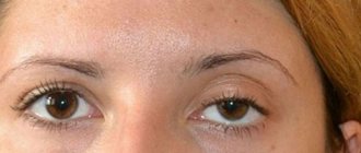
The severity of ptosis is determined by how much the eyelid droops:
- 1st degree is determined when the upper eyelid covers the pupil from above by 1/3,
- 2nd degree - when the upper eyelid is lowered onto the pupil by 2/3,
- 3rd degree - when the upper eyelid almost completely hides the pupil.
The degree of visual impairment depends on the severity of ptosis: from a slight decrease in vision to its complete loss.
What can it be confused with?
The following pathologies of the visual organs can be mistakenly mistaken for ptosis:
- dermatochalasis, due to which excess skin of the upper eyelids is the cause of pseudoptosis or ordinary ptosis;
- ipsilateral hypotrophy, which is expressed in drooping of the upper eyelid following the eyeball. If a person fixes his gaze with the hypotrophied eye, while covering the healthy eye, pseudoptosis will disappear;
- the eyelids are poorly supported by the eyeball due to a decrease in the volume of the orbital contents, which is typical for patients with false eyes, microphthalmos, phthisis of the eyeball and enophthalmos;
- contralateral eyelid retraction, which can be determined by comparing the levels of the upper eyelids. It should be taken into account that covering the cornea with the upper eyelid by two millimeters is the norm;
- brow ptosis, caused by excess skin in the brow area, which can occur with facial nerve palsy. This pathology can be determined by raising the eyebrow using your fingers.
Sudden loss of vision: causes
From time to time it happens that the vision becomes dark, and it seems that vision is leaving. This occurs mainly from pressure differences both in the arterial system and inside the cranium.
However, at times, partial or total loss of vision may occur for a long period of time. This usually occurs due to a head injury, eye injury, or other illness. Loss of vision for an ordinary person who has never encountered this problem is a real grief.
Many people panic and begin to worry, which causes an exacerbation of diseases of the nervous system and further aggravates the situation. Therefore, it is best not to panic during the onset of sudden vision loss. It’s better to have an idea about the warning symptoms of sudden vision loss.
Signs may be subtle and hidden.
It is important to keep in mind that all illnesses have a cause. And when a sudden loss of vision occurs, it is best to think about the reason for what is happening, and not panic and break your nerves.
Factors Contributing to Unexpected Blindness
— Loss of vision (amorosis in Latin) can be a consequence of deposits on the lens or disruption of the functioning of the lens. Diseases of the eye retina also cause sudden blindness.
Whatever the reason, people with sudden deterioration of vision should be immediately placed under therapy.
Data collected in the first minutes after the onset of sudden blindness will help establish the correct diagnosis.
— A sudden deterioration in vision in one eye can be a consequence of retinal disease, as well as a broken eye nerve. The reason for this is improper blood flow in the retina.
Most often, it feels like part of the vision has been covered with some kind of canvas. So in patients with a similar illness, temporary weakness or complete temporary failure of one or two limbs is observed.
The duration of loss of limb function can last from one hour to a day.
- In almost ninety percent of cases, a sudden deterioration in vision occurs due to improper functioning of the heart; a retinal embolism occurs due to a plaque inside the aortic arterial system.
Also, sudden blindness in one eye is a symptom of an upcoming stroke. In such situations, it is necessary to immediately examine the patient to prevent a stroke.
It is necessary to take one hundred to three hundred grams of aspirin to prevent a stroke.
— In young people, migraine may be the cause of temporary, sudden blindness in one eye. The effect of migraine on vision occurs through a migraine aura. However, in order to avoid a stroke, it is necessary, with the help of a special examination, to exclude unusual pathologies in the arterial system and cardiovascular system from the list of probabilities.
Neuropathy
Neuropathy of the nerves of the eyes occurs as a result of improper blood flow to the nerve. Due to the lack of blood from the occipital cerebral region to the eye, the organ begins to gradually lose its functionality. This disease manifests itself as visual impairment in one eye and is not accompanied by pain. Symptoms include swelling and abnormal growths around the optic disc.
Very often, ischemic neuropathy is detected in people with arterial pathologies and diabetes. In old age, the cause of ischemic neuropathy can be temporal arteritis. A symptom of this disease is the absence of blood pulsation in the artery passing through the temple.
It is much less likely to encounter such a type of ischemic neuropathy as ischemic neuropathy of the occipital lobe. The cause of this type of neuropathy is anemia and low pressure in the blood vessels. Due to an excessively sharp drop in blood pressure, an attack of the optic disc nerve occurs.
There is also a known disease that results in a sudden deterioration of vision, this is toxic neuropathy. The cause of toxic neuropathy is a high content of methyl, ethyl, and carbon monoxide in the blood. Thus, this disease can develop with excessive alcohol consumption.
Intracranial pressure
Loss of vision, especially sudden loss, can be a consequence of increased intracranial pressure. This is accompanied by short-term losses of visual abilities, which usually occur when changing body position.
Loss of vision in this case continues for two to three minutes. With this disease, intravenous administration of methylprednisolone into the patient’s body will help.
Since the disease can cause serious complications, it is necessary to consult a specialist.
Infarction of blood vessels in the posterior part of the brain as a factor in vision loss
Acute sudden blindness is a consequence of an infarction of the blood vessels in the posterior parts of the brain. The so-called basilar artery becomes blocked, or the pressure of the blood vessels sharply decreases, resulting in rupture of the blood vessels.
With this type of disease the following may occur:
- dizziness, - double vision.
Blindness in this case is called cortical and differs from bilateral blindness, which occurs when the optic nerve is damaged, the reaction of the pupils to external stimuli is preserved. In many situations, patients deny the onset of visual impairment and state that the lights are off and it is very dark around.
Hysteria as a cause of vision loss
Sudden blindness also has a psychogenic background and is a consequence of acute psychosis expressed in hysteria. Patients based on hysteria, in many cases who are young ladies, declare that the world has plunged into darkness. Psychosomatic symptoms will go away after calming down; it is necessary to monitor the patient and avoid nervousness and stressful situations.
Just as with an infarction of the posterior lobes of the brain, with hysteria the reaction of the pupils is absolutely normal. There are no table symptoms. Due to the calm and even overly relaxed state of consciousness of patients with hysteria, it is very difficult to detect this illness before the onset of blindness.
Causes of the disease
Let us examine in detail the reasons for which ptosis occurs.
Innate
Congenital ptosis occurs in children due to underdevelopment or even absence of the muscle that should be responsible for raising the eyelid. Congenital ptosis sometimes occurs together with strabismus.
When ptosis treatment is not treated for a long time, the child may develop amblyopia (lazy eye syndrome). Congenital ptosis is most often unilateral.
One of the most common eye diseases is pterygium. Read on to learn about its correct diagnosis, treatment and prevention.
We continue our analysis of eyelid diseases! The news contains all the main causes of stye on the eye.
Acquired
Acquired ptosis develops for several reasons and is divided into:
- aponeurotic ptosis , which is associated with the weakening or stretching of the aponeurosis of the muscle that should raise the upper eyelid. This type includes senile ptosis, which is one of the processes during natural aging of the body, ptosis that appears after eye surgery.
- neurogenic ptosis associated with damage to the nervous system after diseases (stroke, multiple sclerosis, etc.) and injuries. Ptosis can appear with paralysis of the sympathetic cervical nerve, since it is the muscle that innervates the levator pallidum. Along with ptosis, constriction of the pupil (or miosis) and retraction of the eyeball (or enophthalmos) occur. A syndrome that combines these symptoms is called Horner's syndrome.
- with mechanical ptosis, the cause is mechanical damage to the eyelid by foreign bodies. Athletes are at risk because eye injuries are quite common.
- false ptosis (apparent ptosis), which appears with excess skin folds on the upper eyelid, as well as hypotonia of the eyeball.
Determining the cause of ptosis is an important task for the doctor, since the surgical treatment of acquired and congenital ptosis is significantly different.
An interesting fragment from the program “Live Healthy” about ptosis of the upper eyelid
Lost Eye
The Crimean army fought actively. So, on August 4 (July 24, O.S.), 1774, a large Turkish landing force landed near Alushta, near the village of Shumy. Russian troops immediately entered into battle with the enemy. After a hot battle, the Russians pushed back the numerically superior Turks, who began to retreat.
Assault on Ochakov
Source: pinterest.ru
Colonel Kutuzov led the pursuit of the fleeing enemy. It must be said that Mikhail Illarionovich was distinguished by great courage and courage. He personally led his soldiers into battle more than once. This was the case in this case as well.
The retreating Turks actively fired back, and when the fate of the battle was already decided, one of the stray bullets hit Kutuzov in the temple. The bullet pierced the nasopharyngeal sinus and exited through the right eye socket. Regimental doctors considered the wound fatal, but the young colonel miraculously pulled out. This is how our famous Mikhail Kutuzov lost his right eye. But that is not all.
Symptoms of the disease
One of the main manifestations of ptosis is a directly drooping upper eyelid.
The following symptoms of ptosis are distinguished:
- inability to blink or close the eye completely,
- irritation of the eyes due to the fact that there is no way to close them,
- increased eye fatigue for the same reason
- possible double vision due to decreased vision,
- the action becomes habitual when a person sharply throws his head back or tenses his forehead and eyebrow muscles in order to open his eye as much as possible and lift the drooping upper eyelid,
- strabismus and amblyopia may occur if treatment is not started on time.
One eye sees worse in a child
Children's visual organs form and develop with age. Decreased visual acuity can occur due to excessive strain on the eyes. This is due to prolonged reading, watching TV, and playing games on the computer. Therefore, the doctor prescribes restrictions on such actions. Parents often teach their children to use modern gadgets from an early age.
Interesting videos, games, cartoons cause delight and fascination in a child. Therefore, excessive loads occur, which negatively affect the organs of vision.
Another reason may be small particles getting into the eyes. For example: sand, dust particles. This is accompanied not only by poor vision. Uncontrollable lacrimation, redness, and burning appear. All symptoms disappear after removing the foreign object. In such cases, you will need to be examined by an ophthalmologist.
Diagnosis of the disease
When identifying a drooping eyelid, which is noticeable even with the naked eye, doctors need to determine the cause of the disease in order to prescribe treatment.
The ophthalmologist measures the height of the eyelid, studies the symmetry of the position of the eyes, eye movements, and the strength of the muscle that should raise the eyelid. When diagnosing, be sure to pay attention to the possible presence of amblyopia and strabismus.
In those patients who have acquired ptosis during life, the muscles that lift the eyelid are quite elastic and elastic, so they can completely close the eye when their gaze is lowered.
With congenital ptosis, the eye cannot close completely even with the gaze lowered to the maximum, and the upper eyelid makes movements of very small amplitude. This often helps diagnose the cause of the disease.
The importance of determining the cause of ptosis is that with congenital and acquired ptosis, different parts of the visual analyzer suffer (with congenital ptosis, the muscle that lifts the eyelid itself, and with acquired ptosis, its aponeurosis). Accordingly, the operation will be performed on different parts of the eyelid.
Notes from an ophthalmologist on the treatment of retinal angiopathy. You will learn about effective, comprehensive treatment for this disease.
The article (link) describes methods for solving the problem of a twitching eye.
What to do if your eyes itch? The answer is not so simple -
Blurred image
When the eye sees blurry, you should definitely consult a doctor. It is important to conduct a full diagnosis to determine the cause of such disorders. Only after this the doctor will be able to choose a method for eliminating them. Such manifestations may be a sign of ophthalmic diseases:
- uveitis;
- any type of glaucoma;
- vascular spasms;
- cataract;
- pathological changes in the retina;
- vitreous opacification.
All these diseases require quality treatment. Delayed diagnosis leads to a severe and chronic stage. Therapy in such cases is longer and more complex.
Treatment of the disease
Neither congenital nor acquired ptosis goes away on its own over time and always requires surgery. It is better to start treatment as early as possible to increase the chances of maintaining vision, because ptosis is not only an aesthetic and cosmetic defect.
The operation is performed by an ophthalmic surgeon under local anesthesia, with the exception of children, sometimes under general anesthesia. The operation takes from half an hour to 2 hours.
Until surgery is scheduled, you can hold the eyelid open during the day with an adhesive tape to prevent strabismus or amblyopia in children.
If acquired ptosis appears due to some disease, then in addition to the ptosis itself, it is necessary to simultaneously treat the provoking disease.
For example, with neurogenic ptosis, the underlying disease is treated, UHF procedures, galvanization are prescribed, and only if there is no result, surgical treatment is prescribed.
The operation to eliminate acquired ptosis is carried out as follows:
- remove a small strip of skin from the upper eyelid,
- then the orbital septum is cut,
- cut the aponeurosis of the muscle that should be responsible for raising the upper eyelid,
- the aponeurosis is shortened by removing part of it and sutured to the cartilage of the eyelid (or tarsal plate) just below,
- The wound is sutured with a cosmetic continuous suture.
During surgery to eliminate congenital ptosis, the surgeon’s actions are as follows:
- also remove a thin strip of skin from the eyelid,
- cut the orbital septum,
- isolate the muscle itself, which should be responsible for raising the eyelid,
- perform muscle plication, i.e. put several stitches on it to shorten it,
- The wound is sutured with a cosmetic continuous suture.
When congenital ptosis of the upper eyelid is severe, the levator palpebral muscle is attached to the frontalis muscle, thereby the eyelid will be controlled by tension of the frontalis muscles.
When the operation is completed, a bandage is applied to the operated eyelid, which can be removed after 2-4 hours.
There is usually no pain during or after surgery. Sutures are removed 4-6 days after surgery.
Bruising, swelling and other effects of surgery usually disappear within a week. The cosmetic effect of the treatment remains unchanged for life.
Surgery to treat ptosis can cause the following side effects:
- pain in the eyelid area and decreased sensitivity;
- incomplete closure of the eyelids;
- dry eyes;
These symptoms in most cases disappear on their own within a few weeks after surgery and do not require any treatment. Some patients may experience subtle asymmetry of the upper eyelids, inflammation and bleeding of the postoperative wound. The cost of surgery to treat ptosis in Russian clinics ranges from 15 to 30 thousand rubles.
Retinal detachment
A similar pathology occurs in cases where the photosensitive membrane in the eyeball is disconnected from the choroid. Such a process is accompanied by a decrease in vision, the appearance of a veil before the eye, the flickering of “lightning”, “flashes”, “sparks”, “flies”, etc. Diagnosis of the disease is carried out using tonometry, perimetry, visometry, ophthalmoscopy, biomicroscopy, ultrasound of the eye, as well as electrophysiological studies. Treatment is carried out surgically or using laser methods.
There are various causes of retinal detachment. Thus, pathology can be caused by thinning of this layer, eye injuries, tumor and inflammatory diseases of the organs of vision, heredity and other factors.
Other pathologies
A sharp deterioration in vision can be a consequence of not only eye diseases. Often its causes are various ailments of internal organs. Among them:
- Toxic neuropathy. In case of intoxication of the body with alcohol substitutes or products resulting from the breakdown of methyl alcohol, partial loss of vision sometimes occurs.
- Intervertebral hernias and osteochondrosis of the cervical spine. With the development of degenerative disorders in the area of the spinal canal, compression of the vessels occurs. This causes disruption of the blood supply to the eyes.
- Pituitary tumor. With neoplasms, the site of which is this endocrine gland, compression of the optic nerves occurs and the quality of visual perception decreases.
- Diabetes. With this endocrine disease, metabolic disorders occur and the preconditions for diabetic retinopathy arise with the formation of a large number of capillaries in the retina.
- Hypertension. This disease negatively affects the capillary network and disrupts the transport of oxygen to the retina.
- Traumatic brain injury. In cases where a fracture or injury occurred in an area located at the base of the skull, or in the visual center, the person’s visual ability will be immediately impaired.
- Retrobulbar neuritis. This disease accompanies an inflammatory process occurring in the nerve endings. Among the main symptoms of the disease are decreased vision, flashing “sparks” and “floaters” before the eyes, pain and burning sensation in them. The disease affects either one eye or both at the same time.
If the diseases listed above are diagnosed, the doctor will prescribe their treatment, which will eliminate the symptoms of the pathology, including decreased vision.