The causes and treatment of hemorrhage in the eye can vary. Therefore, you should not neglect the pathology that has arisen and let everything take its course. Only a traumatic situation or a specific disease could cause such a violation. Based on the reasons, treatment for hemorrhage in the vitreous body of the eye should be developed, since improper exposure or use of drugs not intended for this purpose can lead to the development of complications and even blindness. In this work, consider how dangerous such a disorder is and what are the causes and treatment of hemorrhage in the cornea of the eye.
What is an eye hemorrhage?
Reference! Bleeding in the eye is a common phenomenon, which mainly occurs due to disruption of the integrity of the blood vessels.
Such disorders, depending on the cause, nature and severity, can have different sizes and look like bright red spots on the surface of the outer shell of the eyeball.
If this pathology is of non-traumatic origin, a person in most cases does not experience any pain , although in some cases the patient complains of a feeling of the presence of a foreign body in the eye and a feeling of burning and stinging.
Often such a disorder is accompanied by a decrease in visual acuity, which can be temporary or permanent (this also depends on the cause).
Minor hemorrhages, provided that they are not felt in any way by the patient, may not require treatment , and if they do not increase over time, then treatment may not be necessary (such hemorrhages go away on their own over time).
But it is better, even with minor formations of this nature to consult an ophthalmologist , and it is even more necessary to do this if the bleeding occupies a large area.
Taking medications
Bleeding in the eye tissue can occur as a result of taking medications intended to thin the blood. They are usually prescribed to patients suffering from thrombophlebitis, varicose veins, and arterial hypertension.
If your eyes become red while taking such medications, it is important to consult a doctor promptly. The patient probably needs to temporarily discontinue or replace the prescribed medication.
Source: depositphotos.com
Symptoms
First of all, hemorrhage is determined visually:
- with hyphema, the entire protein becomes red;
- with hemophthalmia, a burgundy spot is localized;
- in case of injury, a vascular network with bruising is formed.
Local angioprotector
The following manifestations are possible:
- flashes before the eyes;
- pain;
- blurred image;
- feeling of a foreign body.
For retinal hemorrhage:
- change in sharpness and acuity of visual function;
- black “flies”;
- inability to look around freely;
- double vision.
What to do if the sclera changes color? – The causes of constantly red whites of the eyes are described in the article.
Conjunctivitis can give rise to a problem
Anatomy of the visual organ
A dangerous symptom or temporary manifestation of fatigue is red spots under the eyes.
Types of hemorrhages
Important! Depending on the nature, type and cause of such formations in the eye, such disorders can be classified as follows:
- Subconjunctival . The cause of such disorders is damage to the walls of blood vessels. Most often, such disorders can appear suddenly and without any serious reasons, so treatment in this case may not be prescribed.
- Hemophthalmos . It is diagnosed when a blood clot forms inside the vitreous body of the eye, and the patient complains of decreased quality of vision (blurred areas form in front of the eyes). Hemophthalmos can be partial or complete, and in the second case there is loss of vision, which, in the absence of timely and adequate treatment, can be irreversible.
- Hyphema. Hemorrhage into the anterior chamber of the eyeball. The main reason for this disorder is various eye injuries, which cause pain and blurred vision. Medical assistance is required, since the healing and regeneration of eye tissue in such cases can be slowed down due to dysfunction of the protective and restorative systems of the visual organs.
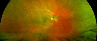
- Retinal . Appear as a result of damage to blood vessels located in the retina. The most dangerous type of hemorrhage, which even with small blood formations can lead to loss of vision.
Retinal formations, in turn, are further divided into:
- preretinal (formed between the vitreous membrane and the retina and exceeds the optic disc in size);
- intraretinal (look like small circles or streaks and are localized in the retina in areas of accumulation of retinal vessels);
- subretinal (they look like contourless spots and are formed between the layer of nerve fibers and the pigment cover, as a rule, they arise when newly formed vessels grow).
Keep in mind! Regardless of the type of hemorrhage, they quickly resolve if they are small in size and generally do not pose a threat to vision.
Significant hemorrhages are eliminated by instilling ophthalmic drops, and the largest formations can only be removed through surgery - for this purpose, laser coagulation is performed.
Eye or head injuries
In case of a severe head injury, it is imperative to visit an ophthalmologist, even if the victim does not notice any deterioration in vision. The fact is that with traumatic brain injuries, hemorrhages into the internal tissues of the eyes are possible. Redness of the proteins does not occur, but the consequences can be very serious (up to retinal detachment and complete loss of vision).
Quite often, injuries to the eye apparatus itself occur - due to the ingress of small solid particles (dust, grains of sand), chemical or thermal burns, etc. Violation of the integrity of the capillaries in such a situation cannot be avoided. If the damage is minor, the discomfort may quickly disappear, but it will still be better if the affected eye is examined by a specialist.
Source: depositphotos.com
Hemophthalmos
Most often, hemorrhages into the vitreous body occur in persons with retinal detachments or ruptures, diabetic retinopathy, acute circulatory disorders in the central retinal artery (due to thrombosis, embolism, atherosclerosis). This pathology most often affects older people.
Symptoms of hemophthalmia:
- decreased visual acuity;
- floating threads, dark spots, cobwebs in the field of view;
- the appearance of a red veil before the eyes;
- disappearance of the reflex from the fundus during ophthalmoscopy.
The disease often leads to fibrosis and destruction of the vitreous, which leads to severe visual impairment. Visual acuity may be reduced to hundredths or even light perception with correct (or incorrect) light projection. In this case, only vitrectomy, an operation to remove the vitreous, can help the patient. It should be noted that surgery is effective only in the absence of structural damage to the retina. In severe retinopathy, even surgery does not always help restore vision.
Hemophthalmos should be distinguished from pinpoint hemorrhages into the vitreous body. As a rule, they resolve after some time and do not lead to vision loss. The most unpleasant consequence of such hemorrhages is the flashing of flies before the eyes, causing a certain discomfort to the person.
Common Causes
Hemorrhages in the eye occur for various reasons, but in most cases it occurs under the influence of the following factors:
- Changes in blood pressure. With such surges, the vessels may not withstand the tension and burst, while violations of their integrity may occur in different areas.
- Infectious diseases in which nausea, vomiting and fever are observed. In these cases, overstraining of blood vessels and their rupture in the weakest areas are also observed.
- Diabetes . With this disease, the structure of blood vessels is destroyed, which begin to burst even with minimal physical exertion. And the abundance of damage in different areas can lead to serious visual impairment.
- Poor blood clotting caused by related pathologies.
- Conjunctivitis, keratitis and other ophthalmological pathologies, accompanied by profuse lacrimation.
- Mechanical damage to the eyeball , including those arising during eye surgery.
- The development of tumors in the area of the organs of vision , exerting excessive pressure on the vessels and capillaries.
- Decreased elasticity of the walls of blood vessels , which develops with insufficient levels of vitamins A and C in the body.
- Excessive alcohol consumption and smoking , leading to the expansion and contraction of the walls of blood vessels, which cannot withstand such changes and rupture.
- The development of bleeding , which is a side effect of taking certain medications.
Note! Also, blood vessels can become overstrained when working at a computer for a long time or reading texts written in small print.
If you give your eyes timely rest during such activities, such formations can be avoided, but with weakened blood vessels, minor hemorrhages are still inevitable.
Symptoms and classification
There are no nerve fibers in the internal environments of the organs of vision; when they are damaged, there are no standard clinical manifestations: discomfort, pain, itching. The only sign of the disease is a sharp drop in the level of vision, up to absolute blindness.
Clinical features of the disease make it possible to divide hemophthalmos:
- Total type - when filled with biological fluid more than three quarters. Similar problems arise after trauma to the visual organs. Symptoms are manifested by an almost complete absence of vision, with preservation of sensitivity to light. The patient cannot distinguish objects located in the field of view or freely navigate in space;
- In subtotal - the internal cavity is filled with blood from one third to three quarters. It is provoked by diabetic damage to the blood vessels of the fiber. The patient sees with the affected eye vague outlines of things and silhouettes of people;
- Partial – when less than a third of the vitreous glass is filled with blood. It is one of the frequently recorded types of hemorrhage; it occurs under the influence of diabetes mellitus, retinal detachment, and hypertension. Characterized by flashing black dots, reddish stripes in front of the eyes or nebula.
This form affects both organs as an exception; mainly one eyeball is affected. The main goal of therapy is observation without specific treatment; partial forms disappear spontaneously.
If primary symptomatic manifestations of hemorrhage in the scleral area appear, the patient should immediately seek medical help. Timely identification of the source of the deviation will allow you to prescribe an adequate course of therapy and prevent the formation of complications.
How to treat and what to do in case of hemorrhage?
Subconjunctival hemorrhages do not require therapeutic or drug treatment.
But at the same time, the ophthalmologist can prescribe medications that relieve painful symptoms.
If swelling or inflammation occurs in the area of capillary ruptures, appropriate medications may be prescribed.
If the cause of hemorrhages is infectious ophthalmological pathologies, it is possible to prescribe antibacterial solutions.
Remember! In any case, such treatment of hemorrhage in the eye lasts no more than two weeks and is characterized by favorable prognoses that do not imply the development of any complications.
The absence of painful symptoms suggests instillation of ophthalmic drops if the hemorrhage does not resolve for at least a week.
The most common of these drugs are:
- Visine. Decongestant and vasoconstrictor drug for local use. It is characterized by rapid action and is not absorbed into the systemic circulation, eliminating the development of side effects. It has proven itself well as a drug against the development of allergic reactions that provoke ruptures of blood vessels in the eyes.
- Emoxipin. A drug that activates the regenerative mechanisms of the vascular system. The product also strengthens the walls of blood vessels, reducing their permeability. Additionally, it stimulates blood circulation processes and ensures access of a sufficient amount of oxygen to the vessels, as a result of which the resorption of formations occurs in the shortest possible time.
- Taufon . Taufon is a drug similar in mechanism of action to emoxipine, which helps restore intraocular pressure and reduces the risk of vascular rupture. In the event of hemorrhage formation, it promotes rapid healing, having a positive effect on the regenerative systems of the eye.
What drops to drip
Hemorrhages in the eye area often require the use of special drugs that eliminate the pathological process and restore the normal state of the visual organs. Frequently prescribed medications include drops:
- Vixipin.
- Albucid.
- Taufon.
- Potassium iodide.
- Emoxipin.
- Diclofenac.
- Hyphenation.
Medicines in drip form are indicated for patients whose hemorrhage does not resolve within 6–7 days and is accompanied by other negative symptoms (pain, development of an infectious process).
Vixipin
This domestic drug is recommended for patients in whom hemorrhage occurred in the anterior chamber of the eye or in the sclera. The product containing methylethylpyridinol hydrochloride as the main component is prescribed no earlier than 18 years of age.
The medicine helps reduce capillary permeability, strengthen vascular walls, stabilize the cell membrane, and normalize blood viscosity. The hyaluronic acid present in the solution ensures complete hydration of the cornea, eliminates discomfort, and improves the tolerability of the drug.
Instillations are carried out 2-3 times a day, introducing 1-2 drops of medication into the affected eye. The duration of the course depends on the severity of symptoms, type of diagnosis and can take from 3 to 30 days. You can buy Vixipin for 198–215 rubles.
Albucid
The active ingredient of this product is sulfacetamide. The main action of Albucid is antimicrobial, bacteriostatic, anti-inflammatory. The medicine is considered safe and can be used even during the neonatal period.
The drug helps fight hemorrhages in the eye area caused by various factors. Adult patients are prescribed a 30% solution, and a 20% solution is prescribed for children. The medication must be instilled 3–5 times during the day, 2–3 drops each. The frequency of use is reduced as symptoms weaken. The average price of the drug in the Russian Federation is 55–62 rubles.
Taufon
The main ingredient of Taufon is the metabolic compound taurine. The medicine, produced in Russia, is intended for the treatment of persons over 18 years of age. The product accelerates the healing of damage in the eye area, improves the functioning of cell membranes, promotes better metabolic and energy processes, and better conductivity of nerve fibers.
The medication is administered intraconjunctivally, 1 drop four times a day. For hemorrhages in the organs of vision, a course with this remedy can last about a month. You can buy the drug for 112–130 rubles.
Potassium iodide
The domestic eye drop product contains potassium iodide as a base. Instillations with potassium iodide are resorted to in the development of various ophthalmological pathologies, including hemorrhages in the eyeball area.
Thanks to instillation, the following results can be obtained:
- increase lipoprotein concentration;
- reduce blood viscosity;
- expand the lumens of the vascular walls.
It is recommended to use this remedy for the treatment of children no earlier than 2 years of age.
A 3% solution is intended for injection into the eye area. The solution should be instilled into the conjunctival sac, 2 drops, 3-4 times a day. The average duration of therapy is 10–15 days. Eye drops cost about 150 rubles.
Emoxipin
The medicine produced in the Russian Federation includes methylethylpyridinol. The medication is prescribed to patients with intraocular hemorrhages, thrombosis in the central vein of the retina, and glaucoma. The drug promotes effective protection of the organs of vision after laser coagulation and sunburn.
To undergo treatment with this drug, the patient must reach the age of majority (18 years).
The solution should be used once a day, in a volume of 0.2–0.5 ml in the area of the affected eye. The duration of treatment may take from 10 to 30 days. The drug has a moderate cost - about 144 rubles.
Diclofenac
This product is produced by pharmaceutical companies in different countries - Russia, Germany, India, Slovenia. The main ingredient of the instillation solution is sodium diclofenac (1 mg per 1 ml). The medicine belongs to the group of non-steroidal anti-inflammatory drugs.
The drug is widely used by ophthalmologists for various eye disorders, including hemorrhages. To perform instillations, a 0.1% solution is used. The dosage and frequency of administration are determined by the doctor individually. Typically, patients are prescribed 1-2 drops of medication into the problematic organ of vision, up to 5 injections during the day.
Diclofenac eye drops are used to a limited extent for the treatment of children under 12 years of age and the elderly. The price of the product is affordable - 45–55 rubles. per bottle with 10 ml of solution.
Hyphenleuse
This domestic drug, which helps with hemorrhages in the eye area, can be bought for 40–47 rubles. The medicine contains hypromellose and auxiliary components. The main property of Defislez is the protection of the corneal epithelium.
Instillations with this medication can be carried out no earlier than 8 years. The frequency of instillation of the solution reaches 4–8 times a day. A single dose for bleeding in the eye area is 2 drops. Treatment is carried out over 2–3 weeks.
Use of ointments
Today, you can purchase various products at the pharmacy that will help you quickly get rid of bruises and swelling under the eye. Such drugs can be purchased without a prescription:
- "Troxevasin";
- "Venitan";
- "Heparin ointment";
- "Bruise off."
These drugs help reduce pain at the site of impact and promote resorption of the hematoma in a short period. They need to be used several times a day in small quantities per application.
Do not use ointments on an open wound. They can only be effective in cases of bruising and swelling without bleeding wounds. To use these medications to treat children, you need to consult a doctor in advance about this possibility. Not all products are approved for use by children under 12 years of age.
Features of hemorrhage in the eye in newborns
You should know! In newborns, hemorrhages in the eyes are more common than in adult patients or older children.
This is due to the presence of certain additional factors:
- use of dinoprostan during complicated childbirth;
- use of a vacuum extractor , necessary in some cases for the same reasons;
- performing a CT scan while the baby is in the womb.
All this has a negative impact on the vessels, which are not yet strong enough to withstand such loads.
Physical overexertion
The eye capillaries are very sensitive to physical stress. Ruptures of the walls of blood vessels occur in athletes who are too keen on strength training. Also at risk are people who are engaged in predominantly sedentary work and are poorly adapted to physical work: they may develop hemorrhages in the sclera even when bending the body sharply or trying to lift a relatively light load.
In women, the capillaries of the eyes are often injured during childbirth due to enormous physical and nervous stress. The reasons for the redness of the whites of the eyes in a small child when he cries for a very long time and loudly are similar.
Source: depositphotos.com
Disease prevention
It is impossible to guarantee protection against the formation of hemorrhages in the eye, since the number of predisposing factors is too large.
But such violations can be avoided by following the following general recommendations:
- limit alcohol consumption and reduce the number of cigarettes smoked;
- monitor the state of the immune system and, if necessary, take immunostimulating drugs, as well as add healthy foods containing vitamins and beneficial microelements to the diet;
- avoid sudden movements and heavy physical exertion if there are problems with blood vessels, especially avoid sudden movements of the head and bending forward;
- patients with diabetes mellitus should be especially careful about monitoring blood glucose levels;
- in case of hypertension, take measures to normalize blood pressure;
- undergo regular examinations with an ophthalmologist.