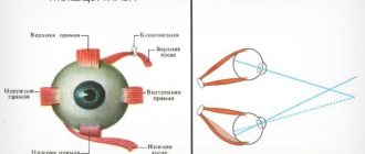
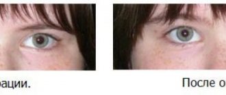
Strabismus is one of the most common pathologies of the visual organs. The condition is very complex and unpleasant, negatively affects a person’s quality of life, and requires an integrated approach to treatment. Doctors usually prescribe physiotherapy and the use of drops. The special Synoptophore device is highly effective in treating strabismus. Regular exercises with it improve the general condition of the retina and restore binocular vision. Synoptophore can also be used to improve the functionality of the visual system as a whole.
What is a synoptophore, its purpose in ophthalmology
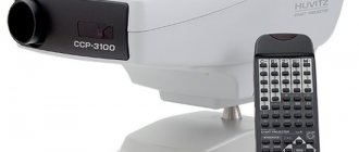
Synoptophore (also known as synoptiscope) is a diagnostic ophthalmological device used to clarify the diagnosis and complex treatment of strabismus. Its use makes it possible to correct disorders of the motor function of the eye, calculate the exact angle of deviation of vision from the norm, detect scotomas (so-called “blind” areas that are in no way connected with the peripheral boundaries of the visual system), and obtain reliable information about fusion reserves and the general condition of the retina. This article will tell you about the treatment of strabismus in adults.
The synoptiscope is a modern analogue of the ambiloscope (with its help, binocular vision was previously determined and strabismus was diagnosed). These devices are available in most modern clinics (but private, not public), since without them it is extremely problematic to diagnose and treat strabismus. The device can be used to treat children without age restrictions, and to treat strabismus if there are contraindications for corrective surgery. Read about the causes of alternating, concomitant strabismus in children and adults here.
The synoptophore makes it easier to perform orthoptic exercises. The goal of the training is to restore simultaneous vision, treat with cattle, and increase the mobility of the eyeballs.
conclusions
- Identifying strabismus is a complex process. To make a full diagnosis, you need to undergo a series of medical tests and collect a complete medical history of the patient.
- When identifying pathology, congenital and acquired factors are taken into account, because In most cases, strabismus is a hereditary disease.
- To correctly prescribe treatment, you need to determine the type and angle of strabismus. For this, the following methods can be used: identification of the strabismus angle according to Hirshberg, study with Synoptophore, Belostotsky-Friedman color test, Worth test, raster haploscopy.
Areas of use
The synoptiscope is used for diagnosis and treatment of strabismus (heterophoria, convergent, divergent), and correction of binocular vision disorders in children and patients who cannot undergo surgery.
The device can be used for preventive purposes to perform appropriate exercises.
All features of the device:
- With regular training, scotomas disappear completely.
- Binocular vision is normalized.
- The fusion reserves of the eye develop and increase.
- The mobility of the eyeballs increases.
- The eye muscles develop evenly (important when correcting strabismus).
- Binocular vision is strengthened and stabilized.
- With the help of Sinopftovor, you can easily understand how well the patient’s eyes are adapted to binocular vision. In parallel with the general diagnosis, the doctor assesses the general condition of the retina and its functioning, and selects a therapeutic regimen if there are certain abnormalities.
Sinopfluor is also used to diagnose astigmatism, myopia, and hypermetropia.
What to look for when making a diagnosis
When determining the diagnosis, it is necessary to pay attention to the medical history, namely:
- The time when strabismus appeared indicates the etiology. The earlier it occurs, the greater the likelihood of surgical intervention. With late onset, the chances of an accommodative component increase.
- Angle variability is an essential criterion, since the periodic appearance of strabismus makes it possible to understand that binocular vision has been preserved.
In the case of alternating strabismus, symmetrical visual acuity is assumed in each eyeball.
- The general condition or abnormal development is of great importance. For example, attention is drawn to the frequency of strabismus in a child suffering from cerebral palsy.
- The specialist needs to check the birth history, become familiar with the indicators during pregnancy, the baby’s weight at birth, pathologies during intrauterine development, or during childbirth.
- Hereditary history also has a strong influence, since in most cases this disease is a congenital pathology.
- As part of testing sensory functions, the level of stability of binocular vision is determined, its acuity, and the presence or absence of bifoveal fusion are revealed. Attention is drawn to the functional scotoma of suppression, the nature of diplopia, and fusion reserves.
When checking motor functions, the doctor analyzes the degree of mobility of each eyeball, characterizes the deviation, and determines the complexity of disturbances in the work of the extraocular muscle of each eye separately.
Principle of operation
The operating principle of the device is extremely simple. It uses paired pictures, which are illuminated using special lamps. Alternately turning off and on the lamps allows the patient to focus on a specific image; switching of devices can be done regularly, taking into account the device, automatically or manually. When the patient focuses his vision on the pictures, the ocular muscles begin to develop more actively and become involved in the work process. Since muscles can change at any age, synoptiscopes are used to correct strabismus, including in adults. Read about eye surgery to correct strabismus in children and adults here.
During training, the load on the eyes is distributed evenly, so the development of complications is excluded.
Strabismus is difficult to treat, so you will have to work with Synoptophore regularly and for quite a long time. In addition to exercises in the clinic, home workouts are recommended. You can buy Synoptophore without any problems for independent use. Be sure to carefully read the instructions so as not to harm yourself.
Please note that different test objects are used during therapy:
- for combination;
- for merging;
- for stereoscopy.
When diagnosing strabismus, each eye must look at the pictures in turn using one test, after which the direction of the optical axis is checked.
If the position of the axis is parallel, the picture will be blurry, and a diagnosis of strabismus is made (when vision is normal, the image is clear). People with visual impairments begin to see the image clearly after changing the angle of the optical axis. During home sessions with Synoptophore, you need to use test pictures from different angles, without going beyond the range of 45 degrees (approximately). To restore normal visual dynamics, the image is made either paler or brighter. The action is available in manual and auto mode.
How will the procedures be carried out?
After the prescribed treatment, the child’s parents bring him to the center so that he does not worry and gets acquainted with the situation and the equipment. Next time, you can leave the baby on his own during the procedures. As practice shows, without the presence of parents, children behave responsibly and find their bearings faster. Classes can be attended by referral during the specified days. After a couple of procedures, the child will already have excellent orientation and begin treatment with devices with minimal help from a nurse.
It is better to start treating a child from an early age, usually from 4-5 years old, but it is possible earlier. But not every child can sit at each device for several minutes. The treatment itself is completely painless and leads to improved vision and correction of strabismus in almost 70% of all cases. At the end of such procedures, the child can be sent to school or for other errands, because after them there will be no need to carry out any manipulations, you will not have to abstain from anything.
Device structure
A synoptiscope is a device that has a complex structure and a well-functioning operating scheme. It includes:
- a pair of pipes, movable, equipped with an adjustment function;
- lenses to which the patient directs his gaze when working with the device;
- two mirrors that reflect images into the lenses in the desired position;
- special sockets that are located opposite the mirrors and allow you to install images selected for correction;
- lamps that illuminate pictures and allow the patient to view them through lenses.
Diagram of the device for a better understanding of the principle of its operation.
Each device comes with cards with pictures, which are located in the slots. The pictures are divided into two parts, and each part represents half of one whole. Thanks to this division, the doctor can determine the severity of the disease. In the Synoptophore, each eye is shown its own picture, then the images are combined. Taking into account the distance at which the objects merged, we can talk about the severity of the disease. Read what penalization is in ophthalmology at this link.
Technology of use, is it possible to use the device at home?
Let's consider the basic principles of using the device.
When diagnosing
After connecting the device, the patient positions the tubes so that the images line up.
You need to move the eyepieces yourself or monitor their movements and inform the doctor about all alignments.
Tube scales demonstrate each eye's deviation from normal. The sum of the divisions determines the calculation of the subjective squint angle. To determine the objective angle, you need to create lighting for the drawings. The position of the eyes indicates the current angle of strabismus, the immobility of the eyeballs indicates that the axes of the visual organs are directed correctly. If the axes with the eyepieces do not coincide, adjustment movements develop - towards the temple, nose, down or up. If there are adjustment movements, the doctor turns on the eyepieces manually; the tubes should move unnoticed by the patient when the lighting is turned off. When all movements stop, the angles will need to be summed up - this will be the objective angle of strabismus. This material will tell you why strabismus occurs in children and adults.
During treatment
A treatment session on Synoptophore should begin with the installation of test objects. We need objects for combining, merging, and stereoscopy. To determine the degree of strabismus, the eye is allowed to see 1/2 of the pattern. If the optical axes are directed parallel, the picture will merge into one whole, and a person will see the picture without distortion.
Home hardware treatment involves placing patterns at an angle of ±45°.
To increase the effectiveness of therapy and completely restore vision, change the light intensity. You can add vibrations and flashing lights, but only after consulting a specialist. The device allows you to customize the backlight and adjust vibrations automatically or manually.
Homework involves the use of glasses. A child or adult sits in front of the device, then the distance between the pupils is adjusted, and alternating irritation of the eyes with a light pulse begins. In situations where this scheme of work is not effective enough, merging of test objects is prescribed. First, large pictures merge, then small patterns. A pronounced therapeutic effect can be achieved after 2-4 dozen sessions. They are carried out daily, each lasting about 15 minutes.
Fusion is used to develop fusion reserves. The patient looks at the whole picture, then the tubes are separated, the image is separated and brought together again. You need to constantly hold the whole picture with your eyes. Fusion reserves refer to the mechanisms that are responsible for combining monocular images into a single whole.
Principles of treating strabismus using synoptophore
Since the synoptophore is a device that is used not only for diagnosis, but also for the treatment of strabismus, many doctors recommend purchasing a device for home use. This will ensure continuity of treatment and increase patient comfort, which will promote adherence to therapy. True, the difficulty often lies in the fact that the price of synoptiscopes starts from 50 thousand rubles, which for some families is an unaffordable amount.
Since not everyone has the financial means to purchase a device, many clinics offer orthoptic therapy sessions using a synoptophore.
Even before starting home treatment using the device, you must consult a doctor. The specialist will introduce the parents (if we are talking about a child) or the patient himself to the principles of operation of the synoptophore, establish the interpupillary distance that will need to be transferred to the device, and draw conclusions about the severity of the disease.
At home, when practicing with a synoptiscope, it is recommended to use glasses. It is better to give preference to an automatic device, which will independently carry out the light rarefaction of the visual system.
Changing the light intensity and oscillating the device often helps improve the effectiveness of therapy. As with turning the backlight on and off, the oscillation can be either automatic or manual, depending on the device.
To increase the fusion reserves of the visual organs, the fusion technique is widely used. Following it, the patient first looks at the whole picture, and then the eyepieces move to the side, separating the image. The patient's task is to maintain a solid image with his eyes, regardless of the retraction of the eyepieces.
Operating principle of the device with pictures
If strabismus does not respond to light therapy, then after consultation with a doctor it can be replaced with therapy using the method of fusion of test objects. This approach usually produces results after 30-40 daily sessions of at least 15 minutes each. Therapy begins with large images, gradually changing them to small patterns.
