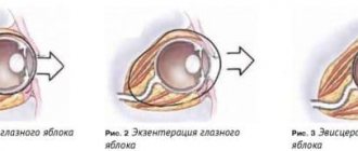
Indications for examination
Biomicroscopy is prescribed for diagnosis:
- pathologies of the conjunctiva of various etiologies, incl. provoked by allergic reactions, inflammatory phenomena;
- eyelid injuries;
- inflammation, tumors of the eyelids;
- abnormal conditions of the sclera: keratitis, corneal dystrophy, etc.;
- inflammation and other pathologies of the iris;
- glaucoma;
- cataracts;
- the presence of foreign bodies in the cornea;
- endocrine diseases, due to which complications appear on the organs of vision.
This type of examination is used to monitor the effectiveness of therapy for eye diseases and preparation for surgical interventions.
Biomicroscopy can evaluate:
- health of the eyelids and conjunctival membrane;
- the condition of the cornea, its structure and thickness;
- the amount of fluid circulating in the anterior chamber of the eye;
- depth of the anterior chamber;
- transparency of the iris and lens;
- Parameters of the vitreous body - transparency, the presence of cloudy areas, blood.
The advantage of the examination is its ability to detect various eye pathologies in the early stages. The doctor can also accurately determine the degree of moisture in the eye and the amount of water in the anterior chamber, which is important for the early diagnosis of ophthalmological pathologies. During the examination, the doctor gets a clear picture of the eye.
There are practically no contraindications for the procedure. In exceptional cases, the study in question is not carried out if the patient has acute mental pathologies or aggression. Eye diagnostics are not carried out in patients who are intoxicated under the influence of drugs or alcohol. Restrictions are associated with the patient’s inability to sit motionless for a long time.
How does the procedure work?
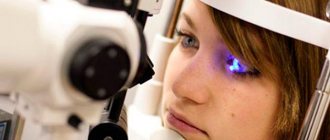
To check the ocular structures, the patient sits opposite the ophthalmologist, in front of the slit lamp. It is necessary to fix the chin and forehead on special supports. The doctor is on the other side of the lamp. The specialist sets the lighting and the width of the light beam. After this, a slit lamp beam is directed into the patient’s eye.
Looking through a microscope, the doctor identifies abnormalities in the functioning of the eye. Although the eye examination is painless, the patient may experience discomfort in the eyes from bright light and watery eyes. The procedure lasts about 10 minutes. The less the patient blinks, the faster and better the ophthalmologist will perform the examination.
If the fundus is examined, the patient is instilled with mydriatic drops that dilate the pupil. If the cornea is examined, dye is first instilled, and then regular eye drops are used to wash away the dye from unaffected areas. The damaged areas are stained for a short time, allowing the ophthalmologist to draw conclusions.
Some patients are afraid of bright light, so before biomicroscopy their eyes are instilled with an anesthetic. It is even more difficult to perform biomicroscopy on a small child. Children's eyes are checked while they sleep. In this case, the child is positioned horizontally so that he does not accidentally move.
Biomicroscopy is contraindicated in patients who are intoxicated, as well as with mental disorders accompanied by unpredictable behavior.
What is the methodology based on?
This examination is performed using a slit lamp. It includes a light source, a stereoscopic microscope and a lens. The device also has a slit diaphragm, which creates the lighting slits necessary for examining the eye.
The body of a special microscope has a device that allows you to magnify the image several tens of times. Above the microscope there is a negative lens of 60 diopters, which allows you to examine the fundus of the eye. Diagnostics of the organ of vision is carried out in complete darkness. This is the only way to create the necessary contrast between the bright lamp and the darkened areas of the eye.
By focusing the light, the doctor is able to see all surfaces of the organ of vision and its structures. When an inflammatory process or clouding is detected, the ophthalmologist sees the depth and localization of the changed area.
If light is focused onto the lens of the eye, the lens, the ophthalmologist sees a biconvex body. It may contain pathological stripes and opacities, which indicate the development of the initial stage of cataracts.
An ophthalmologist also has the opportunity to study the retina, the optic nerve head. Diagnostics of the vitreous allows one to detect characteristic symptoms of inflammatory processes in the eyeball. Biomicroscopy allows you to characterize the condition of the iris and conjunctiva.
Advantages of biomicroscopy:
- non-contact examination;
- accuracy of the results obtained;
- the ability to examine the eye at different depths;
- efficiency;
- possibility of conducting research on an outpatient basis:
- low cost;
- almost complete absence of restrictions;
- can be performed for any eye pathology.
Biomicroscopy: basic concepts
Biomicroscopy is an examination of the internal state of the eyeball using a medical device called a slit lamp. Includes a wide range of complex imaging techniques for pathologies of varying origin, texture, color, transparency, size and depth.
The slit lamp allows for detailed microscopic examination of the eye
A slit lamp is an instrument consisting of a high-intensity light source that can be focused to direct a thin strip of light into the eye through various filters that provide the location and size of the slit. It is used in combination with a biomicroscope, which, together with the illuminator, is mounted on one coordinate table. The lamp facilitates inspection of the anterior and posterior segments of the human eye, which include:
- eyelid;
- sclera;
- conjunctiva;
- iris;
- natural lens (lens);
- cornea;
- vitreous body;
- retina and optic nerve.
The slit lamp is equipped with a diaphragm that forms a slit up to 14 mm in width and height. A binocular microscope includes two eyepieces and an objective (magnifying lens), the optical power of which can be adjusted using a dial that changes the magnification factor. The range of gradual increase is from 10 to 25 times. With an additional eyepiece - up to 50-70x.
Binocular slit lamp examination provides stereoscopic, magnified images of ocular structures in detail, allowing anatomical diagnoses to be made for a variety of ocular conditions. The second, hand-held lens is used to examine the retina.
Biomicroophthalmoscopy is aimed at determining the state of the above structures and optical environments by creating a contrast between illuminated and unlit areas. The necessary conditions are provided by a microscope equipped with two eyepieces, magnifying the image tens of times. It is equipped with a lighting system consisting of a lamp that provides a narrow beam of light and light filters.
For a full examination with a biomicroscope, there are various methods of slit lamp illumination. There are six types of basic lighting options:
- Diffuse illumination - examination through a wide aperture using glass or a diffuser as a filter. It is used for a general examination to detect the localization of pathological changes.
- Direct focal illumination is the most commonly used method, which involves observation using an optical slit or direct focal beam. A thin or medium width slit is directed and focused on the cornea. This type of illumination is effective for determining the spatial depth of ocular structures.
- Specular reflection, or reflected illumination, is a phenomenon similar to the image visible on the sunny surface of a lake. Used to assess the endothelial contour of the cornea (its inner surface). To achieve a mirror effect, the tester directs a narrow beam of light to the eye from the temple side at an angle of about 25-30 degrees to the cornea. A bright area of specular reflection will be visible on the corneal epithelium (outer surface).
- Transillumination (transillumination), or examination in reflected (transmitted) light. In some cases, illumination with an optical slit does not provide sufficient information or is simply impossible. Transillumination is used to examine transparent or translucent structures - the lens, the cornea - by reflecting rays from deeper tissues. To do this, highlight the background of the object under study.
- Indirect lighting - a light beam passing through translucent fabrics is scattered, simultaneously highlighting individual places. Used to identify pathologies of the iris.
- Scleral Scattering - With this type of lighting, a wide beam of light is directed at the limbal area of the cornea (the edge of the cornea, where it meets the sclera) at an angle of 90 degrees to it to create a light scattering effect. In this case, a certain halo appears under the cornea, which illuminates its anomalies from the inside.
The slit lamp makes it possible to study the structural parts of the cornea:
- epithelium;
- endothelium;
- posterior border plate;
- stroma.
Read also Stye on the eye - causes, symptoms and treatment at home
And also - determine the thickness of the transparent outer shell, its blood supply, the presence of inflammation and edema, and other changes caused by injury or dystrophy. The study allows you to study in detail the condition of scars, if they exist: their size, adhesions with surrounding tissues. Biomicroscopy reveals tiny solid deposits on the back surface of the cornea.
If a pathology of the cornea is suspected, the doctor will additionally prescribe confocal microscopy - a method of assessing the morphological changes of this organ using a special microscope with a magnification of 500 times. It allows you to study in detail the layer-by-layer structure of the corneal epithelium.
During biomicroscopy of the lens, the doctor examines the optical section for possible clouding of its substance. Determines the location of the pathological process, which often begins precisely at the periphery, the state of the nucleus and capsule. When examining the lens, almost any type of lighting can be used. But the most common are the first two: diffuse and direct focal lighting. They are usually carried out in this order. The first type of lighting allows you to evaluate the general appearance of the capsule and see foci of pathology, if any. But for a clearer understanding of where exactly the “breakdown” occurred, it is necessary to resort to direct focal lighting.
Examining the vitreous body using a slit lamp is a challenging task that not every novice in ophthalmology can handle. The vitreous body has a jelly-like consistency and lies quite deep. Therefore, it weakly reflects light rays.
Biomicroscopy of the vitreous body requires acquired skill
In addition, the study is hampered by a narrow pupil. An important condition for high-quality biomicroscopy of the vitreous body is preliminary medicinal mydriasis (pupil dilation). The room where the inspection is carried out should be as dark as possible, and the area under study, on the contrary, should be quite brightly lit. This will provide the necessary contrast, since the vitreous body is a weakly refractive optical medium that slightly reflects light. The doctor uses mostly direct focal lighting. When examining the posterior parts of the vitreous body, it is possible to study in reflected light, where the fundus of the eye plays the role of a reflective screen.
Focusing light on the fundus allows one to examine the retina and optic nerve head in an optical section. Early detection of neuritis or swelling of the nerve (congestive papilla), retinal tears helps in the diagnosis of glaucoma, prevents optic nerve atrophy and decreased vision.
Read also: How to protect yourself from vision loss
The slit lamp will also help determine the depth of the anterior chamber of the eye, detect cloudy changes in moisture and possible impurities of pus or blood. A wide selection of lighting types thanks to special filters allows you to clearly study the vessels, detect areas of atrophy and tissue ruptures. Biomicroscopy of translucent and opaque tissues of the eyeball (for example, conjunctiva, iris) is less informative.
Slit lamp device: video
Types of biomicroscopy
There are different types of eye biomicroscopy depending on the combination of lighting options:
- with direct focused light - in order to study the transparency of the optical medium of the organ of vision and identify areas of turbidity in it;
- with reflected light - to detect edema and the presence of foreign bodies;
- with indirect focused light - for detailed detection of changes;
- indirect diaphonoscopic examination is used to detect the exact location of pathological disorders in the structures of the eyeball.
Biomicroscopy of endothelium
It is carried out using a precision microscope connected to a computer. This device makes it possible to examine with microscopic maximum clarity all layers of the cornea, and especially its inner layer - the endothelium. Thus, already in the early stages, it is possible to determine any pathological changes in the cornea. Therefore, the following groups of people need to regularly undergo such diagnostics:
- using contact lenses;
- after various eye surgeries;
- for diabetics.
Carrying out biomicroscopy
When performing eye biomicroscopy, the patient should sit opposite the ophthalmologist and place his chin on a fixed stand in the ophthalmological lamp. This device creates a narrow beam of light that hits the eye directly.
If it is necessary to examine the vitreous body, a small amount of solution is instilled into the eye to increase the diameter of the pupil. In most cases, Tropicamide is used for this.
When examining the integrity of the cornea, a dye is injected into the eye. It interacts with the tissues of the eye and is then washed off. If there are affected areas of the eye, then particles of the coloring substance remain on them.
During an eye examination, the doctor turns off the light in a dark room. After this, he sits opposite the patient on the side of the ophthalmic lamp. During the procedure, the brightness of the light directed into the eyes is adjusted so that all affected areas can be covered.
The patient must remain motionless during the diagnostic process. As much as possible, he should refrain from blinking or do so less frequently. If it is impossible to maintain a certain position of the eyes, anesthetic drops are instilled into them.
The duration of biomicroscopy of the anterior segment of the eye takes no more than 10 minutes. After this, the person does not need restorative measures. Any side effects from drugs that are instilled into the eyes have a short-term effect.
For children under three years of age, the eye biomicroscopy procedure is performed only in a supine position. Drugs are instilled into the eyes to reduce the sensitivity of the cornea.
Decoding the results
After completing the diagnostic measures, depending on the readings of the diagnostic device, the doctor assesses the condition of the eye, and if pathology is present, he draws up a general picture of the examination.
In glaucoma, the central zone of the cornea is slightly clouded due to the presence of proteins (proteins) on its surface. An expansion of the scleral openings and a pronounced increase in the choroid are detected.
With cataracts, clouding of the peripheral areas is detected. The size of the optical section of the eye is significantly reduced. So-called watery cracks appear. If the doctor is dealing with advanced cataracts, then the beam is completely reflected from the affected lens, since the latter does not completely transmit light.
If there are foreign particles in the eyes, the blood vessels in the eye dilate significantly. Foreign bodies are detected in the eyes as yellowish dots. The procedure helps determine the depth of the foreign body.
With keratitis, proliferation of blood vessels occurs. There are small blisters on the outer layer of the cornea. If purulent inflammation occurs, then there is a defect in the central part of the stratum corneum. In advanced cases, an ulcer appears at the site of the defect.
If an ulcer appears in the thickness of the iris, this indicates the development of the congenital disease coloboma. When tumors occur, adjacent structures in the eyes become displaced. Proliferation of the choroid is detected.
Relevance of the problem
Visual acuity—the ability to distinguish between letters, numbers and objects at a distance—is essential for many tasks, from recognizing a friend in a room to driving a car. Researchers previously assumed that visual acuity is primarily determined by the optics of the eye and the anatomy of the retina. Unlike a stationary camera, human eyes are constantly moving, analyzing new fragments of the external environment using the retina.
Impairments in eye movement and vision often coexist in some disorders. For example, eye movements are poorly recorded in patients with visual impairments such as dyslexia and Parkinson's disease.
Useful video
More useful information about ophthalmoscopy can be found in these videos:
Biomicroscopy is an examination that allows you to detect various eye pathologies. It is painless, highly informative and has no side effects.
Author's rating
Author of the article
Alexandrova O.M.
Articles written
2031
about the author
Was the article helpful?
Rate the material on a five-point scale!
If you have any questions or want to share your opinion or experience, write a comment below.
Materials and methods of examination
To determine whether fixed eye movements affect visual acuity, Rucci and Intoy studied how these tiny eye movements affect a person's performance on one of the most common visual acuity assessments: the Snellen chart. The Snellen eye chart consists of 11 lines of block letters, with each line showing an increasing number of letters in a decreasing size. During a vision test, a person is asked to read letters in lines. If a person has normal visual acuity, he is able to see 8 lines on a diagram from a distance of 600 meters.
To measure visual acuity in the absence of fixed eye movements, the researchers stabilized a Snellen chart on the observer's retina, constantly updating the display according to eye movements, counteracting the effects of movements. That is, unlike normal viewing, where visual input changes with eye movements, the scientists ensured that the Snellen chart image remained stationary on the retina.