The vitreous body (vitreum, VT) is a gelatinous substance that makes up approximately two-thirds of the volume of the eye.
Located between the lens and the retina in the front of the eye, in the back of the eye.
The gel-like clear substance is composed of collagen, hyaluronic acid and water and plays a role in modulating eye growth during development.
Vitreous hemorrhage is defined as the presence of blood in the vitreous cavity, which is the space lined by the posterior retina and anterior ciliary body, zonular fibers and posterior capsule of the lens. The prevalence of the pathological condition is 7 cases per 100 thousand.
In most cases, normal vessels bleed only in the setting of trauma or PVD. Conversely, patients with a history of chronic retinal ischemia (eg, diabetic retinopathy) may develop abnormal fragile vessels (ie, neovascularization) with a tendency to bleed.
Causes
Diabetic retinopathy (DR) is considered one of the most common causes of intraocular hemorrhage (IOC). DR affects 5.3 million people and is the most common cause of this pathological condition in the vitreum, accounting for 31–54% of all cases.
Vascular damage in diabetic retinopathy causes chronic cerebral ischemia, promoting the release of angiogenic factors from cells and leading to the growth of new blood vessels (ie, neovascularization).
Other common proliferative retinopathy:
- Occlusion of the central retinal vein accounts for 4–16% of all CT hemorrhages. It is the second most common retinal vascular disorder after diabetic retinopathy. The pathogenesis of venous occlusion is multifactorial, including arterial disease, mechanical compression, degeneration of the venous walls, and hypercoagulability leading to venous occlusion. Venous stasis damages the venules (small blood vessels) that allow blood to flow into the vitreum.
- Sickle cell retinopathy is a rare cause of hemophthalmos. Accounts for 0.2–6% of all cases. It causes a characteristic fan-shaped neovascularization in front of the retina, which bleeds into the vitreous.
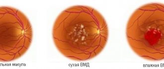
- Age-related macular degeneration (AMD) is the culprit in 0.6–4% of cases. As AMD progresses, Bruch's membrane, which is the innermost layer of the uvea, begins to break down. The breakdown products stimulate angiogenesis of the underlying choroid. New vessels may penetrate Bruch's membrane and bleed into the retina and vitreum.
Direct eye trauma is a common cause of CT hemorrhage in people under 40 years of age. In the setting of penetrating trauma, blood extravasates into the CT from adjacent tissues or injures intraocular structures along the path of the foreign body. As a result, hemophthalmos occurs.
Terson syndrome is a rare cause of CT hemorrhage. It is responsible for less than 1% of all cases. Terson syndrome occurs when hemophthalmos is associated with subarachnoid or intracerebral hemorrhage.
The following diseases can cause hemophthalmos:
- age-related macular degeneration (“wet” AMD) (2%);
- Coat disease;
- ocular ischemic syndrome;
- macroaneurysm (2%);
- hypertensive retinopathy;
- venous stasis retinopathy;
- retinal arterial tortuosity;
- cataract extraction with or without IOL;
- secondary IOL implantation;
- trabeculectomy;
- hypertensive crisis;
- swan syndrome (neovascularization of cataract wound);
- posterior vitreous detachment (8%);
- retinal tear (30%);
- complications of retinal surgery;
- malignant melanoma of the choroid;
- retinoblastoma;
- cavernous hemangioma;
- vasculitis;
- infectious choridopathy;
- Behçet's disease;
- Ile disease;
- endophthalmitis;
- uveitis;
- syphilitic retinitis;
- idiopathic intracranial hypertension;
- thrombocytopenia;
- leukemia;
- von Willebrand's disease.
Some drugs, in particular anticoagulants, also contribute to the development of pathology.
Risk group
The risk group includes persons with insulin-dependent/non-insulin-dependent diabetes and hypertension at the decompensation stage. Also, the risk of hemophthalmos increases in patients with damage to the heart muscle caused by an acute circulatory disorder or a history of cerebrovascular accident.
Most often, people over 40 years of age suffer from vitreous hemorrhage. The disease can develop in infants with shaking syndrome.
What is hemophthalmos?
Most of the eyeball is occupied by the vitreous body (vitreum), a gel-like transparent substance that conducts light. The structure does not have nerve endings or blood vessels, but is closely associated with the inner membrane of the eye that has them - the retina. Rupture of its capillary network entails hemorrhage into the vitreum (formation of hemophthalmos).
Hemorrhages can resolve, but this process is extremely slow and is successfully completed only with minor hemorrhage. In most cases, the disease requires consultation with an ophthalmologist and treatment, since the pathogenesis will only worsen over time and can lead to retinal detachment or even blindness.
Classification
Ophthalmologists classify the disease as follows:
- Partial hemophthalmos OD - develops due to hemorrhages of damaged vessels. Therapy is possible at home, but on the condition that the patient gets more rest and follows all the ophthalmologist’s recommendations. The partial form develops in patients with DR, atherosclerosis, hereditary hemoglobinopathy and against the background of retinal detachment or rupture. With this form of the disease, the affected person sees cobwebs and shadows before the eyes, black dots and stripes in the field of vision.
- Total form - blood occupies ¾ of the vitreum volume. The patient sees only at the level of light perception. He is not able to distinguish objects and independently navigate in space.
- Subtotal form - the blood volume in the CT ranges from ⅓–⅔; this form of the disease is characteristic of severe penetrating injuries. Often occurs against the background of diabetes mellitus. This form is characterized by dark spots that cover most of the field of vision. The patient is able to distinguish the outlines of objects and silhouettes.
Main causes and categories of pathology
Among the causes of hemophthalmos it should be noted:
- penetrating type injury to the eyeball;
- contusions;
- severe eye injury;
- hemorrhagic glaucoma;
- arterial hypertension;
- inflammation of various origins of blood vessels or the retina;
- atherosclerosis;
- diseases of the cardiovascular system.
Hemophthalmos is divided into several types:
- partial – appears when blood enters from damaged vessels, blood fills the space by no more than 1/3;
- subtotal hemophthalmos – occurs after injury, the volume of hemorrhage exceeds half of the visual organ;
- total hemophthalmos - blood completely saturates the organ of vision.
If a disease occurs, it is important to promptly consult a doctor for medical help in order to avoid complete blindness, prevent total hemophthalmos, and save the eye.
Symptoms
The clinical picture for this condition in the ST appears immediately. Patients usually notice the following signs:
- blurred visual perception;
- blurred vision;
- flickering shadows from the side of the damaged organ;
- less often, patients see everything in red shades.
Depending on the cause, the clinical picture varies. Patients describe photopsia. Visual acuity varies.
The density and location of hemophthalmos determines the severity of symptoms. In the total form of pathology (the most severe), patients distinguish only between light and darkness, and the ability to navigate in space is lost.
Photo
Diagnostics
All patients with suspected disease should undergo visual acuity examination, slit-lamp biomicroscopy, and fundus examination. It is important to rule out retinal rupture or detachment.
Even patients with a history of stage 3 diabetic retinopathy can develop a retinal tear.
If a foreign body is suspected, a scan or CT scan should be considered. There is benefit in fluorescein angiography in cases of indolent bleeding of unknown origin, neovascularization, or venous occlusion.
Additionally, laboratory tests are carried out:
- general and biochemical blood test;
- analysis of blood clotting indicators;
- blood sugar test
Are there possible complications of hemophthalmos?
In the absence of proper treatment, the blood in the cavity of the eyeball turns into a hematoma, which grows with connective tissue. Vitreous fibrosis forms. In addition to visual impairment due to the opacity of this tissue, the patient develops retraction of the retina into the vitreous, which is fraught with complete retinal detachment and complete loss of vision.
However, during the operation, adverse consequences are also possible, but they are much less common than with untreated hemophthalmia. Possible complications of vitrectomy include the development of glaucoma and cataracts, damage to the posterior surface of the lens, and iatrogenic bleeding.
Treatment
Treatment of a pathological condition is based on the cause of its occurrence. The last two stages of the disease are treated exclusively in the hospital, the patient is placed in an inpatient department. The patient must remain in bed with the obligatory application of a sterile bandage.
In case of partial form, therapy is conservative; in case of subtotal and total form, surgical intervention may be required. For example, vitrectomy.
If you are bleeding, do not engage in physical activity or take blood thinning medications. Medicines are prescribed to stop bleeding. This is Dicion, calcium chloride, Etamsylate. After the patient is discharged from the hospital, treatment continues. The patient is prescribed medications to prevent recurrence of hemophthalmos.
These are medications with vitamins and calcium. For example, Vikasol, Dicion.
During therapy, an ultrasound of the eyes is performed. If the examination shows clots, enzyme agents are prescribed. They are administered by injection under the lower eyelid.
Additionally, medications are prescribed that strengthen the vascular walls - Riboflavin, Ascorbic acid, Pyridoxine. For partial and total hemophthalmia, retinoprotectors are prescribed. These products protect the retina from damage.
When not to worry
Of course, nothing good can come from a hemorrhage in the eye, but you don’t need to go to an ambulance in all situations.
For example, this symptom appears when wearing contact lenses regularly. Blood indicates that they are chosen incorrectly and cause discomfort. If the mucous membrane is constantly irritated, then in addition to the sensation of a foreign object in the eye, the integrity of small vessels is disrupted. As a result of this, the patient may notice blood, but it will resolve quickly enough if you simply stop wearing contact lenses.
In addition, red spots in the eyes may appear during childbirth. When pushing, women experience enormous stress and bruising in the ocular apparatus is one of the most harmless consequences. Hard work, lack of proper rest and grueling training can also cause bruising. In this case, to eliminate the disorder you will only need to limit physical activity and get plenty of rest.
The condition of the capillaries can worsen with a jump in blood pressure. This is often seen in older people. Long air travel also has a negative impact. In order to normalize your health, it is enough to monitor your blood pressure and wait a few days.
When talking about the reasons, it should be noted that hemorrhage is often found in patients who suffer from allergies or an infectious disease. Ophthalmologists especially often diagnose the disorder in people who have a strong, strained cough. The pathology will disappear immediately after the specialist cures the underlying disease.
Forecast
The prognosis and management of patients depends on the underlying pathology. Patients at low risk of recurrent bleeding have a good prognosis for restoration of baseline visual acuity.
Patients with hemorrhages secondary to diabetic retinopathy or AMD have a worse prognosis. It depends on the volume of hemophthalmos.
If the vitreum is not completely filled with blood, the outcome is favorable; if it is ⅛–¼, there is a high risk of retinal detachment; if it is ¼–¾, the outcome is questionable. In 95% of cases, the pathological condition causes complete blindness with subsequent disability.